Tag Archives: nanopores

Nanopore-tal enables cells to talk to computers?

An August 25, 2021 news item on ScienceDaily announced research that will allow more direct communication between cells and computers,

Genetically encoded reporter proteins have been a mainstay of biotechnology research, allowing scientists to track gene expression, understand intracellular processes and debug engineered genetic circuits.

But conventional reporting schemes that rely on fluorescence and other optical approaches come with practical limitations that could cast a shadow over the field’s future progress. Now, researchers at the University of Washington and Microsoft have created a “nanopore-tal” into what is happening inside these complex biological systems, allowing scientists to see reporter proteins in a whole new light.

The team introduced a new class of reporter proteins that can be directly read by a commercially available nanopore sensing device. The new system ― dubbed “Nanopore-addressable protein Tags Engineered as Reporters” or “NanoporeTERs” ― can detect multiple protein expression levels from bacterial and human cell cultures far beyond the capacity of existing techniques.

An August 12, 2021 University of Washington news release (also on EurekAlert but published August 24, 2021), which originated the news item, provides more detail (Note: Links have been removed),

“NanoporeTERs offer a new and richer lexicon for engineered cells to express themselves and shed new light on the factors they are designed to track. They can tell us a lot more about what is happening in their environment all at once,” said co-lead author Nicolas Cardozo, a doctoral student with the UW Molecular Engineering and Sciences Institute. “We’re essentially making it possible for these cells to ‘talk’ to computers about what’s happening in their surroundings at a new level of detail, scale and efficiency that will enable deeper analysis than what we could do before.”

For conventional labeling methods, researchers can track only a few optical reporter proteins, such as green fluorescent protein, simultaneously because of their overlapping spectral properties. For example, it’s difficult to distinguish between more than three different colors of fluorescent proteins at once. In contrast, NanoporeTERs were designed to carry distinct protein “barcodes” composed of strings of amino acids that, when used in combination, allow at least ten times more multiplexing possibilities. 

These synthetic proteins are secreted outside of a cell into the surrounding environment, where researchers can collect and analyze them using a commercially available nanopore array. Here, the team used the Oxford Nanopore Technologies MinION device. 

The researchers engineered the NanoporeTER proteins with charged “tails” so that they can be pulled into the nanopore sensors by an electric field. Then the team uses machine learning to classify the electrical signals for each NanoporeTER barcode in order to determine each protein’s output levels.

“This is a fundamentally new interface between cells and computers,” said senior author Jeff Nivala, a UW research assistant professor in the Paul G. Allen School of Computer Science & Engineering. “One analogy I like to make is that fluorescent protein reporters are like lighthouses, and NanoporeTERs are like messages in a bottle. 

“Lighthouses are really useful for communicating a physical location, as you can literally see where the signal is coming from, but it’s hard to pack more information into that kind of signal. A message in a bottle, on the other hand, can pack a lot of information into a very small vessel, and you can send many of them off to another location to be read. You might lose sight of the precise physical location where the messages were sent, but for many applications that’s not going to be an issue.”

As a proof of concept, the team developed a library of more than 20 distinct NanoporeTERs tags. But the potential is significantly greater, according to co-lead author Karen Zhang, now a doctoral student in the UC Berkeley-UCSF bioengineering graduate program.

“We are currently working to scale up the number of NanoporeTERs to hundreds, thousands, maybe even millions more,” said Zhang, who graduated this year from the UW with bachelor’s degrees in both biochemistry and microbiology. “The more we have, the more things we can track.

“We’re particularly excited about the potential in single-cell proteomics, but this could also be a game-changer in terms of our ability to do multiplexed biosensing to diagnose disease and even target therapeutics to specific areas inside the body. And debugging complicated genetic circuit designs would become a whole lot easier and much less time-consuming if we could measure the performance of all the components in parallel instead of by trial and error.”

These researchers have made novel use of the MinION device before, when they developed a molecular tagging system to replace conventional inventory control methods. That system relied on barcodes comprising synthetic strands of DNA that could be decoded on demand using the portable reader. 

This time, the team went a step farther.

“This is the first paper to show how a commercial nanopore sensor device can be repurposed for applications other than the DNA and RNA sequencing for which they were originally designed,” said co-author Kathryn Doroschak, a computational biologist at Adaptive Biotechnologies who completed this work as a doctoral student at the Allen School. “This is exciting as a precursor for nanopore technology becoming more accessible and ubiquitous in the future. You can already plug a nanopore device into your cell phone. I could envision someday having a choice of ‘molecular apps’ that will be relatively inexpensive and widely available outside of traditional genomics.”

Additional co-authors of the paper are Aerilynn Nguyen at Northeastern University and Zoheb Siddiqui at Amazon, both former UW undergraduate students; Nicholas Bogard at Patch Biosciences, a former UW postdoctoral research associate; Luis Ceze, an Allen School professor; and Karin Strauss, an Allen School affiliate professor and a senior principal research manager at Microsoft. This research was funded by the National Science Foundation, the National Institutes of Health and a sponsored research agreement from Oxford Nanopore Technologies. 

Here’s a link to and a citation for the paper,

Multiplexed direct detection of barcoded protein reporters on a nanopore array by Nicolas Cardozo, Karen Zhang, Kathryn Doroschak, Aerilynn Nguyen, Zoheb Siddiqui, Nicholas Bogard, Karin Strauss, Luis Ceze & Jeff Nivala. Nature Biotechnology (2021) DOI: https://doi.org/10.1038/s41587-021-01002-6 Published: 12 August 2021

This paper is behind a paywall.

The how and why of nanopores

An August 19, 2021 Universidade NOVA de Lisboa ITQB NOVA press release (also on EurekAlert) explains what nanopores are while describing research into determining how their locations can be controlled,

At the simplest of levels, nanopores are (nanometre-sized) holes in an insulating membrane. The hole allows ions to pass through the membrane when a voltage is applied, resulting in a measurable current. When a molecule passes through a nanopore it causes a change in the current, this can be used to characterize and even identify individual molecules. Nanopores are extremely powerful single-molecule biosensing devices and can be used to detect and sequence DNA, RNA, and even proteins. Recently, it has been used in the SARS-CoV-2 virus sequencing.  

Solid-state nanopores are an extremely versatile type of nanopore formed in ultrathin membranes (less than 50 nanometres), made from materials such as silicon nitride (SiNx). Solid-state nanopores can be created with a range of diameters and can withstand a multitude of conditions (discover more about solid-state nanopore fabrication techniques here). One of the most appealing techniques with which to fabricate nanopores is Controlled Breakdown (CBD). This technique is quick, reduces fabrication costs, does not require specialized equipment, and can be automated.

CBD is a technique in which an electric field is applied across the membrane to induce a current. At some point, a spike in the current is observed, signifying pore formation. The voltage is then quickly reduced to ensure the fabrication of a single, small nanopore.

The mechanisms underlying this process have not been fully elucidated thus an international team involving ITQB NOVA decided to further investigate how electrical conduction through the membrane occurs during breakdown, namely how oxidation and reduction reactions (also called redox reactions, they imply electron loss or gain, respectively) influence the process. To do this, the team created three devices in which the electric field is applied to the membrane (a silicon-rich SiNx membrane) in different ways: via metal electrodes on both sides of the membrane; via electrolyte solutions on both sides of the membrane; and via a mixed device with a metal electrode on one side and an electrolyte solution on the other.

Results showed that redox reactions must occur at the membrane-electrolyte interface, whilst the metal electrodes circumvent this need. The team also demonstrated that, because of this phenomenon, nanopore fabrication could be localized to certain regions by performing CBD with metal microelectrodes on the membrane surface. Finally, by varying the content of silicon in the membrane, the investigators demonstrated that conduction and nanopore formation is highly dependent on the membrane material since it limits the electrical current in the membrane.

“Controlling the location of nanopores has been of interest to us for a number of years”, says James Yates. Pedro Sousa adds that “our findings suggest that CBD can be used to integrate pores with complementary micro or nanostructures, such as tunneling electrodes or field-effect sensors, across a range of different membrane materials.”  These devices may then be used for the detection of specific molecules, such as proteins, DNA, or antibodies, and applied to a wide array of scenarios, including pandemic surveillance or food safety.

This project was developed by a research team led by ITQB NOVA’s James Yates and has received funding from the European Research Council (ERC) under the European Union’s Horizon 2020 research and innovation program (grant agreement No 724300 and 875525). Co-author Pedro Miguel Sousa is also from ITQB NOVA. The other consortium members are from the University of Oxford, Oak Ridge National Laboratory, Imperial College London and Queen Mary University of London. The authors would like to thank Andrew Briggs for providing financial support.

Here’s a link to and a citation for the paper,

Understanding Electrical Conduction and Nanopore Formation During Controlled Breakdown by Jasper P. Fried, Jacob L. Swett, Binoy Paulose Nadappuram, Aleksandra Fedosyuk, Pedro Miguel Sousa, Dayrl P. Briggs, Aleksandar P. Ivanov, Joshua B. Edel, Jan A. Mol, James R. Yates. Small DOI: https://doi.org/10.1002/smll.202102543 First published: 01 August 2021

This paper is open access.

Nanopores and a new technique for desalination

There’s been more than one piece here about water desalination and purification and/or remediation efforts and at least one of them claims to have successfully overcome issues such as reverse osmosis energy needs which are hampering adoption of various technologies. Now, researchers at the University of Illinois at Champaign Urbana have developed another new technique for desalinating water while reverse osmosis issues according to a Nov. 11, 2015 news item on Nanowerk (Note: A link has been removed) ,

University of Illinois engineers have found an energy-efficient material for removing salt from seawater that could provide a rebuttal to poet Samuel Taylor Coleridge’s lament, “Water, water, every where, nor any drop to drink.”

The material, a nanometer-thick sheet of molybdenum disulfide (MoS2) riddled with tiny holes called nanopores, is specially designed to let high volumes of water through but keep salt and other contaminates out, a process called desalination. In a study published in the journal Nature Communications (“Water desalination with a single-layer MoS2 nanopore”), the Illinois team modeled various thin-film membranes and found that MoS2 showed the greatest efficiency, filtering through up to 70 percent more water than graphene membranes. [emphasis mine]

I’ll get to the professor’s comments about graphene membranes in a minute. Meanwhile, a Nov. 11, 2015 University of Illinois news release (also on EurekAlert), which originated the news item, provides more information about the research,

“Even though we have a lot of water on this planet, there is very little that is drinkable,” said study leader Narayana Aluru, a U. of I. professor of mechanical science and engineering. “If we could find a low-cost, efficient way to purify sea water, we would be making good strides in solving the water crisis.

“Finding materials for efficient desalination has been a big issue, and I think this work lays the foundation for next-generation materials. These materials are efficient in terms of energy usage and fouling, which are issues that have plagued desalination technology for a long time,” said Aluru, who also is affiliated with the Beckman Institute for Advanced Science and Technology at the U. of I.

Most available desalination technologies rely on a process called reverse osmosis to push seawater through a thin plastic membrane to make fresh water. The membrane has holes in it small enough to not let salt or dirt through, but large enough to let water through. They are very good at filtering out salt, but yield only a trickle of fresh water. Although thin to the eye, these membranes are still relatively thick for filtering on the molecular level, so a lot of pressure has to be applied to push the water through.

“Reverse osmosis is a very expensive process,” Aluru said. “It’s very energy intensive. A lot of power is required to do this process, and it’s not very efficient. In addition, the membranes fail because of clogging. So we’d like to make it cheaper and make the membranes more efficient so they don’t fail as often. We also don’t want to have to use a lot of pressure to get a high flow rate of water.”

One way to dramatically increase the water flow is to make the membrane thinner, since the required force is proportional to the membrane thickness. Researchers have been looking at nanometer-thin membranes such as graphene. However, graphene presents its own challenges in the way it interacts with water.

Aluru’s group has previously studied MoS2 nanopores as a platform for DNA sequencing and decided to explore its properties for water desalination. Using the Blue Waters supercomputer at the National Center for Supercomputing Applications at the U. of I., they found that a single-layer sheet of MoS2 outperformed its competitors thanks to a combination of thinness, pore geometry and chemical properties.

A MoS2 molecule has one molybdenum atom sandwiched between two sulfur atoms. A sheet of MoS2, then, has sulfur coating either side with the molybdenum in the center. The researchers found that creating a pore in the sheet that left an exposed ring of molybdenum around the center of the pore created a nozzle-like shape that drew water through the pore.

“MoS2 has inherent advantages in that the molybdenum in the center attracts water, then the sulfur on the other side pushes it away, so we have much higher rate of water going through the pore,” said graduate student Mohammad Heiranian, the first author of the study. “It’s inherent in the chemistry of MoS2 and the geometry of the pore, so we don’t have to functionalize the pore, which is a very complex process with graphene.”

In addition to the chemical properties, the single-layer sheets of MoS2 have the advantages of thinness, requiring much less energy, which in turn dramatically reduces operating costs. MoS2 also is a robust material, so even such a thin sheet is able to withstand the necessary pressures and water volumes.

The Illinois researchers are establishing collaborations to experimentally test MoS2 for water desalination and to test its rate of fouling, or clogging of the pores, a major problem for plastic membranes. MoS2 is a relatively new material, but the researchers believe that manufacturing techniques will improve as its high performance becomes more sought-after for various applications.

“Nanotechnology could play a great role in reducing the cost of desalination plants and making them energy efficient,” said Amir Barati Farimani, who worked on the study as a graduate student at Illinois and is now a postdoctoral fellow at Stanford University. “I’m in California now, and there’s a lot of talk about the drought and how to tackle it. I’m very hopeful that this work can help the designers of desalination plants. This type of thin membrane can increase return on investment because they are much more energy efficient.”

Here’s a link to and a citation for the paper,

Water desalination with a single-layer MoS2 nanopore by Mohammad Heiranian, Amir Barati Farimani, & Narayana R. Aluru. Nature Communications 6, Article number: 8616 doi:10.1038/ncomms9616 Published 14 October 2015

Graphene membranes

In a July 13, 2015 essay on Nanotechnology Now, Tim Harper provides an overview of the research into using graphene for water desalination and purification/remediation about which he is quite hopeful. There is no mention of an issue with interactions between water and graphene. It should be noted that Tim Harper is the Chief Executive Officer of G20, a company which produces a graphene-based solution (graphene oxide sheets), which can desalinate water and can purify/remediate it. Tim is a scientist and while you might have some hesitation given his fiscal interests, his essay is worthwhile reading as he supplies context and explanations of the science.

Bacteria is shocked, I tell you, shocked

Casablanca (1942, black and white, Hollywood movie) lovers may recognize the paraphrase of just one of the many famous lines in the movie. However, this ‘shocking’ news has more to do with preventing bacteria from congregating on surfaces according to a Jan. 12, 2014 news item by Alexander Chilton on Azonano (Note: Links have been removed),

Researchers at Rensselaer Polytechnic Institute and Cornell University have devised a new nanoscale surface which uses an electrochemical anodization process in order to prevent the surface attachment of bacteria.

The research published in the Biofouling journal focuses on the formation of nanoscale pores which alter the surface energy and electrical charge of a metal surface. A repulsive force is exerted on the bacterial cells, which prevents the attachment of bacteria and the formation of a biofilm. The size of the nanoscale pores formed can be as small as 15 nanometers.

The application of the anodization process to aluminum created a nanoporous surface, known as alumina. This surface proved effective in preventing the attachment of two popular bacterial species: Listeria monocytogenes and Escherichia coli O157:H7.

Krishna Ramanujan’s Jan. 9, 2015 article for Cornell University’s Chronicle explains why the scientists are excited about the anodization technique,

“It’s probably one of the lowest-cost possibilities to manufacture a nanostructure on a metallic surface,” said Carmen Moraru, associate professor of food science and the paper’s senior author. …

Finding low-cost solutions to limiting bacterial attachments is key, especially in biomedical and food processing applications.  …

Anodized metals could be used to prevent buildups of biofilms – slick communities of bacteria that adhere to surfaces and are tricky to remove – in biomedical clean rooms and in equipment parts that are hard to reach or clean, Moraru said.

There are other strategies for limiting bacterial attachment to surfaces, including chemicals and bactericides, but these have limited applications, especially when it comes to food processing, Moraru said. With food processing, surfaces must meet food safety guidelines and be inert to food that they may contact.

Here’s a link to and a citation for the paper,

Alumina surfaces with nanoscale topography reduce attachment and biofilm formation by Escherichia coli and Listeria spp. by Guoping Feng, Yifan Cheng, Shu-Yi Wang, Lillian C. Hsu, Yazmin Feliz, Diana A. Borca-Tasciuc, Randy W. Worobo, & Carmen I. Moraru. Biofouling: The Journal of Bioadhesion and Biofilm Research Volume 30, Issue 10, 2014 pages 1253-1268 DOI: 10.1080/08927014.2014.976561 Published online: 27 Nov 2014

This article is open access.

Gummy bears and an antiparticle story

Gummy bear on the experimental set-up – To avoid influences of the colour, the scientists only examined red gummy bears using positrons. Photo: Wenzel Schürmann / TUM

Gummy bear on the experimental set-up – To avoid influences of the colour, the scientists only examined red gummy bears using positrons. Photo: Wenzel Schürmann / TUM

Gelatin is commonly used as a delivery system for drugs. It’s particularly effective for timed release of medications, in part, due to tiny pores. According to a Dec. 29, 2014 news item on Nanowerk, researchers at the Technische Universität München (TUM) have found a way to measure these pores using gummy bears in a bid to improve gelatin’s effectiveness as a delivery system (Note: A link has been removed),

Gelatin is used in the pharmaceutical industry to encapsulate active agents. It protects against oxidation and overly quick release. Nanopores in the material have a significant influence on this, yet they are difficult to investigate. In experiments on gummy bears, researchers at Technische Universität München (TUM) have now transferred a methodology to determine the free volume of gelatin preparations (“The Free Volume in Dried and H2O-Loaded Biopolymers Studied by Positron Lifetime Measurements”).

A Dec. ??, 2014 TUM press release, which originated the news item, describes the research in more detail,

Custom-tailored gelatin preparations are widely used in the pharmaceutical industry. Medications that do not taste good can be packed into gelatin capsules, making them easier to swallow. Gelatin also protects sensitive active agents from oxidation. Often the goal is to release the medication gradually. In these cases slowly dissolving gelatin is used.

Nanopores in the material play a significant role in all of these applications. “The larger the free volume, the easier it is for oxygen to penetrate it and harm the medication, but also the less brittle the gelatin,” says PD Dr. Christoph Hugenschmidt, a physicist at TU München.

However, characterizing the size and distribution of these free spaces in the unordered biopolymer is difficult. A methodology adapted by the Garching physicists Christoph Hugenschmidt and Hubert Ceeh provides relief. “Using positrons as highly mobile probes, the volume of the nanopores can be determined, especially also in unordered systems like netted gelatins,” says Christoph Hugenschmidt.

Positrons are the antiparticles corresponding to electrons. They can be produced in the laboratory in small quantities, as in this experiment, or in large volumes at the Heinz Maier Leibnitz Research Neutron Source (FRM II) of the TU München. If a positron encounters an electron they briefly form an exotic particle, the so-called positronium. Shortly after it annihilates to a flash of light.

To model gelatin capsules that slowly dissolve in the stomach, the scientists bombarded red gummy bears in various drying stages with positrons. Their measurements showed, that in dry gummy bears the positroniums survive only 1.2 nanoseconds on average while in soaked gummy bears it takes 1.9 nanoseconds before they are annihilated. From the lifetime of the positroniums the scientists can deduce the number and size of nanopores in the material.

Here’s a link to and a citation for the paper,

The Free Volume in Dried and H2O-Loaded Biopolymers Studied by Positron Lifetime Measurements by Christoph Hugenschmidt and Hubert Ceeh. J. Phys. Chem. B, 2014, 118 (31), pp 9356–9360 DOI: 10.1021/jp504504p Publication Date (Web): July 21, 2014
Copyright © 2014 American Chemical Society

This paper is behind a paywall but there is another, freely available, undated paper on the topic (Note: the July 2014 published paper is cited there).

Drying Gummi Bears Reduce Anti-Matter Lifetime by Christoph Hugenschmidt und Hubert Ceeh.

Enjoy!

Physics, nanopores, viruses, and DNA

A June 17, 2014 news item on Azonano describes a project which could help scientists decode strands of DNA at top speeds,

Nanopores may one day lead a revolution in DNA sequencing. By sliding DNA molecules one at a time through tiny holes in a thin membrane, it may be possible to decode long stretches of DNA at lightning speeds. Scientists, however, haven’t quite figured out the physics of how polymer strands like DNA interact with nanopores. Now, with the help of a particular type of virus, researchers from Brown University have shed new light on this nanoscale physics.

“What got us interested in this was that everybody in the field studied DNA and developed models for how they interact with nanopores,” said Derek Stein, associate professor of physics and engineering at Brown [Brown University, US] who directed the research. “But even the most basic things you would hope models would predict starting from the basic properties of DNA — you couldn’t do it. The only way to break out of that rut was to study something different.”

A June 16, 2014 Brown University news release (also on EurekAlert), which originated the news item, describes the problems with nanopores,

The concept behind nanopore sequencing is fairly simple. A hole just a few billionths of a meter wide is poked in a membrane separating two pools of salty water. An electric current is applied to the system, which occasionally snares a charged DNA strand and whips it through the pore — a phenomenon called translocation. When a molecule translocates, it causes detectable variations in the electric current across the pore. By looking carefully at those variations in current, scientists may be able to distinguish individual nucleotides — the A’s, C’s, G’s and T’s coded in DNA molecules.

The first commercially available nanopore sequencers may only be a few years away, but despite advances in the field, surprisingly little is known about the basic physics involved when polymers interact with nanopores. That’s partly because of the complexities involved in studying DNA. In solution, DNA molecules form balls of random squiggles, which make understanding their physical behavior extremely difficult.

For example, the factors governing the speed of DNA translocation aren’t well understood. Sometimes molecules zip through a pore quickly; other times they slither more slowly, and nobody completely understands why.

One possible explanation is that the squiggly configuration of DNA causes each molecule to experience differences in drag as they’re pulled through the water toward the pore. “If a molecule is crumpled up next to the pore, it has a shorter distance to travel and experiences less drag,” said Angus McMullen, a physics graduate student at Brown and the study’s lead author. “But if it’s stretched out then it would feel drag along the whole length and that would cause it to go slower.”

The news release then goes on to detail a possible solution to the problem of why DNA translocation varies in speed. Answering this question about DNA translocation could lead to faster and more accurate nanopore sequencing,

The drag effect is impossible to isolate experimentally using DNA, but the virus McMullen and his colleagues studied offered a solution.

The researchers looked at fd, a harmless virus that infects e. coli bacteria. Two things make the virus an ideal candidate for study with nanpores. First, fd viruses are all identical clones of each other. Second, unlike squiggly DNA, fd virus is a stiff, rod-like molecule. Because the virus doesn’t curl up like DNA does, the effect of drag on each one should be essentially the same every time.

With drag eliminated as a source of variation in translocation speed, the researchers expected that the only source of variation would be the effect of thermal motion. The tiny virus molecules constantly bump up against the water molecules in which they are immersed. A few random thermal kicks from the rear would speed the virus up as it goes through the pore. A few kicks from the front would slow it down.

The experiments showed that while thermal motion explained much of the variation in translocation speed, it didn’t explain it all. Much to the researchers’ surprise, they found another source of variation that increased when the voltage across the pore was increased.

“We thought that the physics would be crystal clear,” said Jay Tang, associate professor of physics and engineering at Brown and one of the study’s co-authors. “You have this stiff [virus] with well-defined diameter and size and you would expect a very clear-cut signal. As it turns out, we found some puzzling physics we can only partially explain ourselves.”

The researchers can’t say for sure what’s causing the variation they observed, but they have a few ideas.

“It’s been predicted that depending on where [an object] is inside the pore, it might be pulled harder or weaker,” McMullen said. “If it’s in the center of the pore, it pulls a little bit weaker than if it’s right on the edge. That’s been predicted, but never experimentally verified. This could be evidence of that happening, but we’re still doing follow up work.

The new approach using a virus answered questions while leading to new insights and possibilities (from the news release),

A better understanding of translocation speed could improve the accuracy of nanopore sequencing, McMullen says. It would also be helpful in the crucial task of measuring the length of DNA strands. “If you can predict the translocation speed,” McMullen said, “then you can easily get the length of the DNA from how long its translocation was.”

The research also helped to reveal other aspects of the translocation process that could be useful in designing future devices. The study showed that the electrical current tends to align the viruses head first to the pore, but on occasions when they’re not lined up, they tend to bounce around on the edge of the pore until thermal motion aligns them to go through. However, when the voltage was turned too high, the thermal effects were suppressed and the virus became stuck to the membrane. That suggests a sweet spot in voltage where headfirst translocation is most likely.

None of this is observable directly — the system is simply too small to be seen in action. But the researchers could infer what was happening by looking at slight changes in the current across the pore.

“When the viruses miss, they rattle around and we see these little bumps in the current,” Stein said. “So with these little bumps, we’re starting to get an idea of what the molecule is doing before it slides through. Normally these sensors are blind to anything that’s going on until the molecule slides through.”

That would have been impossible to observe using DNA. The floppiness of the DNA molecule allows it to go through a pore in a folded configuration even if it’s not aligned head-on. But because the virus is stiff, it can’t fold to go through. That enabled the researchers to isolate and observe those contact dynamics.

“These viruses are unique,” Stein said. “They’re like perfect little yardsticks.”

In addition to shedding light on basic physics, the work might also have another application. While the fd virus itself is harmless, the bacteria it infects — e. coli — is not. Based on this work, it might be possible to build a nanopore device for detecting the presence of fd, and by proxy, e. coli. Other dangerous viruses — Ebola and Marburg among them — share the same rod-like structure as fd.

“This might be an easy way to detect these viruses,” Tang said. “So that’s another potential application for this.”

Here’s a link to and a citation for the paper,

Stiff filamentous virus translocations through solid-state nanopores by Angus McMullen, Hendrick W. de Haan, Jay X. Tang, & Derek Stein. Nature Communications 5, Article number: 4171 doi:10.1038/ncomms5171 Published 16 June 2014

This paper is behind a paywall.

“It is more important to have beauty in one’s equations than to have them fit experiment” and nano protection against nerve agents

Michael Berger’s Nov. 7, 2012 Nanowerk Spotlight article about nanoporous adsorbents and protection against toxic nerve agents features Dr. Piotr Kowalczyk, a Senior Research Fellow at the Nanochemistry Research Institute at Curtin University of Technology in Australia, quoting English theoretical physicist, Paul Dirac,

“Some of my colleagues asked me if I believe in our theoretical results” says Kowalczyk. “The great physicist Paul Dirac used to say: ‘This result is too beautiful to be false; it is more important to have beauty in one’s equations than to have them fit experiment’.”

“And I truly believe that our theoretical results have to be correct – within the assumed model of nanopores – because they are so simple and beautiful” he concludes.

Kowalczyk is discussing some of  his latest work on protection against toxic nerve agents (Note: I have removed a link),

In a paper published in the October 31, 2012 online edition of Physical Chemistry Chemical Physics (“Screening of Carbonaceous Nanoporous Materials for Capture of Nerve Agents”), an international team led by Kowalczyk and Alexander V Neimark, a professor at Rutgers University, together with scientists from the Physicochemistry of Carbon Materials Research Group at Nicolaus Copernicus University in Poland, is shedding new light on the selection of an optimal nanomaterial for capturing highly volatile nerve agents.

Berger’s article gives some context for this research,

Protection against nerve agents – such as tabun, sarin, soman, VX, and others – is a major terrorism concern of security experts. Nerve agents, which attack the nervous system of the human body, are clear and colorless or slightly colored liquids and may have no odor or a faint, sweetish smell. They evaporate at various rates and are denser than air. Current methods to detect nerve agents include surface acoustic wave sensors; conducting polymer arrays; vector machines; and the most simple: color change paper sensors. Most of these systems have have certain limitations including low sensitivity and slow response times.

You can find more detail about nanopores and toxic nerve agents in Berger’s article.

Nanopore instruments, femtomolar concentrations, Ireland, and New Zealand

It was the word femtomolar that did it for me. While I have somehow managed to conceptualize the nanoscale, the other scales (pico, femto, atto, zetto, and yocto) continue to  elude me. If my experience with the ‘nanoscale ‘ is any guide, the only solution will be to find as much information as I can on these other ones and immerse myself in them. With that said, here’s more from the July 19, 2012 Izon press release,

Researchers at the Lee Bionanosciences Laboratory at UCD [University College Dublin] School of Chemistry and Chemical Biology in Dublin have demonstrated the detection and measurement of biological analytes down to femtomolar concentration levels using an off the shelf qNano instrument. This ultra low level biodetection capability has implications for biomedical research and clinical development as trace amounts of a biological substance in a sample can now be detected amd quantfied using standard commercially available equipment.

Platt [Dr Mark Platt] and colleagues’ [Professor Gil Lee and Dr Geoff Willmott] method for femtomolar-level detection uses magnetic particle systems and can be applied to any biological particle or protein for which specific aptamers or antibodies exist. Resistive pulse sensing, the underlying technology of the qNano [Izon product], was used to monitor individual and aggregated rod-shaped nanoparticles as they move through tunable pores in elastomeric membranes.

Dr Platt says, “The strength of using the qNano is the ability to interrogate individual particles through a nanopore. This allowed us to establish a very sensitive measurement of concentration because we could detect the interactions occurring down to individual particle level.

”The unique and technically innovative approach of the authors was to detect a molecule’s presence by a process that results in end on end or side by side aggregation of rod shaped nickel-gold particles. The rods were designed so that the aptamers could be attached to one end only.

“By comparing particles of similar dimensions we demonstrated that the resistive pulse signal is fundamentally different for rod and sphere-shaped particles, and for rod shaped particles of different lengths. We could exploit these differences in a new agglutina¬tion assay to achieve these low detection levels,” says Dr Platt.

In the agglutination assay particles with a particular aspect ratio can be distinguished by two measurements: the measured drop in current as particles traverse the pore (∆ip), which reveals the particle’s size; and the full width at half maximum (FWHM) duration of the resistive pulse, which relates to the particle’s speed and length. Limits of detection down to femtomolar levels were thus able to be demonstrated.

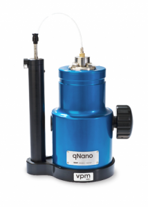
I’m a little unclear as to what qNano actually is. I did find this description on the qNano product page,

qNano uses unique nanopore-based detection to enable the physical properties of a wide range of particle types to be measured with unsurpassed accuracy.

Detailed Multi-Parameter Analysis.

Particle-by-particle measurement through qNano enables detailed determination of:

Nanopore-based detection allows thousands of particles to be measured individually, providing far greater detail and accuracy than light-based techniques.

Applications & Particle Types

A wide range of biological and synthetic particle types, spanning 50 nm – 10 μm, can be measured, across a broad range of research fields.

qNano Package

qNano is sold as a full system ready for use including the base instrument, variable pressure module, fluid cell and a starter kit of nanopores, buffer solution and standard particle sets.

Here’s what the product looks like,

qNano (from the Izon website)

As for what this all might mean to those of us who exist at the macroscale (from the Izon press release),

Izon Science will continue to work with Dr Platt at Loughborough, and with University College Dublin and various customers to develop a series of diagnostic kits that can be used with the qNano to identify and measure biomolecules, viruses, and microvesicles.“This is a real milestone for Izon’s technology as being able to measure biomolecules down to these extremely low levels opens up new bio-analysis options for researchers. 10 femtomolar was achieved, which is the equivalent dilution to 1 gram in 3.3 billion litres, or 1 gram in 1300 Olympic sized swimming pools,” says Hans van der Voorn, Executive Chairman of Izon Science.

For those interested in finding out about nanopores, these were mentioned in my July 18, 2012 posting while aptamers were discussed in my interview (Oct. 25, 2011 posting) with Dr. Maria DeRosa who researches them in her Carleton University laboratory (Ottawa, Canada).

Self-assembling, size-specific nanopores or nanotubes mimic nature

I guess you can call this biomimicry or biomimetics as it’s also known. From the  State University of New York at Buffalo  July 17, 2012 news releaseby Charlotte Hsu,

Inspired by nature, an international research team has created synthetic pores that mimic the activity of cellular ion channels, which play a vital role in human health by severely restricting the types of materials allowed to enter cells.

The pores the scientists built are permeable to potassium ions and water, but not to other ions such as sodium and lithium ions.

This kind of extreme selectivity, while prominent in nature, is unprecedented for a synthetic structure, said University at Buffalo chemistry professor Bing Gong, PhD, who led the study.

Here’s how they did it (from the news release),

To create the synthetic pores, the researchers developed a method to force donut-shaped molecules called rigid macrocycles to pile on top of one another. [emphasis mine] The scientists then stitched these stacks of molecules together using hydrogen bonding. The resulting structure was a nanotube with a pore less than a nanometer in diameter.

The July 17, 2012 media advisory by Tona Kunz from the Argonne National Laboratory (one of the partners in this research) describes why creating consistently sized nanopores/nanotubes has been so difficult and offers more information about the macrocycles,

Nanopores and their rolled up version, nanotubes, consist of atoms bonded to each other in a hexagonal pattern to create an array of nanometer-scale openings or channels. This structure creates a filter that can be sized to select which molecules and ions pass into drinking water or into a cell. The same filter technique can limit the release of chemical by-products from industrial processes.

Successes in making synthetic nanotubes from various materials have been reported previously, but their use has been limited because they degrade in water, the pore size of water-resistant carbon nanotubes is difficult to control, and, more critically, the inability to assemble them into appropriate filters.

An international team of researchers, with help of the Advanced Photon Source at Argonne National Laboratory, have succeeded in overcoming these hurdles by building self-assembling, size-specific nanopores. This new capability enables them to engineer nanotubes for specific functions and use pore size to selectively block specific molecules and ions.

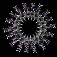
Scientists used groupings of atoms called ridged macrocycles that share a planar hexahenylene ethynylene core that bears six amide side chains. Through a cellular self-assembly process, the macrocycles stack cofacially, or atom on top of atom. Each layer of the macrocycle is held together by bonding among hydrogen atoms in the amide side chains. This alignment creates a uniform pore size regardless of the length of the nanotube. A slight misalignment of even a few macrocycles can alter the pore size and greatly compromise the nanotube’s functionality.

Here’s an image of the macrocycles supplied by the Agronne National Labortory,

A snapshot of a helical stack of macryocycles generated in the computer simulation.

The size specificity is  important if  nanopores/nanotubes are going to be used in medical applications,

The pore sizes can be adjusted to filter molecules and ions according to their size by changing the macroycle size, akin to the way a space can be put into a wedding ring to make it fit tighter. The channels are permeable to water, which aids in the fast transmission of intercellular information. The synthetic nanopores mimic the activity of cellular ion channels used in the human body. The research lays the foundation for an array of exciting new technology, such as new ways to deliver directly into cells proteins or medicines to fight diseases.

The research group’s paper has appeared in Nature Communications as of July 17, 2012, from Hsu’s news release,

The study’s lead authors are Xibin Zhou of Beijing Normal University; Guande Liu of Shanghai Jiao Tong University; Kazuhiro Yamato, postdoctoral scientist at UB; and Yi Shen of Shanghai Jiao Tong University and the Shanghai Institute of Applied Physics, Chinese Academy of Sciences. Other institutions that contributed to the work include the University of Nebraska-Lincoln and Argonne National Laboratory. Frank Bright, a SUNY Distinguished Professor of chemistry at UB, assisted with spectroscopic studies.