Tag Archives: nanoporous gold

Black gold: ultralight, high density nanoporous gold

South Korean researchers have found a way to fabricate a new kind of gold nanoparticle according to a March 28, 2016 news item on ScienceDaily,

A new material is more solid and 30 percent lighter than standard gold, scientists report. In their study, the team investigated grain boundaries in nanocrystalline np-Au and found a way to overcome the weakening mechanisms of this material, thereby suggesting its usefulness.

A March 28, 2016 Ulsan National Institute of Science and Technology (UNIST) press release (also on EurekAlert) by Chorok Oh, which originated the news item, provides more information,

A team of Korean research team, led by Professor Ju-Young Kim (School of Materials Science and Engineering) of Ulsan National Institute of Science and Technology (UNIST), South Korea has recently announced that they have successfully developed a way to fabricate an ultralight, high-dense nanoporous gold (np-Au).

In a new paper, published in Nano Letters on March 22, the team reported that this newly developed material, which they have dubbed “Black Gold” is twice more solid and 30% lighter than standard gold.

According to Prof. Kim, “This particular nanoporous gold has a 100,000 times wider surface when compared to standard gold. Moreover, due to its chemically stablity, it is also harmless to humans.”

The surfaces of np-Au are rough and the metal loses its shine and eventually turns black when they are at sizes less than 100 nanometres (nm). This is the reason that they are called “Black Gold”.

In their study, the team investigated grain boundaries in nanocrystalline np-Au and found a way to overcome the weakening mechanisms of this material, thereby suggesting its usefulness.

The team used a ball milling technique to increase the flexural strength of the three gold-silver precursor alloys. Then, using free corrosion dealloying of silver from gold-silver alloys, they were able to achieve the nanoporous surface. According to the team, “The size of the pores can be controlled by the temperature and concentration of nitrate.” Moreover, they also note that this crack-free nanoporous gold samples are reported to exhibit excellent durability in three-point bending tests.

Prof. Kim’s team notes, “Ball-milled np-Au has a much greater density of two-dimensional defects than annealed and prestrained np-Au, where intergranular fracture is preferred.” They continue, “Therefore, the probable existence of grain boundary opening in the highest tensile region is attributed to the flexural strength of np-Au.”

They suggest that this newly developed technique can be also applied to many other metal, as the np-Au produced by this technique have shown increased strength and durability while still maintaining the good qualities of standard gold.

This means that this technique can be also used in other technologies, like catalytic-converting as observed by platinum, the automobile catalyst and palladium, the hydrogen sensor catalyst.

Here’s a link to and a citation for the paper,

Weakened Flexural Strength of Nanocrystalline Nanoporous Gold by Grain Refinement by Eun-Ji Gwak and Ju-Young Kim. Nano Lett., Article ASAP DOI: 10.1021/acs.nanolett.6b00062 Publication Date (Web): March 16, 2016

Copyright © 2016 American Chemical Society

This paper is behind a paywall.
Sponges made of nanoporous gold and DNA detection

This work from the University of California at Davis seems to represent a step forward for better detection of diseases and pathogens. From a Sept. 4, 2015 news item on ScienceDaily,

Sponge-like nanoporous gold could be key to new devices to detect disease-causing agents in humans and plants, according to UC Davis researchers.

In two recent papers in Analytical Chemistry, a group from the UC Davis Department of Electrical and Computer Engineering demonstrated that they could detect nucleic acids using nanoporous gold, a novel sensor coating material, in mixtures of other biomolecules that would gum up most detectors. This method enables sensitive detection of DNA [deoxyribonucleic acid] in complex biological samples, such as serum from whole blood.

A Sept. 4, 2015 UC Davis news release on EurekAlert, which originated the news item, offers more detail,

“Nanoporous gold can be imagined as a porous metal sponge with pore sizes that are a thousand times smaller than the diameter of a human hair,” said Erkin Şeker, assistant professor of electrical and computer engineering at UC Davis and the senior author on the papers. “What happens is the debris in biological samples, such as proteins, is too large to go through those pores, but the fiber-like nucleic acids that we want to detect can actually fit through them. It’s almost like a natural sieve.”

Rapid and sensitive detection of nucleic acids plays a crucial role in early identification of pathogenic microbes and disease biomarkers. Current sensor approaches usually require nucleic acid purification that relies on multiple steps and specialized laboratory equipment, which limit the sensors’ use in the field. The researchers’ method reduces the need for purification.

“So now we hope to have largely eliminated the need for extensive sample clean-up, which makes the process conducive to use in the field,” Şeker said.

The result is a faster and more efficient process that can be applied in many settings.

The researchers hope the technology can be translated into the development of miniature point-of-care diagnostic platforms for agricultural and clinical applications.

“The applications of the sensor are quite broad ranging from detection of plant pathogens to disease biomarkers,” said Şeker.

For example, in agriculture, scientists could detect whether a certain pathogen exists on a plant without seeing any symptoms. And in sepsis cases in humans, doctors might determine bacterial contamination much more quickly than at present, preventing any unnecessary treatments.

Here are links to and citations for two recent published papers about this work,

Effect of Nanoporous Gold Thin Film Morphology on Electrochemical DNA Sensing by Pallavi Daggumati, Zimple Matharu, and Erkin Şeker. Anal. Chem., 2015, 87 (16), pp 8149–8156 DOI: 10.1021/acs.analchem.5b00846 Publication Date (Web): April 30, 2015

Copyright © 2015 American Chemical Society

Biofouling-Resilient Nanoporous Gold Electrodes for DNA Sensing by Pallavi Daggumati, Zimple Matharu, Ling Wang, and Erkin Şeker. Anal. Chem., 2015, 87 (17), pp 8618–8622 DOI: 10.1021/acs.analchem.5b02969 Publication Date (Web): August 14, 2015

Copyright © 2015 American Chemical Society

These papers are behind a paywall.

Gold and your neurons

Should you need any electrode implants for your neurons at some point in the future, it’s possible they could be coated with gold. Researchers at the Lawrence Livermore National Laboratory (LLNL) and at the University of California at Davis (UC Davis) have discovered that electrodes covered in nanoporous gold could prevent scarring (from a May 5, 2015 news item on Azonano),

A team of researchers from Lawrence Livermore and UC Davis have found that covering an implantable neural electrode with nanoporous gold could eliminate the risk of scar tissue forming over the electrode’s surface.

The team demonstrated that the nanostructure of nanoporous gold achieves close physical coupling of neurons by maintaining a high neuron-to-astrocyte surface coverage ratio. Close physical coupling between neurons and the electrode plays a crucial role in recording fidelity of neural electrical activity.

An April 30, 2015 LLNL news release, which originated the news item, details the scarring issue and offers more information about the proposed solution,

Neural interfaces (e.g., implantable electrodes or multiple-electrode arrays) have emerged as transformative tools to monitor and modify neural electrophysiology, both for fundamental studies of the nervous system, and to diagnose and treat neurological disorders. These interfaces require low electrical impedance to reduce background noise and close electrode-neuron coupling for enhanced recording fidelity.

Designing neural interfaces that maintain close physical coupling of neurons to an electrode surface remains a major challenge for both implantable and in vitro neural recording electrode arrays. An important obstacle in maintaining robust neuron-electrode coupling is the encapsulation of the electrode by scar tissue.

Typically, low-impedance nanostructured electrode coatings rely on chemical cues from pharmaceuticals or surface-immobilized peptides to suppress glial scar tissue formation over the electrode surface, which is an obstacle to reliable neuron−electrode coupling.

However, the team found that nanoporous gold, produced by an alloy corrosion process, is a promising candidate to reduce scar tissue formation on the electrode surface solely through topography by taking advantage of its tunable length scale.

“Our results show that nanoporous gold topography, not surface chemistry, reduces astrocyte surface coverage,” said Monika Biener, one of the LLNL authors of the paper.

Nanoporous gold has attracted significant interest for its use in electrochemical sensors, catalytic platforms, fundamental structure−property studies at the nanoscale and tunable drug release. It also features high effective surface area, tunable pore size, well-defined conjugate chemistry, high electrical conductivity and compatibility with traditional fabrication techniques.

“We found that nanoporous gold reduces scar coverage but also maintains high neuronal coverage in an in vitro neuron-glia co-culture model,” said Juergen Biener, the other LLNL author of the paper. “More broadly, the study demonstrates a novel surface for supporting neuronal cultures without the use of culture medium supplements to reduce scar overgrowth.”

Here’s a link to and a citation for the paper,

Nanoporous Gold as a Neural Interface Coating: Effects of Topography, Surface Chemistry, and Feature Size by Christopher A. R. Chapman, Hao Chen, Marianna Stamou, Juergen Biener, Monika M. Biener, Pamela J. Lein, and Erkin Seker. ACS Appl. Mater. Interfaces, 2015, 7 (13), pp 7093–7100 DOI: 10.1021/acsami.5b00410 Publication Date (Web): February 23, 2015

Copyright © 2015 American Chemical Society

This paper is behind a paywall.

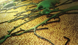
The researchers have provided this image to illustrate their work,

The image depicts a neuronal network growing on a novel nanotextured gold electrode coating. The topographical cues presented by the coating preferentially favor spreading of neurons as opposed to scar tissue. This feature has the potential to enhance the performance of neural interfaces. Image by Ryan Chen/LLNL.

The image depicts a neuronal network growing on a novel nanotextured gold electrode coating. The topographical cues presented by the coating preferentially favor spreading of neurons as opposed to scar tissue. This feature has the potential to enhance the performance of neural interfaces. Image by Ryan Chen/LLNL.