Now in its twelfth year, the international conference “Living Machines”, organised by Istituto Italiano di Tecnologia (Italian Institute of Technology, IIT), returns to Italy and comes to Genoa for the first time, from 10 to 13 July. Around one hundred experts from all over the world are expected, and they will present their achievements in the field of bio-inspired science and technology. The conference will take place in an exceptional venue, the Acquario di Genova (Genoa Aquarium), which, having reached its 30th birthday, is the ideal location at which to bring together various subject areas, from biology to artificial intelligence and robotics, with a focus on sustainability and environmental protection.
The scientific organiser of the event is Barbara Mazzolai, Associate Director for Robotics and head of the Bioinspired Soft Robotics Lab at IIT, along with Fabian Meder, researcher in the Bioinspired Soft Robotics Lab group and co-chair of the conference programme.
The conference will include two events open to the public: an exhibition area, which will be accessible from 11 to 13 July in the afternoon (from 2 to 4.30 pm); and a scientific café, which will take place on the 12 July at 5 pm. The conference will be an opportunity for international guests to appreciate the region’s beauty and talents, and it will also include the participation of students from the Niccolò Paganini Conservatory of Music. In addition, a satellite event of the conference will be the ISPA – Italian Sustainability Photo Award – exhibition, which will open at Palazzo Ducale on 10 July at 6 p.m.
The “Living Machines” conference is the landmark event for the international scientific community which bases its research on living organisms, such as human beings and other animal species – terrestrial, marine, and airborne – in addition to plants, fungi, and bacteria, in order to create so-called “living machines”, in other words, forms of technology capable of replicating their structure and mechanisms of operation.
“The conference is rooted in the union between robotics and neuroscience, using man and other animal species as a model for the study of intelligence and control systems,” said Barbara Mazzolai, Associate Director for Robotics at IIT. “This year the conference will focus on the role of biomimicry in the creation of robots that are more sustainable, with applications for the challenges of environmental protection and human health. Discussions will revolve around the development of robots with a lower energy impact, made using recyclable and biodegradable materials, and that can be used in emergency situations or extreme environments, such as deep sea, soil, space, or environmental disasters, but also for precision agriculture, environmental surveillance, infrastructure monitoring, human care and medical-surgical assistance.”
In the conference programme, experts will take part in a first day of parallel workshop and tutorial sessions (on 10 July), during which the topics of bioinspiration and biohybrid technology in the fields of medicine and the marine environment will be addressed. This first day will be followed by three days of plenary sessions, featuring talks by internationally-renowned scientists. More specifically: Oussama Khatib, one of the pioneers of robotics and director of the Robotics Laboratory at Stanford University; Marco Dorigo, professor at the Université Libre de Bruxelles and one of the pioneers of collective intelligence; Peter Fratzl, director of the Max Planck Institute of Colloids and Interfaces, working on research into osteoporosis and tissue regeneration; Eleni Stavrinidou, coordinator of the “Electronic Plants” group at Linköping University and an expert in bioelectronic and biohybrid systems; Olga Speck, Principal Researcher at the University of Freiburg, specialising in biomimetic materials and the regenerative capabilities of plants; and Kyu-Jin Cho, director of the Research Centre for Soft Robotics and the Biorobotics Laboratory at Seoul National University, one of the world’s leading experts on soft robotics.
For conference participants only, the programme includes: a visit to the Acquario, guided by the facility’s scientific staff, who will illustrate the work and practices needed for the protection and conservation of marine species and the undergoing research projects; an exhibition area for prototypes and products by research groups and companies operating in this field; and a dinner at Villa Lo Zerbino, with a musical contribution by students from the Niccolò Paganini Conservatory.
Open to the general public, on 12 July from 5 p.m. to 6 p.m. there will be a round table entitled “Living Machines: The Origin and the Future” chaired by science journalist Nicola Nosengo, Chief Editor of Nature Italy. Speakers will include Cecilia Laschi from the National University of Singapore, Vickie Webster-Wood from Carnegie Mellon University, Thomas Speck from the University of Freiburg and Paul Verschure from Radboud University Nijmegen.
A satellite initiative of the conference will be the exhibition for ISPA, the Italian Sustainability Photo Award, which will open at Palazzo Ducale on 10 July at 6.00 p.m. ISPA is the photographic award created by the Parallelozero agency in cooperation with the main sponsor PIMCO, to raise public awareness of environmental, social, and governance sustainability issues, encapsulated in the acronym ESG. The works of the winning photographers and finalists in the last three editions will be on display in Genoa: a selection of images that depict the emblematic stories of Italy, a nation moving towards a more sustainable future, a visual narrative that makes it easier to understand the country’s progress in research and innovation.
The organisations supporting the event include, in addition to the principal organiser Istituto Italiano di Tecnologia (Italian Institute of Technology), the international Convergent Science Network [emphasis mine], the Office of Naval Research, Radboud University Nijmegen, and the Living, Adaptive and Energy-autonomous Materials Systems Cluster of Excellence in Freiburg.
I was particularly struck by this quote, “The conference is rooted in the union between robotics and neuroscience [emphasis mine], using man and other animal species as a model for the study of intelligence and control systems,” from Barbara Mazzolai as I have an as yet unpublished post for a UNESCO neurotechnology event coming up on July 13, 2023. These events come on the heels of a May 16, 2023 Canadian Science Policy Centre panel discussion on responsible neurotechnology (see my May 12, 2023 posting).
The brilliant and often iridescent colours that we see in some species of birds, beetles and butterflies arise from a regular arrangement of nanostructures that scatter selective wavelengths of light more strongly to generate colour. These colours are called structural colours, which usually range from blues to greens, and even magenta. However, vibrant or saturated reds are elusive and notably absent from the structural colour range in both natural and synthetic realms.
To achieve highly saturated reds, the material needs to absorb light from all wavelengths shorter than ~600 nm and reflect the remaining longer wavelengths, doing both as completely as possible. This sharp transition from absorption to reflection was prescribed theoretically by none other than Erwin Schrödinger of quantum theory fame. However, the physics of resonators tell us that high-order optical resonances in blue will also occur as soon as we have a fundamental resonance in red. This combination of blue and red thus results in the magenta observed in nature. It is therefore challenging to achieve the Schrödinger’s red pixel, which would produce the most saturated red in the world. Current nanoantenna-based approaches are insufficient to simultaneously satisfy the above conditions.
Researchers from the Agency for Science, Technology and Research’s (A*STAR) Institute of Materials Research and Engineering (IMRE), National University of Singapore (NUS) and Singapore University of Technology and Design (SUTD) have collaborated to design and realise reds at the ultimate limit of saturation as predicted by theory, where the team worked together on conceptualisation methodology, fabrications, characterisations and simulations. This research was published in Science Advances on 23 February 2022.
The design consists of regularly arranged silicon nanoantennas in the shape of ellipses. These produce possibly the most saturated and brightest reds with ~80% reflectance, exceeding the reds in the standard red, green and blue gamut (sRGB) and other well-known red pigments, e.g. cadmium red … .
The nanoantennas support two partially overlapping quasi bound-states-in-the-continuum modes, where the optimal dimensions of the silicon nanoantenna arrays are derived by using a gradient descent algorithm to enable the antennas to achieve sharp spectral edges at red wavelengths. At the same time, high-order modes at blue or green wavelengths are suppressed via engineering the substrate‑induced diffraction channels and the absorption of amorphous silicon.
Potential uses for Schrödinger’s red include developing a polarisation dependent encryption method, with plans to scale up the Schrödinger’s red pixel for applications like functional nanofabrication devices such as optical spectrometers and reflective displays with high colour saturation.
“With this new design that can achieve the most saturated and brightest reds, we can exploit its sensitivity to polarisation and illumination angle on potential applications for information encryption. This proposed concept and design methodology could also be generalised to other Schrödinger’s colour pixels. The highly-saturated red achieved could be potentially scaled up through methods such as deep ultraviolet and nano-imprint lithography, to reach the dimensions of reflective displays based on multilayer film configuration, which could lead to potential applications like compact red filters, highly saturated reflective displays, nonlocal metasurfaces, and miniaturised spectrometers”, said Dr. Dong Zhaogang, Deputy Department Head of Nanofabrication at A*STAR’s IMRE.
“The creation of the record-high saturation and brightness in red opens up possibilities for a plethora of applications related to anti-counterfeiting technologies, high-calibre colour display and more, which were previously perceived as unachievable with structural colour. It showcases a wonderful synergy between conceptual breakthrough, powerful algorithm and advanced nanofabrication”, said Prof. Cheng-Wei Qiu, Dean’s Chair Professor at NUS.
“This work in structural colours goes to show that we can sometimes outdo evolution through clever use of the tools in nanofabrication and accurate optical simulations”, said Prof. Joel Yang, Provost Chair Professor and Associate Professor in Engineering Product Development at SUTD.
Here’s a link to and a citation for the paper,
Schrödinger’s red pixel by quasi-bound-states-in-the-continuum by Zhaogang Dong, Lei Jin, Soroosh Daqiqeh Rezaei, Hao Wang, Yang Chen, Febiana Tjiptoharsono, Jinfa Ho, Sergey Gorelik, Ray Jia Hong Ng, Qifeng Ruan, Cheng-Wei Qiu and Joel K. W. Yang. Science Advances Vol 8, Issue 8 DOI: 10.1126/sciadv.abm4512 Published 23 Feb 2022
This paper is open access.
Math error, colour theory, and perception
An August 10, 2022 news item on phys.org announced a math error made by Erwin Schrödinger and others,
A new study corrects an important error in the 3D mathematical space developed by the Nobel Prize-winning physicist Erwin Schrödinger and others, and used by scientists and industry for more than 100 years to describe how your eye distinguishes one color from another. The research has the potential to boost scientific data visualizations, improve TVs and recalibrate the textile and paint industries.
“The assumed shape of color space requires a paradigm shift,” said Roxana Bujack, a computer scientist with a background in mathematics who creates scientific visualizations at Los Alamos National Laboratory. Bujack is lead author of the paper by a Los Alamos team in the Proceedings of the National Academy of Sciences on the mathematics of color perception.
“Our research shows that the current mathematical model of how the eye perceives color differences is incorrect. That model was suggested by Bernhard Riemann and developed by Hermann von Helmholtz and Erwin Schrödinger—all giants in mathematics and physics—and proving one of them wrong is pretty much the dream of a scientist,” said Bujack.
…
While the Los Alamos National Laboratory work was published in April 2022 (online) and May 2022 (in print), their news announcement doesn’t seem to have been made until August. I can’t be certain but I believe this should have an impact on the work from A*STAR as that team’s paper cites: E. Schrödinger, Theorie der Pigmente von größter Leuchtkraft. Ann. Phys.367, 603–622 (1920).
Modeling human color perception enables automation of image processing, computer graphics and visualization tasks.
“Our original idea was to develop algorithms to automatically improve color maps for data visualization, to make them easier to understand and interpret,” Bujack said. So the team was surprised when they discovered they were the first to determine that the longstanding application of Riemannian geometry, which allows generalizing straight lines to curved surfaces, didn’t work.
To create industry standards, a precise mathematical model of perceived color space is needed. First attempts used Euclidean spaces—the familiar geometry taught in many high schools; more advanced models used Riemannian geometry. The models plot red, green and blue in the 3D space. Those are the colors registered most strongly by light-detecting cones on our retinas, and—not surprisingly—the colors that blend to create all the images on your RGB computer screen.
In the study, which blends psychology, biology and mathematics, Bujack and her colleagues discovered that using Riemannian geometry overestimates the perception of large color differences. That’s because people perceive a big difference in color to be less than the sum you would get if you added up small differences in color that lie between two widely separated shades.
Riemannian geometry cannot account for this effect.
“We didn’t expect this, and we don’t know the exact geometry of this new color space yet,” Bujack said. “We might be able to think of it normally but with an added dampening or weighing function that pulls long distances in, making them shorter. But we can’t prove it yet.”
Here’s a link to and a citation for the paper,
The non-Riemannian nature of perceptual color space by Roxana Bujack, Emily Teti, Jonah Miller, Elektra Caffrey, and Terece L. Turton. Proceedings of the National Academy of Sciences (PNAS) 119 (18) e2119753119 DOI: https://doi.org/10.1073/pnas.2119753119 Published: April 29, 2022
A September 1, 2021 news item on ScienceDaily announces a new type of memristor from Texas A&M University (Texas A&M or TAMU) and the National University of Singapore (NUS)
In a discovery published in the journal Nature, an international team of researchers has described a novel molecular device with exceptional computing prowess.
Reminiscent of the plasticity of connections in the human brain, the device can be reconfigured on the fly for different computational tasks by simply changing applied voltages. Furthermore, like nerve cells can store memories, the same device can also retain information for future retrieval and processing.
“The brain has the remarkable ability to change its wiring around by making and breaking connections between nerve cells. Achieving something comparable in a physical system has been extremely challenging,” said Dr. R. Stanley Williams [emphasis mine], professor in the Department of Electrical and Computer Engineering at Texas A&M University. “We have now created a molecular device with dramatic reconfigurability, which is achieved not by changing physical connections like in the brain, but by reprogramming its logic.”
Dr. T. Venkatesan, director of the Center for Quantum Research and Technology (CQRT) at the University of Oklahoma, Scientific Affiliate at National Institute of Standards and Technology, Gaithersburg, and adjunct professor of electrical and computer engineering at the National University of Singapore, added that their molecular device might in the future help design next-generation processing chips with enhanced computational power and speed, but consuming significantly reduced energy.
Whether it is the familiar laptop or a sophisticated supercomputer, digital technologies face a common nemesis, the von Neumann bottleneck. This delay in computational processing is a consequence of current computer architectures, wherein the memory, containing data and programs, is physically separated from the processor. As a result, computers spend a significant amount of time shuttling information between the two systems, causing the bottleneck. Also, despite extremely fast processor speeds, these units can be idling for extended amounts of time during periods of information exchange.
As an alternative to conventional electronic parts used for designing memory units and processors, devices called memristors offer a way to circumvent the von Neumann bottleneck. Memristors, such as those made of niobium dioxide and vanadium dioxide, transition from being an insulator to a conductor at a set temperature. This property gives these types of memristors the ability to perform computations and store data.
However, despite their many advantages, these metal oxide memristors are made of rare-earth elements and can operate only in restrictive temperature regimes. Hence, there has been an ongoing search for promising organic molecules that can perform a comparable memristive function, said Williams.
Dr. Sreebrata Goswami, a professor at the Indian Association for the Cultivation of Science, designed the material used in this work. The compound has a central metal atom (iron) bound to three phenyl azo pyridine organic molecules called ligands.
“This behaves like an electron sponge that can absorb as many as six electrons reversibly, resulting in seven different redox states,” said Sreebrata. “The interconnectivity between these states is the key behind the reconfigurability shown in this work.”
Dr. Sreetosh Goswami, a researcher at the National University of Singapore, devised this project by creating a tiny electrical circuit consisting of a 40-nanometer layer of molecular film sandwiched between a layer of gold on top and gold-infused nanodisc and indium tin oxide at the bottom.
On applying a negative voltage on the device, Sreetosh witnessed a current-voltage profile that was nothing like anyone had seen before. Unlike metal-oxide memristors that can switch from metal to insulator at only one fixed voltage, the organic molecular devices could switch back and forth from insulator to conductor at several discrete sequential voltages.
“So, if you think of the device as an on-off switch, as we were sweeping the voltage more negative, the device first switched from on to off, then off to on, then on to off and then back to on. I’ll say that we were just blown out of our seat,” said Venkatesan. “We had to convince ourselves that what we were seeing was real.”
Sreetosh and Sreebrata investigated the molecular mechanisms underlying the curious switching behavior using an imaging technique called Raman spectroscopy. In particular, they looked for spectral signatures in the vibrational motion of the organic molecule that could explain the multiple transitions. Their investigation revealed that sweeping the voltage negative triggered the ligands on the molecule to undergo a series of reduction, or electron-gaining, events that caused the molecule to transition between off state and on states.
Next, to describe the extremely complex current-voltage profile of the molecular device mathematically, Williams deviated from the conventional approach of basic physics-based equations. Instead, he described the behavior of the molecules using a decision tree algorithm with “if-then-else” statements, a commonplace line of code in several computer programs, particularly digital games.
“Video games have a structure where you have a character that does something, and then something occurs as a result. And so, if you write that out in a computer algorithm, they are if-then-else statements,” said Williams. “Here, the molecule is switching from on to off as a consequence of applied voltage, and that’s when I had the eureka moment to use decision trees to describe these devices, and it worked very well.”
But the researchers went a step further to exploit these molecular devices to run programs for different real-world computational tasks. Sreetosh showed experimentally that their devices could perform fairly complex computations in a single time step and then be reprogrammed to perform another task in the next instant.
“It was quite extraordinary; our device was doing something like what the brain does, but in a very different way,” said Sreetosh. “When you’re learning something new or when you’re deciding, the brain can actually reconfigure and change physical wiring around. Similarly, we can logically reprogram or reconfigure our devices by giving them a different voltage pulse then they’ve seen before.”
Venkatesan noted that it would take thousands of transistors to perform the same computational functions as one of their molecular devices with its different decision trees. Hence, he said their technology might first be used in handheld devices, like cell phones and sensors, and other applications where power is limited.
Other contributors to the research include Dr. Abhijeet Patra and Dr. Ariando from the National University of Singapore; Dr. Rajib Pramanick and Dr. Santi Prasad Rath from the Indian Association for the Cultivation of Science; Dr. Martin Foltin from Hewlett Packard Enterprise, Colorado; and Dr. Damien Thompson from the University of Limerick, Ireland.
Venkatesan said that this research is indicative of the future discoveries from this collaborative team, which will include the center of nanoscience and engineering at the Indian Institute of Science and the Microsystems and Nanotechnology Division at the NIST.
I’ve highlighted R. Stanley Williams because he and his team at HP [Hewlett Packard] Labs helped to kick off current memristor research in 2008 with the publication of two papers as per my April 5, 2010 posting,
In 2008, two memristor papers were published in Nature and Nature Nanotechnology, respectively. In the first (Nature, May 2008 [article still behind a paywall], a team at HP Labs claimed they had proved the existence of memristors (a fourth member of electrical engineering’s ‘Holy Trinity of the capacitor, resistor, and inductor’). In the second paper (Nature Nanotechnology, July 2008 [article still behind a paywall]) the team reported that they had achieved engineering control.
…
The novel memory device is based on a molecular system that can transition between on and off states at several discrete sequential voltages Courtesy: National University of Singapore
Many electronic devices today are dependent on semiconductor logic circuits based on switches hard-wired to perform predefined logic functions. Physicists from the National University of Singapore (NUS), together with an international team of researchers, have developed a novel molecular memristor, or an electronic memory device, that has exceptional memory reconfigurability.
Unlike hard-wired standard circuits, the molecular device can be reconfigured using voltage to embed different computational tasks. The energy-efficient new technology, which is capable of enhanced computational power and speed, can potentially be used in edge computing, as well as handheld devices and applications with limited power resource.
“This work is a significant breakthrough in our quest to design low-energy computing. The idea of using multiple switching in a single element draws inspiration from how the brain works and fundamentally reimagines the design strategy of a logic circuit,” said Associate Professor Ariando from the NUS Department of Physics who led the research.
The research was first published in the journal Nature on 1 September 2021, and carried out in collaboration with the Indian Association for the Cultivation of Science, Hewlett Packard Enterprise, the University of Limerick, the University of Oklahoma, and Texas A&M University.
Brain-inspired technology
“This new discovery can contribute to developments in edge computing as a sophisticated in-memory computing approach to overcome the von Neumann bottleneck, a delay in computational processing seen in many digital technologies due to the physical separation of memory storage from a device’s processor,” said Assoc Prof Ariando. The new molecular device also has the potential to contribute to designing next generation processing chips with enhanced computational power and speed.
“Similar to the flexibility and adaptability of connections in the human brain, our memory device can be reconfigured on the fly for different computational tasks by simply changing applied voltages. Furthermore, like how nerve cells can store memories, the same device can also retain information for future retrieval and processing,” said first author Dr Sreetosh Goswami, Research Fellow from the Department of Physics at NUS.
Research team member Dr Sreebrata Goswami, who was a Senior Research Scientist at NUS and previously Professor at the Indian Association for the Cultivation of Science, conceptualised and designed a molecular system belonging to the chemical family of phenyl azo pyridines that have a central metal atom bound to organic molecules called ligands. “These molecules are like electron sponges that can offer as many as six electron transfers resulting in five different molecular states. The interconnectivity between these states is the key behind the device’s reconfigurability,” explained Dr Sreebrata Goswami.
Dr Sreetosh Goswami created a tiny electrical circuit consisting a 40-nanometer layer of molecular film sandwiched between a top layer of gold, and a bottom layer of gold-infused nanodisc and indium tin oxide. He observed an unprecedented current-voltage profile upon applying a negative voltage to the device. Unlike conventional metal-oxide memristors that are switched on and off at only one fixed voltage, these organic molecular devices could switch between on-off states at several discrete sequential voltages.
Using an imaging technique called Raman spectroscopy, spectral signatures in the vibrational motion of the organic molecule were observed to explain the multiple transitions. Dr Sreebrata Goswami explained, “Sweeping the negative voltage triggered the ligands on the molecule to undergo a series of reduction, or electron-gaining which caused the molecule to transition between off and on states.”
The researchers described the behavior of the molecules using a decision tree algorithm with “if-then-else” statements, which is used in the coding of several computer programs, particularly digital games, as compared to the conventional approach of using basic physics-based equations.
New possibilities for energy-efficient devices
Building on their research, the team used the molecular memory devices to run programs for different real-world computational tasks. As a proof of concept, the team demonstrated that their technology could perform complex computations in a single step, and could be reprogrammed to perform another task in the next instant. An individual molecular memory device could perform the same computational functions as thousands of transistors, making the technology a more powerful and energy-efficient memory option.
“The technology might first be used in handheld devices, like cell phones and sensors, and other applications where power is limited,” added Assoc Prof Ariando.
The team in the midst of building new electronic devices incorporating their innovation, and working with collaborators to conduct simulation and benchmarking relating to existing technologies.
Other contributors to the research paper include Abhijeet Patra and Santi Prasad Rath from NUS, Rajib Pramanick from the Indian Association for the Cultivation of Science, Martin Foltin from Hewlett Packard Enterprise, Damien Thompson from the University of Limerick, T. Venkatesan from the University of Oklahoma, and R. Stanley Williams from Texas A&M University.
Here’s a link to and a citation for the paper,
Decision trees within a molecular memristor by Sreetosh Goswami, Rajib Pramanick, Abhijeet Patra, Santi Prasad Rath, Martin Foltin, A. Ariando, Damien Thompson, T. Venkatesan, Sreebrata Goswami & R. Stanley Williams. Nature volume 597, pages 51–56 (2021) DOI: https://doi.org/10.1038/s41586-021-03748-0 Published 01 September 2021 Issue Date 02 September 2021
Caption: This audio is oxytocin receptor protein music using the Fantasy Impromptu guided algorithm. Credit: Chen et al. / Heliyon
A September 29, 2021 news item on ScienceDaily describes new research into music as a means of communicating science,
In recent years, scientists have created music based on the structure of proteins as a creative way to better popularize science to the general public, but the resulting songs haven’t always been pleasant to the ear. In a study appearing September 29 [2021] in the journal Heliyon, researchers use the style of existing music genres to guide the structure of protein song to make it more musical. Using the style of Frédéric Chopin’s Fantaisie-Impromptu and other classical pieces as a guide, the researchers succeeded in converting proteins into song with greater musicality.
…
Scientists (Peng Zhang, Postdoctoral Researcher in Computational Biology at The Rockefeller University, and Yuzong Chen, Professor of Pharmacy at National University of Singapore [NUS]) wrote a September 29, 2021 essay for The Conversation about their protein songs (Note: Links have been removed),
…
There are many surprising analogies between proteins, the basic building blocks of life, and musical notation. These analogies can be used not only to help advance research, but also to make the complexity of proteins accessible to the public.
We’re computational biologists who believe that hearing the sound of life at the molecular level could help inspire people to learn more about biology and the computational sciences. While creating music based on proteins isn’t new, different musical styles and composition algorithms had yet to be explored. So we led a team of high school students and other scholars to figure out how to create classical music from proteins.
The musical analogies of proteins
Proteins are structured like folded chains. These chains are composed of small units of 20 possible amino acids, each labeled by a letter of the alphabet.
A protein chain can be represented as a string of these alphabetic letters, very much like a string of music notes in alphabetical notation.
Protein chains can also fold into wavy and curved patterns with ups, downs, turns and loops. Likewise, music consists of sound waves of higher and lower pitches, with changing tempos and repeating motifs.
Protein-to-music algorithms can thus map the structural and physiochemical features of a string of amino acids onto the musical features of a string of notes.
Enhancing the musicality of protein mapping
Protein-to-music mapping can be fine-tuned by basing it on the features of a specific music style. This enhances musicality, or the melodiousness of the song, when converting amino acid properties, such as sequence patterns and variations, into analogous musical properties, like pitch, note lengths and chords.
For our study, we specifically selected 19th-century Romantic period classical piano music, which includes composers like Chopin and Schubert, as a guide because it typically spans a wide range of notes with more complex features such as chromaticism, like playing both white and black keys on a piano in order of pitch, and chords. Music from this period also tends to have lighter and more graceful and emotive melodies. Songs are usually homophonic, meaning they follow a central melody with accompaniment. These features allowed us to test out a greater range of notes in our protein-to-music mapping algorithm. In this case, we chose to analyze features of Chopin’s “Fantaisie-Impromptu” to guide our development of the program.
If you have the time, I recommend reading the essay in its entirety and listening to the embedded audio files.
In recent years, scientists have created music based on the structure of proteins as a creative way to better popularize science to the general public, but the resulting songs haven’t always been pleasant to the ear. In a study appearing September 29 [2021] in the journal Heliyon, researchers use the style of existing music genres to guide the structure of protein song to make it more musical. Using the style of Frédéric Chopin’s Fantaisie-Impromptu and other classical pieces as a guide, the researchers succeeded in converting proteins into song with greater musicality.
Creating unique melodies from proteins is achieved by using a protein-to-music algorithm. This algorithm incorporates specific elements of proteins—like the size and position of amino acids—and maps them to various musical elements to create an auditory “blueprint” of the proteins’ structure.
“Existing protein music has mostly been designed by simple mapping of certain amino acid patterns to fundamental musical features such as pitches and note lengths, but they do not map well to more complex musical features such as rhythm and harmony,” says senior author Yu Zong Chen, a professor in the Department of Pharmacy at National University of Singapore. “By focusing on a music style, we can guide more complex mappings of combinations of amino acid patterns with various musical features.”
For their experiment, researchers analyzed the pitch, length, octaves, chords, dynamics, and main theme of four pieces from the mid-1800s Romantic era of classical music. These pieces, including Fantasie-Impromptu from Chopin and Wanderer Fantasy from Franz Schubert, were selected to represent the notable Fantasy-Impromptu genre that emerged during that time.
“We chose the specific music style of a Fantasy-Impromptu as it is characterized by freedom of expression, which we felt would complement how proteins regulate much of our bodily functions, including our moods,” says co-author Peng Zhang (@zhangpeng1202), a post-doctoral fellow at the Rockefeller University
Likewise, several of the proteins in the study were chosen for their similarities to the key attributes of the Fantasy-Impromptu style. Most of the 18 proteins tested regulate functions including human emotion, cognition, sensation, or performance which the authors say connect to the emotional and expressive of the genre.
Then, they mapped 104 structural, physicochemical, and binding amino acid properties of those proteins to the six musical features. “We screened the quantitative profile of each amino acid property against the quantized values of the different musical features to find the optimal mapped pairings. For example, we mapped the size of amino acid to note length, so that having a larger amino acid size corresponds to a shorter note length,” says Chen.
Across all the proteins tested, the researchers found that the musicality of the proteins was significantly improved. In particular, the protein receptor for oxytocin (OXTR) was judged to have one of the greatest increases in musicality when using the genre-guided algorithm, compared to an earlier version of the protein-to-music algorithm.
“The oxytocin receptor protein generated our favorite song,” says Zhang. “This protein sequence produced an identifiable main theme that repeats in rhythm throughout the piece, as well as some interesting motifs and patterns that recur independent of our algorithm. There were also some pleasant harmonic progressions; for example, many of the seventh chords naturally resolve.”
The authors do note, however, that while the guided algorithm increased the overall musicality of the protein songs, there is still much progress to be made before it resembles true human music.
“We believe a next step is to explore more music styles and more complex combinations of amino acid properties for enhanced musicality and novel music pieces. Another next step, a very important step, is to apply artificial intelligence to jointly learn complex amino acid properties and their combinations with respect to the features of various music styles for creating protein music of enhanced musicality,” says Chen.
###
Research supported by the National Key R&D Program of China, the National Natural Science Foundation of China, and Singapore Academic Funds.
What I find most exciting about this conference is the range of countries being represented. At first glance, I’ve found Argentina, Thailand, Senegal, Ivory Coast, Costa Rica and more in a science meeting being held in Canada. Thank you to the organizers and to the organization International Network for Government Science Advice (INGSA)
As I’ve noted many times here in discussing the science advice we (Canadians) get through the Council of Canadian Academies (CCA), there’s far too much dependence on the same old, same old countries for international expertise. Let’s hope this meeting changes things.
The conference (with the theme Build Back Wiser: Knowledge, Policy and Publics in Dialogue) started on Monday, August 30, 2021 and is set to run for four days in Montréal, Québec. and as an online event The Premier of Québec, François Legault, and Mayor of Montréal, Valérie Plante (along with Peter Gluckman, Chair of INGSA and Rémi Quirion, Chief Scientist of Québec; this is the only province with a chief scientist) are there to welcome those who are present in person.
You can find a PDF of the four day programme here or go to the INGSA 2021 website for the programme and more. Here’s a sample from the programme of what excited me, from Day 1 (August 30, 2021),
8:45 | Plenary | Roundtable: Reflections from Covid-19: Where to from here?
Moderator: Mona Nemer – Chief Science Advisor of Canada
Speakers: Joanne Liu – Professor, School of Population and Global Health, McGill University, Quebec, Canada Chor Pharn Lee – Principal Foresight Strategist at Centre for Strategic Futures, Prime Minister’s Office, Singapore Andrea Ammon – Director of the European Centre for Disease Prevention and Control, Sweden Rafael Radi – President of the National Academy of Sciences; Coordinator of Scientific Honorary Advisory Group to the President on Covid-19, Uruguay
9:45 | Panel: Science advice during COVID-19: What factors made the difference?
Moderator:
Romain Murenzi – Executive Director, The World Academy of Sciences (TWAS), Italy
Speakers:
Stephen Quest – Director-General, European Commission’s Joint Research Centre (JRC), Belgium Yuxi Zhang – Postdoctoral Research Fellow, Blavatnik School of Government, University of Oxford, United Kingdom Amadou Sall – Director, Pasteur Institute of Dakar, Senegal Inaya Rakhmani – Director, Asia Research Centre, Universitas Indonesia
One last excerpt, from Day 2 (August 31, 2021),
Studio Session | Panel: Science advice for complex risk assessment: dealing with complex, new, and interacting threats
Moderator: Eeva Hellström – Senior Lead, Strategy and Foresight, Finnish Innovation Fund Sitra, Finland
Speakers: Albert van Jaarsveld – Director General and Chief Executive Officer, International Institute for Applied Systems Analysis, Austria Abdoulaye Gounou – Head, Benin’s Office for the Evaluation of Public Policies and Analysis of Government Action Catherine Mei Ling Wong – Sociologist, LRF Institute for the Public Understanding of Risk, National University of Singapore Andria Grosvenor – Deputy Executive Director (Ag), Caribbean Disaster Emergency Management Agency, Barbados
…
Studio Session | Innovations in Science Advice – Science Diplomacy driving evidence for policymaking
Moderator: Mehrdad Hariri – CEO and President of the Canadian Science Policy Centre, Canada
Speakers: Primal Silva – Canadian Food Inspection Agency’s Chief Science Operating Officer, Canada Zakri bin Abdul Hamid – Chair of the South-East Asia Science Advice Network (SEA SAN); Pro-Chancellor of Multimedia University in Malaysia Christian Arnault Emini – Senior Economic Adviser to the Prime Minister’s Office in Cameroon Florence Gauzy Krieger and Sebastian Goers – RLS-Sciences Network [See more about RLS-Sciences below] Elke Dall and Angela Schindler-Daniels – European Union Science Diplomacy Alliance Alexis Roig – CEO, SciTech DiploHub – Barcelona Science and Technology Diplomacy Hub, Spain
RLS-Sciences works under the framework of the Regional Leaders Summit. The Regional Leaders Summit (RLS) is a forum comprising seven regional governments (state, federal state, or provincial), which together represent approximately one hundred eighty million people across five continents, and a collective GDP of three trillion USD. The regions are: Bavaria (Germany), Georgia (USA), Québec (Canada), São Paulo (Brazil), Shandong (China), Upper Austria (Austria), and Western Cape (South Africa). Since 2002, the heads of government for these regions have met every two years for a political summit. These summits offer the RLS regions an opportunity for political dialogue.
Getting back to the main topic of this post, INGSA has some satellite events on offer, including this on Open Science,
Open Science: Science for the 21st century |
Science ouverte : la science au XXIe siècle
Thursday September 9, 2021; 11am-2pm EST | Jeudi 9 septembre 2021, 11 h à 14 h (HNE).
This event will be in English and French (using simultaneous translation) | Cet événement se déroulera en anglais et en français (traduction simultanée)
In the past 18 months we have seen an unprecedented level of sharing as medical scientists worked collaboratively and shared data to find solutions to the COVID-19 pandemic. The pandemic has accelerated the ongoing cultural shift in research practices towards open science.
This acceleration of the discovery/research process presents opportunities for institutions and governments to develop infrastructure, tools, funding, policies, and training to support, promote, and reward open science efforts. It also presents new opportunities to accelerate progress towards the UN Agenda 2030 Sustainable Development Goals through international scientific cooperation.
At the same time, it presents new challenges: rapid developments in open science often outpace national open science policies, funding, and infrastructure frameworks. Moreover, the development of international standard setting instruments, such as the future UNESCO Recommendation on Open Science, requires international harmonization of national policies, the establishment of frameworks to ensure equitable participation, and education, training, and professional development.
This 3-hour satellite event brings together international and national policy makers, funders, and experts in open science infrastructure to discuss these issues.
…
The outcome of the satellite event will be a summary report with recommendations for open science policy alignment at institutional, national, and international levels.
The event will be hosted on an events platform, with simultaneous interpretation in English and French. Participants will be able to choose which concurrent session they participate in upon registration. Registration is free but will be closed when capacity is reached.
This satellite event takes place in time for an interesting anniversary. The Montreal Neurological Institute (MNI), also known as Montreal Neuro, declared itself as Open Science in 2016, the first academic research institute (as far as we know) to do so in the world (see my January 22, 2016 posting for details about their open science initiative and my December 19, 2016 posting for more about their open science and their decision to not pursue patents for a five year period).
There’s a lot of arsenic in the world and it’s often a factor in making water undrinkable. When that water is used in farming It also pollutes soil and enters food-producing plants. A December 11, 2020 news item on Nanowerk announces research into arsenic detectors in plants,
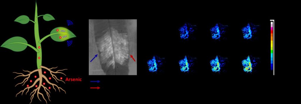
Researchers have developed a living plant-based sensor that can in real-time detect and monitor levels of arsenic, a highly toxic heavy metal, in the soil. Arsenic pollution is a major threat to humans and ecosystems in many Asia Pacific countries.
…
Caption: Non-destructive plant nanobionic sensor embedded within leaves to report arsenic levels within plants to portable electronics, enabling real-time monitoring of arsenic uptake in living plants. Credit: Dr. Tedrick Thomas Salim Lew
Scientists from the Disruptive and Sustainable Technologies for Agricultural Precision (DiSTAP) research group at the Singapore-MIT Alliance for Research and Technology (SMART), MIT’s research enterprise in Singapore, have engineered a novel type of plant nanobionic optical sensor that can detect and monitor, in real time, levels of the highly toxic heavy metal arsenic in the underground environment. This development provides significant advantages over conventional methods used to measure arsenic in the environment and will be important for both environmental monitoring and agricultural applications to safeguard food safety, as arsenic is a contaminant in many common agricultural products such as rice, vegetables, and tea leaves.
…
Arsenic and its compounds are a serious threat to humans and ecosystems. Long-term exposure to arsenic in humans can cause a wide range of detrimental health effects, including cardiovascular disease such as heart attack, diabetes, birth defects, severe skin lesions, and numerous cancers including those of the skin, bladder, and lung. Elevated levels of soil arsenic as a result of anthropogenic activities such as mining and smelting are also harmful to plants, inhibiting growth and resulting in substantial crop losses.
Food crops can absorb arsenic from the soil, leading to contamination of food and produce consumed by humans. Arsenic in underground environments can also contaminate groundwater and other underground water sources, the long-term consumption of which can cause severe health issues. As such, developing accurate, effective, and easy-to-deploy arsenic sensors is important to protect both the agriculture industry and wider environmental safety.
The novel optical nanosensors exhibit changes in their fluorescence intensity upon detecting arsenic. Embedded in plant tissues, with no detrimental effects on the plant, these sensors provide a nondestructive way to monitor the internal dynamics of arsenic taken up by plants from the soil. This integration of optical nanosensors within living plants enables the conversion of plants into self-powered detectors of arsenic from their natural environment, marking a significant upgrade from the time- and equipment-intensive arsenic sampling methods of current conventional methods.
“Our plant-based nanosensor is notable not only for being the first of its kind, but also for the significant advantages it confers over conventional methods of measuring arsenic levels in the below-ground environment, requiring less time, equipment, and manpower,” says Lew. “We envision that this innovation will eventually see wide use in the agriculture industry and beyond. I am grateful to SMART DiSTAP and the Temasek Life Sciences Laboratory (TLL), both of which were instrumental in idea generation and scientific discussion as well as research funding for this work.”
Besides detecting arsenic in rice and spinach, the team also used a species of fern, Pteris cretica, which can hyperaccumulate arsenic. This fern species can absorb and tolerate high levels of arsenic with no detrimental effect — engineering an ultrasensitive plant-based arsenic detector, capable of detecting very low concentrations of arsenic, as low as 0.2 parts per billion. In contrast, the regulatory limit for arsenic detectors is 10 parts per billion. Notably, the novel nanosensors can also be integrated into other species of plants. The researchers say this is the first successful demonstration of living plant-based sensors for arsenic and represents a groundbreaking advancement that could prove highly useful in both agricultural research (e.g., to monitor arsenic taken up by edible crops for food safety) and general environmental monitoring.
Previously, conventional methods of measuring arsenic levels included regular field sampling, plant tissue digestion, extraction, and analysis using mass spectrometry. These methods are time-consuming, require extensive sample treatment, and often involve the use of bulky and expensive instrumentation. The new approach couples nanoparticle sensors with plants’ natural ability to efficiently extract analytes via the roots and transport them. This allows for the detection of arsenic uptake in living plants in real time, with portable, inexpensive electronics such as a portable Raspberry Pi platform equipped with a charge-coupled device camera akin to a smartphone camera.
Co-author, DiSTAP co-lead principal investigator, and MIT Professor Michael Strano adds, “This is a hugely exciting development, as, for the first time, we have developed a nanobionic sensor that can detect arsenic — a serious environmental contaminant and potential public health threat. With its myriad advantages over older methods of arsenic detection, this novel sensor could be a game-changer, as it is not only more time-efficient, but also more accurate and easier to deploy than older methods. It will also help plant scientists in organizations such as TLL to further produce crops that resist uptake of toxic elements. Inspired by TLL’s recent efforts to create rice crops which take up less arsenic, this work is a parallel effort to further support SMART DiSTAP’s efforts in food security research, constantly innovating and developing new technological capabilities to improve Singapore’s food quality and safety.”
The research is carried out by SMART and supported by the National Research Foundation (NRF) Singapore under its Campus for Research Excellence And Technological Enterprise (CREATE) program.
Led by MIT’s Strano and Singapore co-lead principal investigator Professor Chua Nam Hai, DiSTAP is one of the five Interdisciplinary Research Groups (IRGs) in SMART. The DiSTAP program addresses deep problems in food production in Singapore and the world by developing a suite of impactful and novel analytical genetic and biosynthetic technologies. The goal is to fundamentally change how plant biosynthetic pathways are discovered, monitored, engineered, and ultimately translated to meet the global demand for food and nutrients. Scientists from MIT, TTL, Nanyang Technological University, and National University of Singapore are collaboratively developing new tools for the continuous measurement of important plant metabolites and hormones for novel discovery, deeper understanding and control of plant biosynthetic pathways in ways not yet possible, especially in the context of green leafy vegetables; leveraging these new techniques to engineer plants with highly desirable properties for global food security, including high yield density production, drought and pathogen resistance and biosynthesis of high-value commercial products; developing tools for producing hydrophobic food components in industry-relevant microbes; developing novel microbial and enzymatic technologies to produce volatile organic compounds that can protect and/or promote growth of leafy vegetables; and applying these technologies to improve urban farming.
…
Here’s a link to and a citation for the paper,
Plant Nanobionic Sensors for Arsenic Detection by Tedrick Thomas Salim Lew, Minkyung Park, Jianqiao Cui, Michael S. Strano. Advanced Materials DOI: https://doi.org/10.1002/adma.202005683 First published: 26 November 2020
This news comes from the National University of Singapore’s Centre for Quantum Technologies according to a May 4, 2020 news item on Nanowerk (Note: A link has been removed),
Here’s a new chapter in the story of the miniaturisation of machines: researchers in a laboratory in Singapore have shown that a single atom can function as either an engine or a fridge. Such a device could be engineered into future computers and fuel cells to control energy flows.
“Think about how your computer or laptop has a lot of things inside it that heat up. Today you cool that with a fan that blows air. In nanomachines or quantum computers, small devices that do cooling could be something useful,” says Dario Poletti from the Singapore University of Technology and Design (SUTD).
This work gives new insight into the mechanics of such devices. The work is a collaboration involving researchers at the Centre for Quantum Technologies (CQT) and Department of Physics at the National University of Singapore (NUS), SUTD and at the University of Augsburg in Germany. The results were published in the peer-reviewed journal npj Quantum Information (“Single-atom energy-conversion device with a quantum load”).
…
The researchers have included an exceptionally pretty illustration with the press release,
Caption: Experiments with a single-atom device help researchers understand what quantum effects come into play when machinery shrinks to the atomic scale. Credit: Aki Honda / Centre for Quantum Technologies, National University of Singapore
Engines and refrigerators are both machines described by thermodynamics, a branch of science that tells us how energy moves within a system and how we can extract useful work. A classical engine turns energy into useful work. A refrigerator does work to transfer heat, reducing the local temperature. They are, in some sense, opposites.
People have made small heat engines before using a single atom, a single molecule and defects in diamond. A key difference about this device is that it shows quantumness in its action. “We want to understand how we can build thermodynamic devices with just a few atoms. The physics is not well understood so our work is important to know what is possible,” says Manas Mukherjee, a Principal Investigator at CQT, NUS, who led the experimental work.
The researchers studied the thermodynamics of a single barium atom. They devised a scheme in which lasers move one of the atom’s electrons between two energy levels as part of a cycle, causing some energy to be pushed into the atom’s vibrations. Like a car engine consumes petrol to both move pistons and charge up its battery, the atom uses energy from lasers as fuel to increase its vibrating motion. The atom’s vibrations act like a battery, storing energy that can be extracted later. Rearrange the cycle and the atom acts like a fridge, removing energy from the vibrations.
In either mode of operation, quantum effects show up in correlations between the atom’s electronic states and vibrations. “At this scale, the energy transfer between the engine and the load is a bit fuzzy. It is no longer possible to simply do work on the load, you are bound to transfer some heat,” says Poletti. He worked out the theory with collaborators Jiangbin Gong at NUS Physics and Peter Hänggi in Augsburg. The fuzziness makes the process less efficient, but the experimentalists could still make it work.
Mukherjee and colleagues Noah Van Horne, Dahyun Yum and Tarun Dutta used a barium atom from which an electron (a negative charge) is removed. This makes the atom positively charged, so it can be more easily held still inside a metal chamber by electrical fields. All other air is removed from around it. The atom is then zapped with lasers to move it through a four-stage cycle.
The researchers measured the atom’s vibration after applying 2 to 15 cycles. They repeated a given number of cycles up to 150 times, measuring on average how much vibrational energy was present at the end. They could see the vibrational energy increasing when the atom was zapped with an engine cycle, and decreasing when the zaps followed the fridge cycle.
Understanding the atom-sized machine involved both complicated calculations and observations. The team needed to track two thermodynamic quantities known as ergotropy, which is the energy that can be converted to useful work, and entropy, which is related to disorder in the system. Both ergotropy and entropy increase as the atom-machine runs. There’s still a simple way of looking at it, says first author and PhD student Van Horne, “Loosely speaking, we’ve designed a little machine that creates entropy as it is filled up with free energy, much like kids when they are given too much sugar.”
Here’s a link to and a citation for the paper,
Single-atom energy-conversion device with a quantum load by Noah Van Horne, Dahyun Yum, Tarun Dutta, Peter Hänggi, Jiangbin Gong, Dario Poletti & Manas Mukherjee. npj Quantum Information volume 6, Article number: 37 (2020) Published: 01 May 2020
I have briefly speculated about the importance of touch elsewhere (see my July 19, 2019 posting regarding BlocKit and blockchain; scroll down about 50% of the way) but this upcoming news bit and the one following it put a different spin on the importance of touch.
Robots and prosthetic devices may soon have a sense of touch equivalent to, or better than, the human skin with the Asynchronous Coded Electronic Skin (ACES), an artificial nervous system developed by a team of researchers at the National University of Singapore (NUS).
The new electronic skin system achieved ultra-high responsiveness and robustness to damage, and can be paired with any kind of sensor skin layers to function effectively as an electronic skin.
The innovation, achieved by Assistant Professor Benjamin Tee and his team from the Department of Materials Science and Engineering at the NUS Faculty of Engineering, was first reported in prestigious scientific journal Science Robotics on 18 July 2019.
Faster than the human sensory nervous system
“Humans use our sense of touch to accomplish almost every daily task, such as picking up a cup of coffee or making a handshake. Without it, we will even lose our sense of balance when walking. Similarly, robots need to have a sense of touch in order to interact better with humans, but robots today still cannot feel objects very well,” explained Asst Prof Tee, who has been working on electronic skin technologies for over a decade in hope of giving robots and prosthetic devices a better sense of touch.
Drawing inspiration from the human sensory nervous system, the NUS team spent a year and a half developing a sensor system that could potentially perform better. While the ACES electronic nervous system detects signals like the human sensor nervous system, it is made up of a network of sensors connected via a single electrical conductor, unlike the nerve bundles in the human skin. It is also unlike existing electronic skins which have interlinked wiring systems that can make them sensitive to damage and difficult to scale up.
Elaborating on the inspiration, Asst Prof Tee, who also holds appointments in the NUS Department of Electrical and Computer Engineering, NUS Institute for Health Innovation & Technology (iHealthTech), N.1 Institute for Health and the Hybrid Integrated Flexible Electronic Systems (HiFES) programme, said, “The human sensory nervous system is extremely efficient, and it works all the time to the extent that we often take it for granted. It is also very robust to damage. Our sense of touch, for example, does not get affected when we suffer a cut. If we can mimic how our biological system works and make it even better, we can bring about tremendous advancements in the field of robotics where electronic skins are predominantly applied.”
ACES can detect touches more than 1,000 times faster than the human sensory nervous system. For example, it is capable of differentiating physical contacts between different sensors in less than 60 nanoseconds – the fastest ever achieved for an electronic skin technology – even with large numbers of sensors. ACES-enabled skin can also accurately identify the shape, texture and hardness of objects within 10 milliseconds, ten times faster than the blinking of an eye. This is enabled by the high fidelity and capture speed of the ACES system.
The ACES platform can also be designed to achieve high robustness to physical damage, an important property for electronic skins because they come into the frequent physical contact with the environment. Unlike the current system used to interconnect sensors in existing electronic skins, all the sensors in ACES can be connected to a common electrical conductor with each sensor operating independently. This allows ACES-enabled electronic skins to continue functioning as long as there is one connection between the sensor and the conductor, making them less vulnerable to damage.
Smart electronic skins for robots and prosthetics
ACES’ simple wiring system and remarkable responsiveness even with increasing numbers of sensors are key characteristics that will facilitate the scale-up of intelligent electronic skins for Artificial Intelligence (AI) applications in robots, prosthetic devices and other human machine interfaces.
“Scalability is a critical consideration as big pieces of high performing electronic skins are required to cover the relatively large surface areas of robots and prosthetic devices,” explained Asst Prof Tee. “ACES can be easily paired with any kind of sensor skin layers, for example, those designed to sense temperatures and humidity, to create high performance ACES-enabled electronic skin with an exceptional sense of touch that can be used for a wide range of purposes,” he added.
For instance, pairing ACES with the transparent, self-healing and water-resistant sensor skin layer also recently developed by Asst Prof Tee’s team, creates an electronic skin that can self-repair, like the human skin. This type of electronic skin can be used to develop more realistic prosthetic limbs that will help disabled individuals restore their sense of touch.
Other potential applications include developing more intelligent robots that can perform disaster recovery tasks or take over mundane operations such as packing of items in warehouses. The NUS team is therefore looking to further apply the ACES platform on advanced robots and prosthetic devices in the next phase of their research.
For those who like videos, the researchers have prepared this,
Here’s a link to and a citation for the paper,
A neuro-inspired artificial peripheral nervous system for scalable electronic skins by Wang Wei Lee, Yu Jun Tan, Haicheng Yao, Si Li, Hian Hian See, Matthew Hon, Kian Ann Ng, Betty Xiong, John S. Ho and Benjamin C. K. Tee. Science Robotics Vol 4, Issue 32 31 July 2019 eaax2198 DOI: 10.1126/scirobotics.aax2198 Published online first: 17 Jul 2019:
This paper is behind a paywall.
Picking up a grape and holding his wife’s hand
This story comes from the Canadian Broadcasting Corporation (CBC) Radio with a six minute story embedded in the text, from a July 25, 2019 CBC Radio ‘As It Happens’ article by Sheena Goodyear,
…
The West Valley City, Utah, real estate agent [Keven Walgamott] lost his left hand in an electrical accident 17 years ago. Since then, he’s tried out a few different prosthetic limbs, but always found them too clunky and uncomfortable.
Then he decided to work with the University of Utah in 2016 to test out new prosthetic technology that mimics the sensation of human touch, allowing Walgamott to perform delicate tasks with precision — including shaking his wife’s hand.
“I extended my left hand, she came and extended hers, and we were able to feel each other with the left hand for the first time in 13 years, and it was just a marvellous and wonderful experience,” Walgamott told As It Happens guest host Megan Williams.
Walgamott, one of seven participants in the University of Utah study, was able to use an advanced prosthetic hand called the LUKE Arm to pick up an egg without cracking it, pluck a single grape from a bunch, hammer a nail, take a ring on and off his finger, fit a pillowcase over a pillow and more.
…
While performing the tasks, Walgamott was able to actually feel the items he was holding and correctly gauge the amount of pressure he needed to exert — mimicking a process the human brain does automatically.
“I was able to feel something in each of my fingers,” he said. “What I feel, I guess the easiest way to explain it, is little electrical shocks.”
Those shocks — which he describes as a kind of a tingling sensation — intensify as he tightens his grip.
“Different variations of the intensity of the electricity as I move my fingers around and as I touch things,” he said.
…
To make that [sense of touch] happen, the researchers implanted electrodes into the nerves on Walgamott’s forearm, allowing his brain to communicate with his prosthetic through a computer outside his body. That means he can move the hand just by thinking about it.
But those signals also work in reverse.
The team attached sensors to the hand of a LUKE Arm. Those sensors detect touch and positioning, and send that information to the electrodes so it can be interpreted by the brain.
…
For Walgamott, performing a series of menial tasks as a team of scientists recorded his progress was “fun to do.”
“I’d forgotten how well two hands work,” he said. “That was pretty cool.”
But it was also a huge relief from the phantom limb pain he has experienced since the accident, which he describes as a “burning sensation” in the place where his hand used to be.
Keven Walgamott had a good “feeling” about picking up the egg without crushing it.
What seems simple for nearly everyone else can be more of a Herculean task for Walgamott, who lost his left hand and part of his arm in an electrical accident 17 years ago. But he was testing out the prototype of a high-tech prosthetic arm with fingers that not only can move, they can move with his thoughts. And thanks to a biomedical engineering team at the University of Utah, he “felt” the egg well enough so his brain could tell the prosthetic hand not to squeeze too hard.
That’s because the team, led by U biomedical engineering associate professor Gregory Clark, has developed a way for the “LUKE Arm” (so named after the robotic hand that Luke Skywalker got in “The Empire Strikes Back”) to mimic the way a human hand feels objects by sending the appropriate signals to the brain. Their findings were published in a new paper co-authored by U biomedical engineering doctoral student Jacob George, former doctoral student David Kluger, Clark and other colleagues in the latest edition of the journal Science Robotics. A copy of the paper may be obtained by emailing robopak@aaas.org.
“We changed the way we are sending that information to the brain so that it matches the human body. And by matching the human body, we were able to see improved benefits,” George says. “We’re making more biologically realistic signals.”
That means an amputee wearing the prosthetic arm can sense the touch of something soft or hard, understand better how to pick it up and perform delicate tasks that would otherwise be impossible with a standard prosthetic with metal hooks or claws for hands.
“It almost put me to tears,” Walgamott says about using the LUKE Arm for the first time during clinical tests in 2017. “It was really amazing. I never thought I would be able to feel in that hand again.”
Walgamott, a real estate agent from West Valley City, Utah, and one of seven test subjects at the U, was able to pluck grapes without crushing them, pick up an egg without cracking it and hold his wife’s hand with a sensation in the fingers similar to that of an able-bodied person.
“One of the first things he wanted to do was put on his wedding ring. That’s hard to do with one hand,” says Clark. “It was very moving.”
Those things are accomplished through a complex series of mathematical calculations and modeling.
The LUKE Arm
The LUKE Arm has been in development for some 15 years. The arm itself is made of mostly metal motors and parts with a clear silicon “skin” over the hand. It is powered by an external battery and wired to a computer. It was developed by DEKA Research & Development Corp., a New Hampshire-based company founded by Segway inventor Dean Kamen.
Meanwhile, the U’s team has been developing a system that allows the prosthetic arm to tap into the wearer’s nerves, which are like biological wires that send signals to the arm to move. It does that thanks to an invention by U biomedical engineering Emeritus Distinguished Professor Richard A. Normann called the Utah Slanted Electrode Array. The array is a bundle of 100 microelectrodes and wires that are implanted into the amputee’s nerves in the forearm and connected to a computer outside the body. The array interprets the signals from the still-remaining arm nerves, and the computer translates them to digital signals that tell the arm to move.
But it also works the other way. To perform tasks such as picking up objects requires more than just the brain telling the hand to move. The prosthetic hand must also learn how to “feel” the object in order to know how much pressure to exert because you can’t figure that out just by looking at it.
First, the prosthetic arm has sensors in its hand that send signals to the nerves via the array to mimic the feeling the hand gets upon grabbing something. But equally important is how those signals are sent. It involves understanding how your brain deals with transitions in information when it first touches something. Upon first contact of an object, a burst of impulses runs up the nerves to the brain and then tapers off. Recreating this was a big step.
“Just providing sensation is a big deal, but the way you send that information is also critically important, and if you make it more biologically realistic, the brain will understand it better and the performance of this sensation will also be better,” says Clark.
To achieve that, Clark’s team used mathematical calculations along with recorded impulses from a primate’s arm to create an approximate model of how humans receive these different signal patterns. That model was then implemented into the LUKE Arm system.
Future research
In addition to creating a prototype of the LUKE Arm with a sense of touch, the overall team is already developing a version that is completely portable and does not need to be wired to a computer outside the body. Instead, everything would be connected wirelessly, giving the wearer complete freedom.
Clark says the Utah Slanted Electrode Array is also capable of sending signals to the brain for more than just the sense of touch, such as pain and temperature, though the paper primarily addresses touch. And while their work currently has only involved amputees who lost their extremities below the elbow, where the muscles to move the hand are located, Clark says their research could also be applied to those who lost their arms above the elbow.
Clark hopes that in 2020 or 2021, three test subjects will be able to take the arm home to use, pending federal regulatory approval.
The research involves a number of institutions including the U’s Department of Neurosurgery, Department of Physical Medicine and Rehabilitation and Department of Orthopedics, the University of Chicago’s Department of Organismal Biology and Anatomy, the Cleveland Clinic’s Department of Biomedical Engineering and Utah neurotechnology companies Ripple Neuro LLC and Blackrock Microsystems. The project is funded by the Defense Advanced Research Projects Agency and the National Science Foundation.
“This is an incredible interdisciplinary effort,” says Clark. “We could not have done this without the substantial efforts of everybody on that team.”
Here’s a link to and a citation for the paper,
Biomimetic sensory feedback through peripheral nerve stimulation improves dexterous use of a bionic hand by J. A. George, D. T. Kluger, T. S. Davis, S. M. Wendelken, E. V. Okorokova, Q. He, C. C. Duncan, D. T. Hutchinson, Z. C. Thumser, D. T. Beckler, P. D. Marasco, S. J. Bensmaia and G. A. Clark. Science Robotics Vol. 4, Issue 32, eaax2352 31 July 2019 DOI: 10.1126/scirobotics.aax2352 Published online first: 24 Jul 2019
This paper is definitely behind a paywall.
The University of Utah researchers have produced a video highlighting their work,
I’ve never heard of a snout weevil before but it seems to be a marvelous creature,
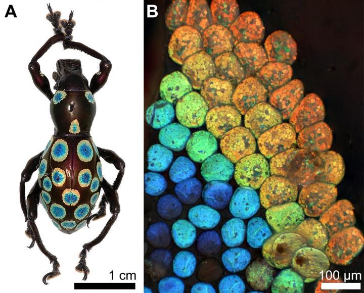
Caption: Left: A photograph of the ‘rainbow’ weevil, with the rainbow-colored spots on its thorax and elytra (wing casings). Right: A microscope image of the rim of a single rainbow spot, showing the different colors of individual scales. Credit: Dr Bodo D Wilts
Researchers from Yale [University]-NUS College and the University of Fribourg in Switzerland have discovered a novel colour-generation mechanism in nature, which if harnessed, has the potential to create cosmetics and paints with purer and more vivid hues, screen displays that project the same true image when viewed from any angle, and even reduce the signal loss in optical fibres.
Yale-NUS College Assistant Professor of Science (Life Science) Vinodkumar Saranathan led the study with Dr Bodo D Wilts from the Adolphe Merkle Institute at the University of Fribourg. Dr Saranathan examined the rainbow-coloured patterns in the elytra (wing casings) of a snout weevil from the Philippines, Pachyrrhynchus congestus pavonius, using high-energy X-rays, while Dr Wilts performed detailed scanning electron microscopy and optical modelling.
They discovered that to produce the rainbow palette of colours, the weevil utilised a colour-generation mechanism that is so far found only in squid, cuttlefish, and octopuses, which are renowned for their colour-shifting camouflage.
P. c. pavonius, or the “Rainbow” Weevil, is distinctive for its rainbow-coloured spots on its thorax and elytra (see attached image). These spots are made up of nearly-circular scales arranged in concentric rings of different hues, ranging from blue in the centre to red at the outside, just like a rainbow. While many insects have the ability to produce one or two colours, it is rare that a single insect can produce such a vast spectrum of colours. Researchers are interested to figure out the mechanism behind the natural formation of these colour-generating structures, as current technology is unable to synthesise structures of this size.
“The ultimate aim of research in this field is to figure out how the weevil self-assembles these structures, because with our current technology we are unable to do so,” Dr Saranathan said. “The ability to produce these structures, which are able to provide a high colour fidelity regardless of the angle you view it from, will have applications in any industry which deals with colour production. We can use these structures in cosmetics and other pigmentations to ensure high-fidelity hues, or in digital displays in your phone or tablet which will allow you to view it from any angle and see the same true image without any colour distortion. We can even use them to make reflective cladding for optical fibres to minimise signal loss during transmission.”
Dr Saranathan and Dr Wilts examined these scales to determine that the scales were composed of a three-dimensional crystalline structure made from chitin (the main ingredient in insect exoskeletons). They discovered that the vibrant rainbow colours on this weevil’s scales are determined by two factors: the size of the crystal structure which makes up each scale, as well as the volume of chitin used to make up the crystal structure. Larger scales have a larger crystalline structure and use a larger volume of chitin to reflect red light; smaller scales have a smaller crystalline structure and use a smaller volume of chitin to reflect blue light. According to Dr Saranathan, who previously examined over 100 species of insects and spiders and catalogued their colour-generation mechanisms, this ability to simultaneously control both size and volume factors to fine-tune the colour produced has never before been shown in insects, and given its complexity, is quite remarkable. “It is different from the usual strategy employed by nature to produce various different hues on the same animal, where the chitin structures are of fixed size and volume, and different colours are generated by orienting the structure at different angles, which reflects different wavelengths of light,” Dr Saranathan explained.
The research was partly supported though the National Centre of Competence in Research “Bio-Inspired Materials” and the Ambizione program of the Swiss National Science Foundation (SNSF) to Dr Wilts, and partly through a UK Royal Society Newton Fellowship, a Linacre College EPA Cephalosporin Junior Research Fellowship, and Yale-NUS College funds to Dr Saranathan. Dr Saranathan is currently part of a research team led by Yale-NUS College Associate Professor of Science Antonia Monteiro, which has recently been awarded a separate Competitive Research Programme (CRP) grant by Singapore’s National Research Foundation (NRF) to examine the genetic basis of the colour-generation mechanism in butterflies. Dr Saranathan and Dr Monteiro are both also from the Department of Biological Sciences at the National University of Singapore (NUS) Faculty of Science. In addition, Dr Saranathan is affiliated with the NUS Nanoscience and Nanotechnology Initiative.
I’m starting to have a collection of postings related to plastic nanoparticles and aquatic life (I have a listing below). The latest originates in Singapore (from a May 31, 2018 news item on ScienceDaily),
Plastic nanoparticles — these are tiny pieces of plastic less than 1 micrometre in size — could potentially contaminate food chains, and ultimately affect human health, according to a recent study by scientists from the National University of Singapore (NUS). They discovered that nanoplastics are easily ingested by marine organisms, and they accumulate in the organisms over time, with a risk of being transferred up the food chain, threatening food safety and posing health risks.
Ocean plastic pollution is a huge and growing global problem. It is estimated that the oceans may already contain over 150 million tonnes of plastic, and each year, about eight million tonnes of plastic will end up in the ocean. Plastics do not degrade easily. In the marine environment, plastics are usually broken down into smaller pieces by the sun, waves, wind and microbial action. These micro- and nanoplastic particles in the water may be ingested by filter-feeding marine organisms such as barnacles, tube worms and sea-squirts.
Using the acorn barnacle Amphibalanus amphitrite as a model organism, the NUS research team demonstrated for the first time that nanoplastics consumed during the larval stage are retained and accumulated inside the barnacle larvae until they reach adulthood.
“We opted to study acorn barnacles as their short life cycle and transparent bodies made it easy to track and visualise the movement of nanoplastics in their bodies within a short span of time,” said Mr Samarth Bhargava, a PhD student from the Department of Chemistry at the NUS Faculty of Science, who is the first author of the research paper.
“Barnacles can be found in all of the world’s oceans. This accumulation of nanoplastics within the barnacles is of concern. Further work is needed to better understand how they may contribute to longer term effects on marine ecosystems,” said Dr Serena Teo, Senior Research Fellow from the Tropical Marine Science Institute at NUS, who co-supervised the research.
Studying the fate of nanoplastics in marine organisms
The NUS research team incubated the barnacle larvae in solutions of their regular feed coupled with plastics that are about 200 nanometres in size with green fluorescent tags. The larvae were exposed to two different treatments: ‘acute’ and ‘chronic’.
Under the ‘acute’ treatment, the barnacle larvae were kept for three hours in a solution that contained 25 times more nanoplastics than current estimates of what is present in the oceans. On the other hand, under the ‘chronic’ treatment, the barnacle larvae were exposed to a solution containing low concentrations of nanoplastics for up to four days.
The larvae were subsequently filtered from the solution, and examined under the microscope. The distribution and movement of the nanoplastics were monitored by examining the fluorescence from the particles present within the larvae over time.
“Our results showed that after exposing the barnacle larvae to nanoplastics in both treatments, the larvae had not only ingested the plastic particles, but the tiny particles were found to be distributed throughout the bodies of the larvae,” said Ms Serina Lee from the Tropical Marine Science Institute at NUS, who is the second author of the paper.
Even though the barnacles’ natural waste removal pathways of moulting and excretion resulted in some removal of the nanoplastics, the team detected the continued presence of nanoplastics inside the barnacles throughout their growth until they reached adulthood.
“Barnacles may be at the lower levels of the food chain, but what they consume will be transferred to the organisms that eat them. In addition, plastics are capable of absorbing pollutants and chemicals from the water. These toxins may be transferred to the organisms if the particles of plastics are consumed, and can cause further damage to marine ecosystems and human health,” said marine biologist Dr Neo Mei Lin from the Tropical Marine Science Institute at NUS, who is one of the authors of the paper.
The team’s research findings were first published online in the journal ACS Sustainable Chemistry & Engineering in March 2018. The study was funded under the Marine Science Research and Development Programme of the National Research Foundation Singapore.
Next steps
The NUS research team seeks to further their understanding of the translocation of nanoparticles within the marine organisms and potential pathways of transfer in the marine ecosystem.
“The life span and fate of plastic waste materials in marine environment is a big concern at the moment owing to the large amounts of plastic waste and its potential impact on marine ecosystem and food security around the world. The team would like to explore such topics in the near future and possibly to come up with pathways to address such problems,” explained Associate Professor Suresh Valiyaveettil from the Department of Chemistry at the NUS Faculty of Science, who co-supervised the research.
The team is currently examining how nanoplastics affect other invertebrate model organisms to understand the impact of plastics on marine ecosystems.