The last time I featured memrisors and a neuronal network it was in an April 22, 2016 posting about Russian research in that field. This latest work comes from the UK’s University of Southampton. From a Sept. 27, 2016 news item on phys.org,
New research, led by the University of Southampton, has demonstrated that a nanoscale device, called a memristor, could be the ‘missing link’ in the development of implants that use electrical signals from the brain to help treat medical conditions.
Monitoring neuronal cell activity is fundamental to neuroscience and the development of neuroprosthetics – biomedically engineered devices that are driven by neural activity. However, a persistent problem is the device being able to process the neural data in real-time, which imposes restrictive requirements on bandwidth, energy and computation capacity.
In a new study, published in Nature Communications, the researchers showed that memristors could provide real-time processing of neuronal signals (spiking events) leading to efficient data compression and the potential to develop more precise and affordable neuroprosthetics and bioelectronic medicines.
Memristors are electrical components that limit or regulate the flow of electrical current in a circuit and can remember the amount of charge that was flowing through it and retain the data, even when the power is turned off.
Lead author Isha Gupta, Postgraduate Research Student at the University of Southampton, said: “Our work can significantly contribute towards further enhancing the understanding of neuroscience, developing neuroprosthetics and bio-electronic medicines by building tools essential for interpreting the big data in a more effective way.”
The research team developed a nanoscale Memristive Integrating Sensor (MIS) into which they fed a series of voltage-time samples, which replicated neuronal electrical activity.
Acting like synapses in the brain, the metal-oxide MIS was able to encode and compress (up to 200 times) neuronal spiking activity recorded by multi-electrode arrays. Besides addressing the bandwidth constraints, this approach was also very power efficient – the power needed per recording channel was up to 100 times less when compared to current best practice.
Co-author Dr Themis Prodromakis, Reader in Nanoelectronics and EPSRC Fellow in Electronics and Computer Science at the University of Southampton said: “We are thrilled that we succeeded in demonstrating that these emerging nanoscale devices, despite being rather simple in architecture, possess ultra-rich dynamics that can be harnessed beyond the obvious memory applications to address the fundamental constraints in bandwidth and power that currently prohibit scaling neural interfaces beyond 1,000 recording channels.”
The Prodromakis Group at the University of Southampton is acknowledged as world-leading in this field, collaborating among others with Leon Chua (a Diamond Jubilee Visiting Academic at the University of Southampton), who theoretically predicted the existence of memristors in 1971.
For anyone who’s interested in better understanding memristors, there’s an interview with Forrest H Bennett III in my April 7, 2010 posting and you can always check Wikipedia.
Stephen Melendez’s June 11, 2016 story about biohackers/bodyhackers/grinders for Fast Company sports a striking image in the banner, an x-ray of a pair hands featuring some mysterious additions to the webbing between thumbs and forefingers (Note: Links have been removed),
Tim Shank can guarantee he’ll never leave home without his keys. Why? His house keys are located inside his body.
Shank, the president of the Minneapolis futurist group TwinCities+, has a chip installed in his hand that can communicate electronically with his front door and tell it to unlock itself. His wife has one, too.
…
In fact, Shank has several chips in his hand, including a near field communication (NFC) chip like the ones used in Apple Pay and similar systems, which stores a virtual business card with contact information for TwinCities+. “[For] people with Android phones, I can just tap their phone with my hand, right over the chip, and it will send that information to their phone,” he says. In the past, he’s also used a chip to store a bitcoin wallet.
Shank is one of a growing number of “biohackers” who implant hardware ranging from microchips to magnets inside their bodies.
Certainly the practice seems considerably more developed since the first time it was mentioned here in a May 27, 2010 posting about a researcher who’d implanted a chip into his body which he then contaminated with a computer virus. In the comments, you’ll find Amal Grafstraa who’s mentioned in the Melendez article at some length, from the Melendez article (Note: Links have been removed),
Some biohackers use their implants in experimental art projects. Others who have disabilities or medical conditions use them to improve their quality of life, while still others use the chips to extend the limits of human perception. …
Experts sometimes caution that the long-term health risks of the practice are still unknown. But many biohackers claim that, if done right, implants can be no more dangerous than getting a piercing or tattoo. In fact, professional body piercers are frequently the ones tasked with installing these implants, given that they possess the training and sterilization equipment necessary to break people’s skin safely.
“When you talk about things like risk, things like putting it in your body, the reality is the risk of having one of these installed is extremely low—it’s even lower than an ear piercing,” claims Amal Graafstra, the founder of Dangerous Things, a biohacking supply company.
Graafstra, who is also the author of the book RFID Toys, says he first had an RFID chip installed in his hand in 2005, which allowed him to unlock doors without a key. When the maker movement took off a few years later, and as more hackers began to explore what they could put inside their bodies, he founded Dangerous Things with the aim of ensuring these procedures were done safely.
“I decided maybe it’s time to wrap a business model around this and make sure that the things people are trying to put in their bodies are safe,” he says. The company works with a network of trained body piercers and offers online manuals and videos for piercers looking to get up to speed on the biohacking movement.
At present, these chips are capable of verifying users’ identities and opening doors. And according to Graafstra, a next-generation chip will have enough on-board cryptographic power to potentially work with credit card terminals securely.
“The technology is there—we can definitely talk to payment terminals with it—but we don’t have the agreements in place with banks [and companies like] MasterCard to make that happen,” he says.
Paying for goods with an implantable chip might sound unusual for consumers and risky for banks, but Graafstra thinks the practice will one day become commonplace. He points to a survey released by Visa last year that found that 25% of Australians are “at least slightly interested” in paying for purchases through a chip implanted in their bodies.
Melendez’s article is fascinating and well worth reading in its entirety. It’s not all keys and commerce as this next and last excerpt shows,
Other implantable technology has more of an aesthetic focus: Pittsburgh biohacking company Grindhouse Wetware offers a below-the-skin, star-shaped array of LED lights called Northstar. While the product was inspired by the on-board lamps of a device called Circadia that Grindhouse founder Tim Cannon implanted to send his body temperature to a smartphone, the commercially available Northstar features only the lights and is designed to resemble natural bioluminescence.
“This particular device is mainly aesthetic,” says Grindhouse spokesman Ryan O’Shea. “It can backlight tattoos or be used in any kind of interpretive dance, or artists can use it in various ways.”
The lights activate in the presence of a magnetic field—one that is often provided by magnets already implanted in the same user’s fingertips. Which brings up another increasingly common piece of bio-hardware: magnetic finger implants. ….
There are other objects that can be implanted in bodies. In one case, an artist, Wafaa Bilal had a camera implanted into the back of his head for a 3rd eye. I mentioned the Iraqi artist in my April 13, 2011 posting titled: Blood, memristors, cyborgs plus brain-controlled computers, prosthetics, and art (scroll down about 75% of the way). Bilal was unable to find a doctor who would perform the procedure so he went to a body-piercing studio. Unfortunately, the posting chronicles his infection and subsequent removal of the camera (h/t Feb. 11, 2011 BBC [British Broadcasting Corporation] news online article).
Observations
It’s been a while since I’ve written about bodyhacking and I’d almost forgotten about the practice relegating it to the category of “one of those trendy ideas that get left behind as interest shifts.” My own interest had shifted more firmly to neuroprosthetics (the integration of prostheses into the nervous system).
I had coined a tag for bodyhacking and neuroprostheses: machine/flesh which covers both those topics and more (e.g. cyborgs) as we continue to integrate machines into our bodies.
Final note
I was reminded of Wafaa Bilal recently when checking out a local arts magazine, Preview: the gallery guide, June/July/August 2016 issue. His work (the 168:01show) is being shown in Calgary, Alberta, Canada at the Esker Foundation from May 27 to August 28, 2016,
168:01 is a major solo exhibition of new and recent work by Iraqi-born, New York-based artist Wafaa Bilal, renowned for his online performances and technologically driven encounters that speak to the impact of international politics on individual lives.
In 168:01, Bilal takes the Bayt al-Hikma, or House of Wisdom, as a starting point for a sculptural installation of a library. The Bayt al-Hikma was a major academic center during the Islamic Golden Age where Muslim, Jewish, and Christian scholars studied the humanities and science. By the middle of the Ninth Century, the House of Wisdom had accumulated the largest library in the world. Four centuries later, a Mongol siege laid waste to all the libraries of Baghdad along with the House of Wisdom. According to some accounts, the library was thrown into the Tigris River to create a bridge of books for the Mongol army to cross. The pages bled ink into the river for seven days – or 168 hours, after which the books were drained of knowledge. Today, the Bayt al-Hikma represents one of the most well-known examples of historic cultural loss as a casualty of wartime.
For this exhibition, Bilal has constructed a makeshift library filled with empty white books. The white books symbolize the priceless cultural heritage destroyed at Bayt al-Hikma as well as the libraries, archives, and museums whose systematic decimation by occupying forces continues to ravage his homeland. Throughout the duration of the exhibition, the white books will slowly be replaced with visitor donations from a wishlist compiled by The College of Fine Arts at the University of Baghdad, whose library was looted and destroyed in 2003. At the end of each week a volunteer unpacks the accumulated shipments, catalogues each new book by hand, and places the books on the shelves. At the end of the exhibition, all the donated books will be sent to the University of Baghdad to help rebuild their library. This exchange symbolizes the power of individuals to rectify violence inflicted on cultural spaces that are meant to preserve and store knowledge for future generations.
In conjunction with the library, Bilal presents a powerful suite of photographs titled The Ashes Series that brings the viewer closer to images of violence and war in the Middle East. In an effort to foster empathy and humanize the onslaught of violent images that inundate Western media during wartime, Bilal has reconstructed journalistic images of the destruction caused by the Iraq War. He writes, “Reconstructing the destructed spaces is a way to exist in them, to share them with an audience, and to provide a layer of distance, as the original photographs are too violent and run the risk of alienating the viewer. It represents an attempt to make sense of the destruction and to preserve the moment of serenity after the dust has settled, to give the ephemeral moment extended life in a mix of beauty and violence.” In the photograph Al-MutanabbiStreet from The Ashes Series, the viewer encounters dilapidated historic and modern buildings on a street covered with layers upon layers of rubble and fragments of torn books. Bilal’s images emanate a slowness that deepens engagement between the viewer and the image, thereby inviting them to share the burden of obliterated societies and reimagine a world built on the values of peace and hope.
The House of Wisdom has been mentioned here a few times perhaps most comprehensively and in the context of the then recent opening of the King Abdullah University for Science and Technology (KAUST; located in Saudi Arabia) in this Sept. 24, 2009 posting (scroll down about 45% of the way).
This research from Singapore could make neuroprosthetics and exoskeletons a little easier to manage as long as you don’t mind having a neural implant. From a Feb. 11, 2016 news item on ScienceDaily,
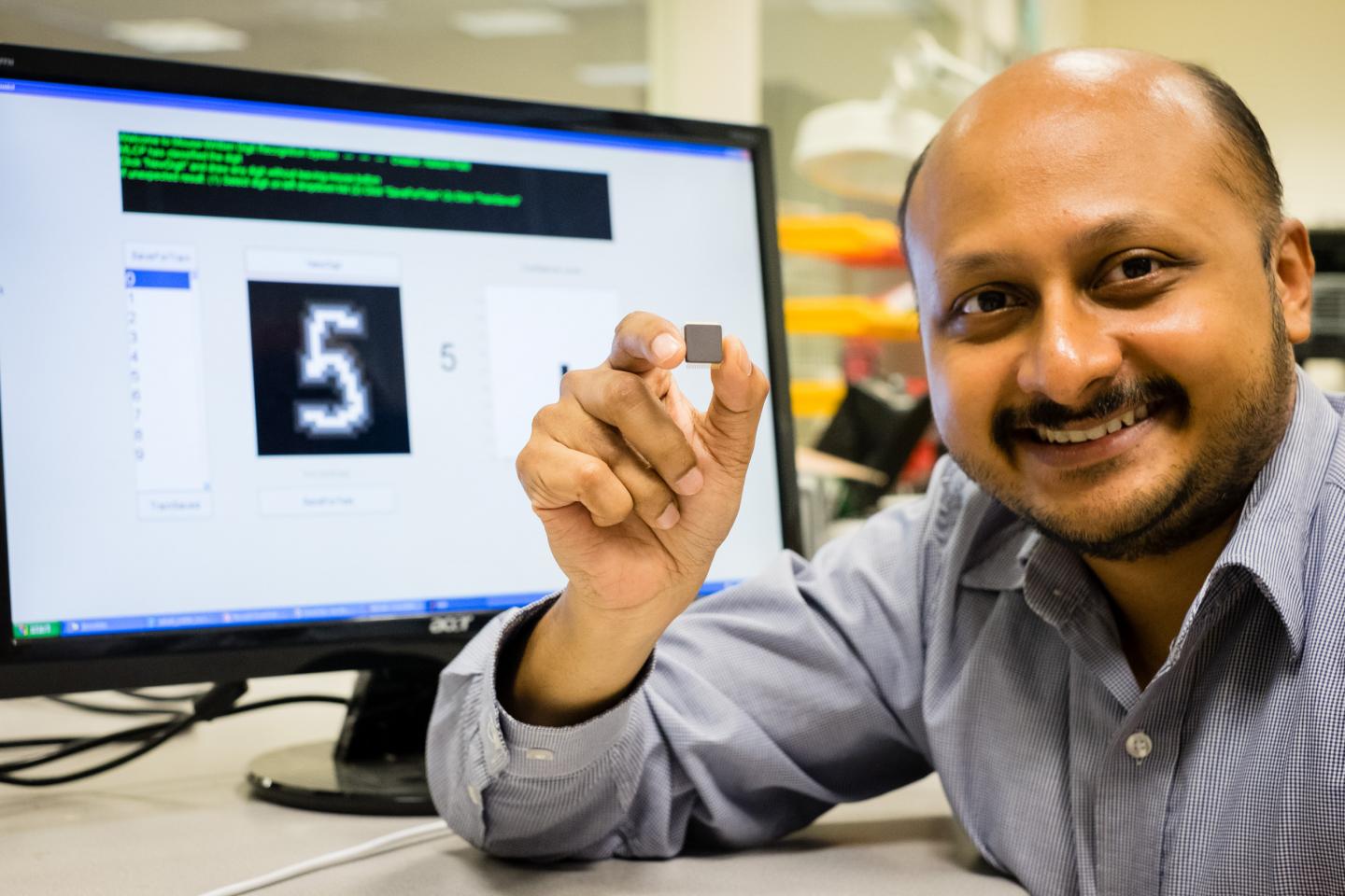
A versatile chip offers multiple applications in various electronic devices, report researchers, suggested that there is now hope that a low-powered, wireless neural implant may soon be a reality. Neural implants when embedded in the brain can alleviate the debilitating symptoms of Parkinson’s disease or give paraplegic people the ability to move their prosthetic limbs.
Caption: NTU Asst Prof Arindam Basu is holding his low-powered smart chip. Credit: NTU Singapore
Scientists at Nanyang Technological University, Singapore (NTU Singapore) have developed a small smart chip that can be paired with neural implants for efficient wireless transmission of brain signals.
Neural implants when embedded in the brain can alleviate the debilitating symptoms of Parkinson’s disease or give paraplegic people the ability to move their prosthetic limbs.
However, they need to be connected by wires to an external device outside the body. For a prosthetic patient, the neural implant is connected to a computer that decodes the brain signals so the artificial limb can move.
These external wires are not only cumbersome but the permanent openings which allow the wires into the brain increases the risk of infections.
The new chip by NTU scientists can allow the transmission of brain data wirelessly and with high accuracy.
Assistant Professor Arindam Basu from NTU’s School of Electrical and Electronic Engineering said the research team have tested the chip on data recorded from animal models, which showed that it could decode the brain’s signal to the hand and fingers with 95 per cent accuracy.
“What we have developed is a very versatile smart chip that can process data, analyse patterns and spot the difference,” explained Prof Basu.
“It is about a hundred times more efficient than current processing chips on the market. It will lead to more compact medical wearable devices, such as portable ECG monitoring devices and neural implants, since we no longer need large batteries to power them.”
Different from other wireless implants
To achieve high accuracy in decoding brain signals, implants require thousands of channels of raw data. To wirelessly transmit this large amount of data, more power is also needed which means either bigger batteries or more frequent recharging.
This is not feasible as there is limited space in the brain for implants while frequent recharging means the implants cannot be used for long-term recording of signals.
Current wireless implant prototypes thus suffer from a lack of accuracy as they lack the bandwidth to send out thousands of channels of raw data.
Instead of enlarging the power source to support the transmission of raw data, Asst Prof Basu tried to reduce the amount of data that needs to be transmitted.
Designed to be extremely power-efficient, NTU’s patented smart chip will analyse and decode the thousands of signals from the neural implants in the brain, before compressing the results and sending it wirelessly to a small external receiver.
This invention and its findings were published last month [December 2015] in the prestigious journal, IEEE Transactions on Biomedical Circuits & Systems, by the Institute of Electrical and Electronics Engineers, the world’s largest professional association for the advancement of technology.
Its underlying science was also featured in three international engineering conferences (two in Atlanta, USA and one in China) over the last three months.
Versatile smart chip with multiple uses
This new smart chip is designed to analyse data patterns and spot any abnormal or unusual patterns.
For example, in a remote video camera, the chip can be programmed to send a video back to the servers only when a specific type of car or something out of the ordinary is detected, such as an intruder.
This would be extremely beneficial for the Internet of Things (IOT), where every electrical and electronic device is connected to the Internet through a smart chip.
With a report by marketing research firm Gartner Inc predicting that 6.4 billion smart devices and appliances will be connected to the Internet by 2016, and will rise to 20.8 billion devices by 2020, reducing network traffic will be a priority for most companies.
Using NTU’s new chip, the devices can process and analyse the data on site, before sending back important details in a compressed package, instead of sending the whole data stream. This will reduce data usage by over a thousand times.
Asst Prof Basu is now in talks with Singapore Technologies Electronics Limited to adapt his smart chip that can significantly reduce power consumption and the amount of data transmitted by battery-operated remote sensors, such as video cameras.
The team is also looking to expand the applications of the chip into commercial products, such as to customise it for smart home sensor networks, in collaboration with a local electronics company.
The chip, measuring 5mm by 5mm can now be licensed by companies from NTU’s commercialisation arm, NTUitive.
Earlier this month there was a Feb. 9, 2016 announcement about a planned human clinical trial in Australia for a new brain-machine interface (neural implant). Before proceeding with the news, here’s what this implant looks like,
Caption: This tiny device, the size of a small paperclip, is implanted in to a blood vessel next to the brain and can read electrical signals from the motor cortex, the brain’s control centre. These signals can then be transmitted to an exoskeleton or wheelchair to give paraplegic patients greater mobility. Users will need to learn how to communicate with their machinery, but over time, it is thought it will become second nature, like driving or playing the piano. The first human trials are slated for 2017 in Melbourne, Australia. Credit: The University of Melbourne.
Melbourne medical researchers have created a new minimally invasive brain-machine interface, giving people with spinal cord injuries new hope to walk again with the power of thought.
The brain machine interface consists of a stent-based electrode (stentrode), which is implanted within a blood vessel next to the brain, and records the type of neural activity that has been shown in pre-clinical trials to move limbs through an exoskeleton or to control bionic limbs.
The new device is the size of a small paperclip and will be implanted in the first in-human trial at The Royal Melbourne Hospital in 2017.
The results published today in Nature Biotechnology show the device is capable of recording high-quality signals emitted from the brain’s motor cortex, without the need for open brain surgery.
Principal author and Neurologist at The Royal Melbourne Hospital and Research Fellow at The Florey Institute of Neurosciences and the University of Melbourne, Dr Thomas Oxley, said the stentrode was revolutionary.
“The development of the stentrode has brought together leaders in medical research from The Royal Melbourne Hospital, The University of Melbourne and the Florey Institute of Neuroscience and Mental Health. In total 39 academic scientists from 16 departments were involved in its development,” Dr Oxley said.
“We have been able to create the world’s only minimally invasive device that is implanted into a blood vessel in the brain via a simple day procedure, avoiding the need for high risk open brain surgery.
“Our vision, through this device, is to return function and mobility to patients with complete paralysis by recording brain activity and converting the acquired signals into electrical commands, which in turn would lead to movement of the limbs through a mobility assist device like an exoskeleton. In essence this a bionic spinal cord.”
Stroke and spinal cord injuries are leading causes of disability, affecting 1 in 50 people. There are 20,000 Australians with spinal cord injuries, with the typical patient a 19-year old male, and about 150,000 Australians left severely disabled after stroke.
Co-principal investigator and biomedical engineer at the University of Melbourne, Dr Nicholas Opie, said the concept was similar to an implantable cardiac pacemaker – electrical interaction with tissue using sensors inserted into a vein, but inside the brain.
“Utilising stent technology, our electrode array self-expands to stick to the inside wall of a vein, enabling us to record local brain activity. By extracting the recorded neural signals, we can use these as commands to control wheelchairs, exoskeletons, prosthetic limbs or computers,” Dr Opie said.
“In our first-in-human trial, that we anticipate will begin within two years, we are hoping to achieve direct brain control of an exoskeleton for three people with paralysis.”
“Currently, exoskeletons are controlled by manual manipulation of a joystick to switch between the various elements of walking – stand, start, stop, turn. The stentrode will be the first device that enables direct thought control of these devices”
Neurophysiologist at The Florey, Professor Clive May, said the data from the pre-clinical study highlighted that the implantation of the device was safe for long-term use.
“Through our pre-clinical study we were able to successfully record brain activity over many months. The quality of recording improved as the device was incorporated into tissue,” Professor May said.
“Our study also showed that it was safe and effective to implant the device via angiography, which is minimally invasive compared with the high risks associated with open brain surgery.
“The brain-computer interface is a revolutionary device that holds the potential to overcome paralysis, by returning mobility and independence to patients affected by various conditions.”
Professor Terry O’Brien, Head of Medicine at Departments of Medicine and Neurology, The Royal Melbourne Hospital and University of Melbourne said the development of the stentrode has been the “holy grail” for research in bionics.
“To be able to create a device that can record brainwave activity over long periods of time, without damaging the brain is an amazing development in modern medicine,” Professor O’Brien said.
“It can also be potentially used in people with a range of diseases aside from spinal cord injury, including epilepsy, Parkinsons and other neurological disorders.”
The development of the minimally invasive stentrode and the subsequent pre-clinical trials to prove its effectiveness could not have been possible without the support from the major funding partners – US Defense Department DARPA [Defense Advanced Research Projects Agency] and Australia’s National Health and Medical Research Council.
So, DARPA is helping fund this, eh? Interesting but not a surprise given the agency’s previous investments in brain research and neuroprosthetics.
For those who like to get their news via video,
Here’s a link to and a citation for the paper,
Minimally invasive endovascular stent-electrode array for high-fidelity, chronic recordings of cortical neural activity by Thomas J Oxley, Nicholas L Opie, Sam E John, Gil S Rind, Stephen M Ronayne, Tracey L Wheeler, Jack W Judy, Alan J McDonald, Anthony Dornom, Timothy J H Lovell, Christopher Steward, David J Garrett, Bradford A Moffat, Elaine H Lui, Nawaf Yassi, Bruce C V Campbell, Yan T Wong, Kate E Fox, Ewan S Nurse, Iwan E Bennett, Sébastien H Bauquier, Kishan A Liyanage, Nicole R van der Nagel, Piero Perucca, Arman Ahnood et al. Nature Biotechnology (2016) doi:10.1038/nbt.3428 Published online 08 February 2016
This paper is behind a paywall.
I wish the researchers in Singapore, Australia, and elsewhere, good luck!
*’Sinagpore’ in head changed to ‘Singapore’ on May 14, 2019.
Gregory Carpenter has written a gripping (albeit somewhat exaggerated) piece for Signal, a publication of the Armed Forces Communications and Electronics Association (AFCEA) about cybersecurity issues and nanomedicine endeavours. From Carpenter’s Jan. 1, 2016 article titled, When Lifesaving Technology Can Kill; The Cyber Edge,
The exciting advent of nanotechnology that has inspired disruptive and lifesaving medical advances is plagued by cybersecurity issues that could result in the deaths of people that these very same breakthroughs seek to heal. Unfortunately, nanorobotic technology has suffered from the same security oversights that afflict most other research and development programs.
Nanorobots, or small machines [or nanobots[, are vulnerable to exploitation just like other devices.
At the moment, the issue of cybersecurity exploitation is secondary to making nanobots, or nanorobots, dependably functional. As far as I’m aware, there is no such nanobot. Even nanoparticles meant to function as packages for drug delivery have not been perfected (see one of the controversies with nanomedicine drug delivery described in my Nov. 26, 2015 posting).
That said, Carpenter’s point about cybersecurity is well taken since security features are often overlooked in new technology. For example, automated banking machines (ABMs) had woefully poor (inadequate, almost nonexistent) security when they were first introduced.
Carpenter outlines some of the problems that could occur, assuming some of the latest research could be reliably brought to market,
The U.S. military has joined the fray of nanorobotic experimentation, embarking on revolutionary research that could lead to a range of discoveries, from unraveling the secrets of how brains function to figuring out how to permanently purge bad memories. Academia is making amazing advances as well. Harnessing progress by Harvard scientists to move nanorobots within humans, researchers at the University of Montreal, Polytechnique Montreal and Centre Hospitalier Universitaire Sainte-Justine are using mobile nanoparticles inside the human brain to open the blood-brain barrier, which protects the brain from toxins found in the circulatory system.
…
A different type of technology presents a risk similar to the nanoparticles scenario. A DARPA-funded program known as Restoring Active Memory (RAM) addresses post-traumatic stress disorder, attempting to overcome memory deficits by developing neuroprosthetics that bridge gaps in an injured brain. In short, scientists can wipe out a traumatic memory, and they hope to insert a new one—one the person has never actually experienced. Someone could relish the memory of a stroll along the French Riviera rather than a terrible firefight, even if he or she has never visited Europe.
As an individual receives a disruptive memory, a cyber criminal could manage to hack the controls. Breaches of the brain could become a reality, putting humans at risk of becoming zombie hosts [emphasis mine] for future virus deployments. …
At this point, the ‘zombie’ scenario Carpenter suggests seems a bit over-the-top but it does hearken to the roots of the zombie myth where the undead aren’t mindlessly searching for brains but are humans whose wills have been overcome. Mike Mariani in an Oct. 28, 2015 article for The Atlantic has presented a thought-provoking history of zombies,
… the zombie myth is far older and more rooted in history than the blinkered arc of American pop culture suggests. It first appeared in Haiti in the 17th and 18th centuries, when the country was known as Saint-Domingue and ruled by France, which hauled in African slaves to work on sugar plantations. Slavery in Saint-Domingue under the French was extremely brutal: Half of the slaves brought in from Africa were worked to death within a few years, which only led to the capture and import of more. In the hundreds of years since, the zombie myth has been widely appropriated by American pop culture in a way that whitewashes its origins—and turns the undead into a platform for escapist fantasy.
The original brains-eating fiend was a slave not to the flesh of others but to his own. The zombie archetype, as it appeared in Haiti and mirrored the inhumanity that existed there from 1625 to around 1800, was a projection of the African slaves’ relentless misery and subjugation. Haitian slaves believed that dying would release them back to lan guinée, literally Guinea, or Africa in general, a kind of afterlife where they could be free. Though suicide was common among slaves, those who took their own lives wouldn’t be allowed to return to lan guinée. Instead, they’d be condemned to skulk the Hispaniola plantations for eternity, an undead slave at once denied their own bodies and yet trapped inside them—a soulless zombie.
I recommend reading Mariani’s article although I do have one nit to pick. I can’t find a reference to brain-eating zombies until George Romero’s introduction of the concept in his movies. This Zombie Wikipedia entry seems to be in agreement with my understanding (if I’m wrong, please do let me know and, if possible, provide a link to the corrective text).
Getting back to Carpenter and cybersecurity with regard to nanomedicine, while his scenarios may seem a trifle extreme it’s precisely the kind of thinking you need when attempting to anticipate problems. I do wish he’d made clear that the technology still has a ways to go.
I don’t often get news releases from Sweden but I do on occasion and, sometimes, they even come in their original Swedish versions. In this case, Lund University sent me an English language version about their latest work making brain implants (neural prostheses) safer and effective. From a Sept. 29, 2015 Lund University news release (also on EurekAlert),
Neurons thrive and grow in a new type of nanowire material developed by researchers in Nanophysics and Ophthalmology at Lund University in Sweden. In time, the results might improve both neural and retinal implants, and reduce the risk of them losing their effectiveness over time, which is currently a problem
By implanting electrodes in the brain tissue one can stimulate or capture signals from different areas of the brain. These types of brain implants, or neuro-prostheses as they are sometimes called, are used to treat Parkinson’s disease and other neurological diseases.
They are currently being tested in other areas, such as depression, severe cases of autism, obsessive-compulsive disorders and paralysis. Another research track is to determine whether retinal implants are able to replace light-sensitive cells that die in cases of Retinitis Pigmentosa and other eye diseases.
However, there are severe drawbacks associated with today’s implants. One problem is that the body interprets the implants as foreign objects, resulting in an encapsulation of the electrode, which in turn leads to loss of signal.
One of the researchers explains the approach adopted by the research team (from the news release),
“Our nanowire structure prevents the cells that usually encapsulate the electrodes – glial cells – from doing so”, says Christelle Prinz, researcher in Nanophysics at Lund University in Sweden, who developed this technique together with Maria Thereza Perez, a researcher in Ophthalmology.
“I was very pleasantly surprised by these results. In previous in-vitro experiments, the glial cells usually attach strongly to the electrodes”, she says.
To avoid this, the researchers have developed a small substrate where regions of super thin nanowires are combined with flat regions. While neurons grow and extend processes on the nanowires, the glial cells primarily occupy the flat regions in between.
“The different types of cells continue to interact. This is necessary for the neurons to survive because the glial cells provide them with important molecules.”
So far, tests have only been done with cultured cells (in vitro) but hopefully they will soon be able to continue with experiments in vivo.
The substrate is made from the semiconductor material gallium phosphide where each outgrowing nanowire has a diameter of only 80 nanometres (billionths of a metre).
This research will not find itself occupying anyone’s brain for some time to come but it is interesting to find out that neural prosthetics have some drawbacks and there is work being done to address them. From an Aug. 10, 2015 news item on Azonano,
Instead of using neural prosthetic devices–which suffer from immune-system rejection and are believed to fail due to a material and mechanical mismatch–a multi-institutional team, including Lohitash Karumbaiah of the University of Georgia’s Regenerative Bioscience Center, has developed a brain-friendly extracellular matrix environment of neuronal cells that contain very little foreign material. These by-design electrodes are shielded by a covering that the brain recognizes as part of its own composition.
Although once believed to be devoid of immune cells and therefore of immune responses, the brain is now recognized to have its own immune system that protects it against foreign invaders.
“This is not by any means the device that you’re going to implant into a patient,” said Karumbaiah, an assistant professor of animal and dairy science in the UGA College of Agricultural and Environmental Sciences. “This is proof of concept that extracellular matrix can be used to ensheathe a functioning electrode without the use of any other foreign or synthetic materials.”
Implantable neural prosthetic devices in the brain have been around for almost two decades, helping people living with limb loss and spinal cord injury become more independent. However, not only do neural prosthetic devices suffer from immune-system rejection, but most are believed to eventually fail because of a mismatch between the soft brain tissue and the rigid devices.
The collaboration, led by Wen Shen and Mark Allen of the University of Pennsylvania, found that the extracellular matrix derived electrodes adapted to the mechanical properties of brain tissue and were capable of acquiring neural recordings from the brain cortex.
“Neural interface technology is literally mind boggling, considering that one might someday control a prosthetic limb with one’s own thoughts,” Karumbaiah said.
The study’s joint collaborators were Ravi Bellamkonda, who conceived the new approach and is chair of the Wallace H. Coulter Department of Biomedical Engineering at the Georgia Institute of Technology and Emory University, as well as Allen, who at the time was director of the Institute for Electronics and Nanotechnology.
“Hopefully, once we converge upon the nanofabrication techniques that would enable these to be clinically translational, this same methodology could then be applied in getting these extracellular matrix derived electrodes to be the next wave of brain implants,” Karumbaiah said.
Currently, one out of every 190 Americans is living with limb loss, according to the National Institutes of Health. There is a significant burden in cost of care and quality of life for people suffering from this disability.
The research team is one part of many in the prosthesis industry, which includes those who design the robotics for the artificial limbs, others who make the neural prosthetic devices and developers who design the software that decodes the neural signal.
“What neural prosthetic devices do is communicate seamlessly to an external prosthesis,” Karumbaiah said, “providing independence of function without having to have a person or a facility dedicated to their care.”
Karumbaiah hopes further collaboration will allow them to make positive changes in the industry, saying that, “it’s the researcher-to-industry kind of conversation that now needs to take place, where companies need to come in and ask: ‘What have you learned? How are the devices deficient, and how can we make them better?'”
One final note, I have written frequently about prosthetics and neural prosthetics, which you can find by using either of those terms and/or human enhancement. Here’s my latest piece, a March 25, 2015 posting.
I was a bit surprised to find that this University of Oregon story was about a patent. Here’s more from a July 28, 2015 news item on Azonano,
Richard Taylor’s vision of using artificial fractal-based implants to restore sight to the blind — part of a far-reaching concept that won an innovation award this year from the White House — is now covered under a broad U.S. patent.
The patent goes far beyond efforts to use the emerging technology to restore eyesight. It covers all fractal-designed electronic implants that link signaling activity with nerves for any purpose in animal and human biology.
Fractals are objects with irregular curves or shapes. “They are a trademark building block of nature,” said Taylor, a professor of physics and director of the Materials Science Institute at the University of Oregon [UO]. “In math, that property is self-similarity. Trees, clouds, rivers, galaxies, lungs and neurons are fractals. What we hope to do is adapt the technology to nature’s geometry.”
Named in U.S. patent 9079017 are Taylor, the UO, Taylor’s research collaborator Simon Brown, and Brown’s home institution, the University of Canterbury in New Zealand.
“We’re very delighted,” Taylor said. “The U.S. Patent and Trademark Office has recognized the novelty and utility of our general concept, but there is a lot to do. We want to get all of the fundamental science sorted out. We’re looking at least another couple of years of basic science before moving forward.”
The patent solidifies the relationship between the two universities, said Charles Williams, associate vice president for innovation at the UO. “This is still in the very early days. This project has attracted national attention, awards and grants.
“We hope to engage the right set of partners to develop the technology over time as the concept moves into potentially vast forms of medical applications,” Williams added. “Dr. Taylor’s interdisciplinary science is a hallmark of the creativity at the University of Oregon and a great example of the international research collaborations that our faculty engage in every day.”
Here’s an image illustrating the ‘fractal neurons’,
Caption: Retinal neurons, outlined in yellow, attach to and follows branches of a fractal interconnect. Such connections, says University of Oregon physicist Richard Taylor, could some day help to treat eye diseases such as macular degeneration. Credit: Courtesy of Richard Taylor
The news release goes on to describe the ‘fractal approach’ to eye implants which is markedly different from the implants entering the marketplace,
Taylor raised the idea of a fractal-based approach to treat eye diseases in a 2011 article in Physics World, writing that it could overcome problems associated with efforts to insert photodiodes behind the eyes. Current chip technology doesn’t allow sufficient connections with neurons.
“The wiring — the neurons — in the retina is fractal, but the chips are not fractal,” Taylor said. His vision, based on research with Brown, is to grow nanoflowers seeded from nanoparticles of metals that self assemble in a natural process, producing fractals that mimic and communicate with neurons.
It is conceivable, Taylor said, that fractal interconnects — as the implants are called in the patent — could be shaped so they network with like-shaped neurons to address narrow needs, such as a feedback loop for the sensation of touch from a prosthetic arm or leg to the brain.
Such implants would overcome the biological rejection of implants with smooth surfaces or those randomly patterned that have been developed in a trial-and-error approach to link to neurons.
Once perfected, he said, the implants would generate an electrical field that would fool a sea of glial cells that insulate and protect neurons from foreign invaders. Fractal interconnects would allow electrical signals to operate in “a safety zone biologically” that avoids toxicity issues.
“The patent covers any generic interface for connecting any electronics to any nerve,” Taylor said, adding that fractal interconnects are not electrodes. “Our interface is multifunctional. The primary thing is to get the electrical field into the system so that reaches the neurons and induces the signal.”
Taylor’s proposal for using fractal-based technology earned the top prize in a contest held by the innovation company InnoCentive. Taylor was honored in April [2015] at a meeting of the White House Office of Science and Technology Policy.
The competition was sponsored by a collaboration of science philanthropies including the Research Corporation for Science Advancement, the Gordon and Betty Moore Foundation, the W.M. Keck Foundation, the Kavli Foundation, the Templeton Foundation and the Burroughs Wellcome Fund.
Communication between man and machine – a fascinating area at the interface of chemistry, biomedicine, and engineering. (Figure: KIT/S. Giselbrecht, R. Meyer, B. Rapp)
German researchers from the Karlsruhe Institute of Technology (KIT), Professor Christof M. Niemeyer and Dr. Stefan Giselbrecht of the Institute for Biological Interfaces 1 (IBG 1) and Dr. Bastian E. Rapp, Institute of Microstructure Technology (IMT) have written a good overview of the current state of cyborgs while pointing out some of the ethical issues associated with this field. From the Jan. 10, 2014 news item on ScienceDaily,
Medical implants, complex interfaces between brain and machine or remotely controlled insects: Recent developments combining machines and organisms have great potentials, but also give rise to major ethical concerns. In a new review, KIT scientists discuss the state of the art of research, opportunities, and risks.
They are known from science fiction novels and films – technically modified organisms with extraordinary skills, so-called cyborgs. This name originates from the English term “cybernetic organism”. In fact, cyborgs that combine technical systems with living organisms are already reality. The KIT researchers Professor Christof M. Niemeyer and Dr. Stefan Giselbrecht of the Institute for Biological Interfaces 1 (IBG 1) and Dr. Bastian E. Rapp, Institute of Microstructure Technology (IMT), point out that this especially applies to medical implants.
In recent years, medical implants based on smart materials that automatically react to changing conditions, computer-supported design and fabrication based on magnetic resonance tomography datasets or surface modifications for improved tissue integration allowed major progress to be achieved. For successful tissue integration and the prevention of inflammation reactions, special surface coatings were developed also by the KIT under e.g. the multidisciplinary Helmholtz program “BioInterfaces”.
Progress in microelectronics and semiconductor technology has been the basis of electronic implants controlling, restoring or improving the functions of the human body, such as cardiac pacemakers, retina implants, hearing implants, or implants for deep brain stimulation in pain or Parkinson therapies. Currently, bioelectronic developments are being combined with robotics systems to design highly complex neuroprostheses. Scientists are working on brain-machine interfaces (BMI) for the direct physical contacting of the brain. BMI are used among others to control prostheses and complex movements, such as gripping. Moreover, they are important tools in neurosciences, as they provide insight into the functioning of the brain. Apart from electric signals, substances released by implanted micro- and nanofluidic systems in a spatially or temporarily controlled manner can be used for communication between technical devices and organisms.
BMI are often considered data suppliers. However, they can also be used to feed signals into the brain, which is a highly controversial issue from the ethical point of view. “Implanted BMI that feed signals into nerves, muscles or directly into the brain are already used on a routine basis, e.g. in cardiac pacemakers or implants for deep brain stimulation,” Professor Christof M. Niemeyer, KIT, explains. “But these signals are neither planned to be used nor suited to control the entire organism – brains of most living organisms are far too complex.”
Brains of lower organisms, such as insects, are less complex. As soon as a signal is coupled in, a certain movement program, such as running or flying, is started. So-called biobots, i.e. large insects with implanted electronic and microfluidic control units, are used in a new generation of tools, such as small flying objects for monitoring and rescue missions. In addition, they are applied as model systems in neurosciences in order to understand basic relationships.
Electrically active medical implants that are used for longer terms depend on reliable power supply. Presently, scientists are working on methods to use the patient body’s own thermal, kinetic, electric or chemical energy.
In their review the KIT researchers sum up that developments combining technical devices with organisms have a fascinating potential. They may considerably improve the quality of life of many people in the medical sector in particular. However, ethical and social aspects always have to be taken into account.
After briefly reading the paper, I can say the researchers are most interested in the science and technology aspects but they do have this to say about ethical and social issues in the paper’s conclusion (Note: Links have been removed),
The research and development activities summarized here clearly raise significant social and ethical concerns, in particular, when it comes to the use of BMIs for signal injection into humans, which may lead to modulation or even control of behavior. The ethical issues of this new technology have been discussed in the excellent commentary of Jens Clausen,33 which we highly recommend for further reading. The recently described engineering of a synthetic polymer construct, which is capable of propulsion in water through a collection of adhered rat cardiomyocytes,77 a “medusoid” also described as a “cyborg jellyfish with a rat heart”, brings up an additional ethical aspect. The motivation of the work was to reverse-engineer muscular pumps, and it thus represents fundamental research in tissue engineering for biomedical applications. However, it is also an impressive, early demonstration that autonomous control of technical devices can be achieved through small populations of cells or microtissues. It seems reasonable that future developments along this line will strive, for example, to control complex robots through the use of brain tissue. Given the fact that the robots of today are already capable of autonomously performing complex missions, even in unknown territories,78 this approach might indeed pave the way for yet another entirely new generation of cybernetic organisms.
Here’s a link to and a citation for the English language version of the paper, which is open access (as of Jan. 10, 2014),
The Chemistry of Cyborgs—Interfacing Technical Devices with Organisms by Dr. Stefan Giselbrecht, Dr. Bastian E. Rapp, & Prof.Dr. Christof M. Niemeyer. Angewandte Chemie International Edition Volume 52, Issue 52, pages 13942–13957, December 23, 2013 Article first published online: 29 NOV 2013 DOI: 10.1002/anie.201307495
For anyone wanting to search this blog for these pieces, try using the term machine/flesh as a tag, as well as, human enhancement, neuroprostheses, cyborgs …
The announcement of Dublin’s nano hosting duties is in a Mar. 14, 2013 news item on Nanowerk (Note: A link has been removed),
The 6th biannual conference, EuroNanoForum 2013, will gather experts and decision-makers of the nanotechnology community to Dublin this June. EuroNanoForum 2013 is the largest nanotechnology conference in Europe and will focus on the impact of nanotechnology in improving people’s lives, especially in the key societal sectors such as health, energy and environment. The event coincides with Nanotech Europe exhibition and the Nanoweek Ireland.
“The conference showcases innovation as a driver of economic growth. New technologies arising from nano-science and their applications are presented and potential new end products are discussed”, describes Herbert von Bose, Director, European Commission, DG Research & Innovation, Industrial Technologies.
The EuroNanoForum March 14, 2013 news release, which originated the news item, can be found here.
The forum organizers have created a Hot Topics page on the conference website (you can register for EuroNanoForum 2013 here) which provides some compelling reasons for attending,
Self-cleaning walls, lightweight airplanes and hydrogen fueled scooters drive the nano-future at EuroNanoForum 2013
We claim that by 2030, Europe will be a frontrunner in sustainable economy. The European Cleantech sector is steadily growing and it is taking a leading position in the global markets. Companies, nations, and international consortia will capitalise on the business opportunity and what we have so far seen is just the tip of a vastly growing iceberg.
In EuroNanoForum 2013 Henning Zoz, the President of the Zoz Group, will present a concept which will revolutionize the refueling infrastructure. In the plenary, Nano in everyday life, he will elaborate on his company’s innovation – small tank cartridges containing nanostructured powder that can store an enormous amount of hydrogen virtually without pressure. With such changeable tanks it is already possible to drive a scooter, at Zoz GmbH in Wenden. The innovation ensures that surplus electricity output from renewable energy sources economically converted into hydrogen can be consumed as transportation-fuel.
…
Cure for cancer and improving hearing implants
Hans Hofstraat, VP of Philips Healthcare, and Patrick Boisseau, the Chairman of the ETP Nanomedicine, will lead the cadre of healthcare specialists in EuroNanoForum 2013. In Dublin we will hear what is the role of nanotechnology in answering the societal challenge of ageing populations. Moreover, will nano make vital medicine available to all people – not only in Europe but worldwide?
Over 60 million citizens in the EU suffer from hearing loss with its associated restrictions. Pascal Senn, Project Coordinator of NanoCi project from University of Bern, will present on the first conference day at the Healthcare session, how their project is developing implants to improve hearing. Using functional nano-materials, including carbon nanotubes, NanoCi aims at developing a cost-efficient and fully implantable neuro-prosthesis with substantially increased sound quality.
…
The Graphene Flagship will sail to EuroNanoForum 2013
The European Commission has chosen Graphene as one of Europe’s first 10-year, 1,000 million euro FET flagships. The mission of the flagship is to take graphene and related layered materials from academic laboratories to society, revolutionize multiple industries and create economic growth and new jobs in Europe. The Graphene flagship is a new form of joint, coordinated research initiative of unprecedented scale. It brings together an academic-industrial consortium aiming at a breakthrough for technological innovation. Involved are Nobel Laureates, top-notch research groups and the next generation industrial leaders.
…
From the start in 2013 the Graphene Flagship will coordinate 126 academic and industrial research groups in 17 European countries with an initial 30-month-budget of 54 million euro. The consortium will be extended with another 20-30 groups through an open call, issued soon after the start of the initiative, just after EuroNanoForum 2013. Will you sail with the ship or be left behind on the shore?
Wish I could be there.
ETA Apr. 22, 2013: Drat! I don’t like it when someone else does it. Well, I like it even less when I do it! I see the EuroNanoforum dates are not mentioned, they are June 18 – 20, 2013.
Researcher Miguel Nicolelis and his colleagues at Duke University have implanted a neuroprosthetic device in the portion of a rat’s brain related to touch that allows the rats to see infrared light. From the Feb. 12, 2013 news release on EurekAlert,
Researchers have given rats the ability to “touch” infrared light, normally invisible to them, by fitting them with an infrared detector wired to microscopic electrodes implanted in the part of the mammalian brain that processes tactile information. The achievement represents the first time a brain-machine interface has augmented a sense in adult animals, said Duke University neurobiologist Miguel Nicolelis, who led the research team.
The experiment also demonstrated for the first time that a novel sensory input could be processed by a cortical region specialized in another sense without “hijacking” the function of this brain area said Nicolelis. This discovery suggests, for example, that a person whose visual cortex was damaged could regain sight through a neuroprosthesis implanted in another cortical region, he said.
Although the initial experiments tested only whether rats could detect infrared light, there seems no reason that these animals in the future could not be given full-fledged infrared vision, said Nicolelis. For that matter, cortical neuroprostheses could be developed to give animals or humans the ability to see in any region of the electromagnetic spectrum, or even magnetic fields. “We could create devices sensitive to any physical energy,” he said. “It could be magnetic fields, radio waves, or ultrasound. We chose infrared initially because it didn’t interfere with our electrophysiological recordings.”
Interestingly, the research was supported by the US National Institute of Mental Health (as per the news release).
The researchers have more to say about what they’re doing,
“The philosophy of the field of brain-machine interfaces has until now been to attempt to restore a motor function lost to lesion or damage of the central nervous system,” said Thomson, [Eric Thomson] first author of the study. “This is the first paper in which a neuroprosthetic device was used to augment function—literally enabling a normal animal to acquire a sixth sense.”
Here’s how they conducted the research,
The mammalian retina is blind to infrared light, and mammals cannot detect any heat generated by the weak infrared light used in the studies. In their experiments, the researchers used a test chamber that contained three light sources that could be switched on randomly. Using visible LED lights, they first taught each rat to choose the active light source by poking its nose into an attached port to receive a reward of a sip of water.
After training the rats, the researchers implanted in their brains an array of stimulating microelectrodes, each roughly a tenth the diameter of a human hair. The microelectrodes were implanted in the cortical region that processes touch information from the animals’ facial whiskers.
Attached to the microelectrodes was an infrared detector affixed to the animals’ foreheads. The system was programmed so that orientation toward an infrared light would trigger an electrical signal to the brain. The signal pulses increased in frequency with the intensity and proximity of the light.
The researchers returned the animals to the test chamber, gradually replacing the visible lights with infrared lights. At first in infrared trials, when a light was switched on the animals would tend to poke randomly at the reward ports and scratch at their faces, said Nicolelis. This indicated that they were initially interpreting the brain signals as touch. However, over about a month, the animals learned to associate the brain signal with the infrared source. They began to actively “forage” for the signal, sweeping their heads back and forth to guide themselves to the active light source. Ultimately, they achieved a near-perfect score in tracking and identifying the correct location of the infrared light source.
To ensure that the animals were really using the infrared detector and not their eyes to sense the infrared light, the researchers conducted trials in which the light switched on, but the detector sent no signal to the brain. In these trials, the rats did not react to the infrared light.
Their finding could have an impact on notions of mammalian brain plasticity,
A key finding, said Nicolelis, was that enlisting the touch cortex for light detection did not reduce its ability to process touch signals. “When we recorded signals from the touch cortex of these animals, we found that although the cells had begun responding to infrared light, they continued to respond to whisker touch. It was almost like the cortex was dividing itself evenly so that the neurons could process both types of information.
This finding of brain plasticity is in contrast with the “optogenetic” approach to brain stimulation, which holds that a particular neuronal cell type should be stimulated to generate a desired neurological function. Rather, said Nicolelis, the experiments demonstrate that a broad electrical stimulation, which recruits many distinct cell types, can drive a cortical region to adapt to a new source of sensory input.
All of this work is part of Nicolelis’ larger project ‘Walk Again’ which is mentioned in my March 16, 2012 posting and includes a reference to some ethical issues raised by the work. Briefly, Nicolelis and an international team of collaborators are developing a brain-machine interface that will enable full mobility for people who are severely paralyzed. From the news release,
The Walk Again Project has recently received a $20 million grant from FINEP, a Brazilian research funding agency to allow the development of the first brain-controlled whole body exoskeleton aimed at restoring mobility in severely paralyzed patients. A first demonstration of this technology is expected to happen in the opening game of the 2014 Soccer World Cup in Brazil.
Expanding sensory abilities could also enable a new type of feedback loop to improve the speed and accuracy of such exoskeletons, said Nicolelis. For example, while researchers are now seeking to use tactile feedback to allow patients to feel the movements produced by such “robotic vests,” the feedback could also be in the form of a radio signal or infrared light that would give the person information on the exoskeleton limb’s position and encounter with objects.
There’s more information including videos about the work with infrared light and rats at the Nicolelis Lab website. Here’s a citation for and link to the team’s research paper,
Meanwhile, the US Food and Drug Administraton (FDA) has approved the first commercial artificial retina, from the Feb. 14, 2013 news release,
The U.S. Food and Drug Administration (FDA) granted market approval to an artificial retina technology today, the first bionic eye to be approved for patients in the United States. The prosthetic technology was developed in part with support from the National Science Foundation (NSF).
The device, called the Argus® II Retinal Prosthesis System, transmits images from a small, eye-glass-mounted camera wirelessly to a microelectrode array implanted on a patient’s damaged retina. The array sends electrical signals via the optic nerve, and the brain interprets a visual image.
The FDA approval currently applies to individuals who have lost sight as a result of severe to profound retinitis pigmentosa (RP), an ailment that affects one in every 4,000 Americans. The implant allows some individuals with RP, who are completely blind, to locate objects, detect movement, improve orientation and mobility skills and discern shapes such as large letters.
The Argus II is manufactured by, and will be distributed by, Second Sight Medical Products of Sylmar, Calif., which is part of the team of scientists and engineers from the university, federal and private sectors who spent nearly two decades developing the system with public and private investment.
Scientists are often compelled to do research in an area inspired by family,
“Seeing my grandmother go blind motivated me to pursue ophthalmology and biomedical engineering to develop a treatment for patients for whom there was no foreseeable cure,” says the technology’s co-developer, Mark Humayun, associate director of research at the Doheny Eye Institute at the University of Southern California and director of the NSF Engineering Research Center for Biomimetic MicroElectronic Systems (BMES). …”
There’s also been considerable government investment,
The effort by Humayun and his colleagues has received early and continuing support from NSF, the National Institutes of Health and the Department of Energy, with grants totaling more than $100 million. The private sector’s support nearly matched that of the federal government.
“The retinal implant exemplifies how NSF grants for high-risk, fundamental research can directly result in ground-breaking technologies decades later,” said Acting NSF Assistant Director for Engineering Kesh Narayanan. “In collaboration with the Second Sight team and the courageous patients who volunteered to have experimental surgery to implant the first-generation devices, the researchers of NSF’s Biomimetic MicroElectronic Systems Engineering Research Center are developing technologies that may ultimately have as profound an impact on blindness as the cochlear implant has had for hearing loss.”
Leaving aside controversies about cochlear implants and the possibility of such controversies with artificial retinas (bionic eyes), it’s interesting to note that this device is dependent on an external camera,
The researchers’ efforts have bridged cellular biology–necessary for understanding how to stimulate the retinal ganglion cells without permanent damage–with microelectronics, which led to the miniaturized, low-power integrated chip for performing signal conversion, conditioning and stimulation functions. The hardware was paired with software processing and tuning algorithms that convert visual imagery to stimulation signals, and the entire system had to be incorporated within hermetically sealed packaging that allowed the electronics to operate in the vitreous fluid of the eye indefinitely. Finally, the research team had to develop new surgical techniques in order to integrate the device with the body, ensuring accurate placement of the stimulation electrodes on the retina.
“The artificial retina is a great engineering challenge under the interdisciplinary constraint of biology, enabling technology, regulatory compliance, as well as sophisticated design science,” adds Liu. [Wentai Liu of the University of California, Los Angeles] “The artificial retina provides an interface between biotic and abiotic systems. Its unique design characteristics rely on system-level optimization, rather than the more common practice of component optimization, to achieve miniaturization and integration. Using the most advanced semiconductor technology, the engine for the artificial retina is a ‘system on a chip’ of mixed voltages and mixed analog-digital design, which provides self-contained power and data management and other functionality. This design for the artificial retina facilitates both surgical procedures and regulatory compliance.”
The Argus II design consists of an external video camera system matched to the implanted retinal stimulator, which contains a microelectrode array that spans 20 degrees of visual field. [emphasis mine] …
“The external camera system-built into a pair of glasses-streams video to a belt-worn computer, which converts the video into stimulus commands for the implant,” says Weiland [USC researcher Jim Weiland], “The belt-worn computer encodes the commands into a wireless signal that is transmitted to the implant, which has the necessary electronics to receive and decode both wireless power and data. Based on those data, the implant stimulates the retina with small electrical pulses. The electronics are hermetically packaged and the electrical stimulus is delivered to the retina via a microelectrode array.”
You can see some footage of people using artificial retinas in the context of Grégoire Cosendai’s TEDx Vienna presentation. As I noted in my Aug. 18, 2011 posting where this talk and developments in human enhancement are mentioned, the relevant material can be seen at approximately 13 mins., 25 secs. in Cosendai’s talk.