Tag Archives: neuroscience

Democratizing science .. neuroscience that is

What is going on with the neuroscience folks? First it was Montreal Neuro opening up its science  as featured in my January 22, 2016 posting,

The Montreal Neurological Institute (MNI) in Québec, Canada, known informally and widely as Montreal Neuro, has ‘opened’ its science research to the world. David Bruggeman tells the story in a Jan. 21, 2016 posting on his Pasco Phronesis blog (Note: Links have been removed),

The Montreal Neurological Institute (MNI) at McGill University announced that it will be the first academic research institute to become what it calls ‘Open Science.’  As Science is reporting, the MNI will make available all research results and research data at the time of publication.  Additionally it will not seek patents on any of the discoveries made on research at the Institute.

Will this catch on?  I have no idea if this particular combination of open access research data and results with no patents will spread to other university research institutes.  But I do believe that those elements will continue to spread.  More universities and federal agencies are pursuing open access options for research they support.  Elon Musk has opted to not pursue patent litigation for any of Tesla Motors’ patents, and has not pursued patents for SpaceX technology (though it has pursued litigation over patents in rocket technology). …

Whether or not they were inspired by the MNI, the scientists at the University of Washington (UW [state]) have found their own unique way of opening up science. From a March 15, 2018 UW news blog posting (also on EurekAlert) by James Urton, Note: Links have been removed,

Over the past few years, scientists have faced a problem: They often cannot reproduce the results of experiments done by themselves or their peers.

This “replication crisis” plagues fields from medicine to physics, and likely has many causes. But one is undoubtedly the difficulty of sharing the vast amounts of data collected and analyses performed in so-called “big data” studies. The volume and complexity of the information also can make these scientific endeavors unwieldy when it comes time for researchers to share their data and findings with peers and the public.

Researchers at the University of Washington have developed a set of tools to make one critical area of big data research — that of our central nervous system — easier to share. In a paper published online March 5 [2018] in Nature Communications, the UW team describes an open-access browser they developed to display, analyze and share neurological data collected through a type of magnetic resonance imaging study known as diffusion-weighted MRI.

“There has been a lot of talk among researchers about the replication crisis,” said lead author Jason Yeatman. “But we wanted a tool — ready, widely available and easy to use — that would actually help fight the replication crisis.”

Yeatman — who is an assistant professor in the UW Department of Speech & Hearing Sciences and the Institute for Learning & Brain Sciences (I-LABS) — is describing AFQ-Browser. This web browser-based tool, freely available online, is a platform for uploading, visualizing, analyzing and sharing diffusion MRI data in a format that is publicly accessible, improving transparency and data-sharing methods for neurological studies. In addition, since it runs in the web browser, AFQ-Browser is portable — requiring no additional software package or equipment beyond a computer and an internet connection.

“One major barrier to data transparency in neuroscience is that so much data collection, storage and analysis occurs on local computers with special software packages,” said senior author Ariel Rokem, a senior data scientist in the UW eScience Institute. “But using AFQ-Browser, we eliminate those requirements and make uploading, sharing and analyzing diffusion-weighted MRI data a simple, straightforward process.”

Diffusion-weighted MRI measures the movement of fluid in the brain and spinal cord, revealing the structure and function of white-matter tracts. These are the connections of the central nervous system, tissue that are made up primarily of axons that transmit long-range signals between neural circuits. Diffusion MRI research on brain connectivity has fundamentally changed the way neuroscientists understand human brain function: The state, organization and layout of white matter tracts are at the core of cognitive functions such as memory, learning and other capabilities. Data collected using diffusion-weighted MRI can be used to diagnose complex neurological conditions such as multiple sclerosis (MS) and amyotrophic lateral sclerosis (ALS). Researchers also use diffusion-weighted MRI data to study the neurological underpinnings of conditions such as dyslexia and learning disabilities.

“This is a widely-used technique in neuroscience research, and it is particularly amenable to the benefits that can be gleaned from big data, so it became a logical starting point for developing browser-based, open-access tools for the field,” said Yeatman.

The AFQ-Browser — the AFQ stands for Automated Fiber-tract Quantification — can receive diffusion-weighted MRI data and perform tract analysis for each individual subject. The analyses occur via a remote server, again eliminating technical and financial barriers for researchers. The AFQ-Browser also contains interactive tools to display data for multiple subjects — allowing a researcher to easily visualize how white matter tracts might be similar or different among subjects, identify trends in the data and generate hypotheses for future experiments. Researchers also can insert additional code to analyze the data, as well as save, upload and share data instantly with fellow researchers.

“We wanted this tool to be as generalizable as possible, regardless of research goals,” said Rokem. “In addition, the format is easy for scientists from a variety of backgrounds to use and understand — so that neuroscientists, statisticians and other researchers can collaborate, view data and share methods toward greater reproducibility.”

The idea for the AFQ-Browser came out of a UW course on data visualization, and the researchers worked with several graduate students to develop and perfect the browser. They tested it on existing diffusion-weighted MRI datasets, including research subjects with ALS and MS. In the future, they hope that the AFQ-Browser can be improved to do automated analyses — and possibly even diagnoses — based on diffusion-weighted MRI data.

“AFQ-Browser is really just the start of what could be a number of tools for sharing neuroscience data and experiments,” said Yeatman. “Our goal here is greater reproducibility and transparency, and a more robust scientific process.”

Here are a couple of images the researchers have used to illustrate their work,

AFQ-Browser.Jason Yeatman/Ariel Rokem Courtesy: University of Washington

Depiction of the left hemisphere of the human brain. Colored regions are selected white matter regions that could be measured using diffusion-weighted MRI: Corticospinal tract (orange), arcuate fasciculus (blue) and cingulum (green).Jason Yeatman/Ariel Rokem

You can find an embedded version of the AFQ-Browser here: http://www.washington.edu/news/2018/03/15/democratizing-science-researchers-make-neuroscience-experiments-easier-to-share-reproduce/ (scroll down about 50 – 55% of the way).

As for the paper, here’s a link and a citation,

A browser-based tool for visualization and analysis of diffusion MRI data by Jason D. Yeatman, Adam Richie-Halford, Josh K. Smith, Anisha Keshavan, & Ariel Rokem. Nature Communicationsvolume 9, Article number: 940 (2018) doi:10.1038/s41467-018-03297-7 Published online: 05 March 2018

Fittingly, this paper is open access.

Yes! Art, genetic modifications, gene editing, and xenotransplantation at the Vancouver Biennale (Canada)

Patricia Piccinini’s Curious Imaginings Courtesy: Vancouver Biennale [downloaded from http://dailyhive.com/vancouver/vancouver-biennale-unsual-public-art-2018/]

Up to this point, I’ve been a little jealous of the Art/Sci Salon’s (Toronto, Canada) January 2018 workshops for artists and discussions about CRISPR ((clustered regularly interspaced short palindromic repeats))/Cas9 and its social implications. (See my January 10, 2018 posting for more about the events.) Now, it seems Vancouver may be in line for its ‘own’ discussion about CRISPR and the implications of gene editing. The image you saw (above) represents one of the installations being hosted by the 2018 – 2020 edition of the Vancouver Biennale.

While this posting is mostly about the Biennale and Piccinini’s work, there is a ‘science’ subsection featuring the science of CRISPR and xenotransplantation. Getting back to the Biennale and Piccinini: A major public art event since 1988, the Vancouver Biennale has hosted over 91 outdoor sculptures and new media works by more than 78 participating artists from over 25 countries and from 4 continents.

Quickie description of the 2018 – 2020 Vancouver Biennale

The latest edition of the Vancouver Biennale was featured in a June 6, 2018 news item on the Daily Hive (Vancouver),

The Vancouver Biennale will be bringing new —and unusual— works of public art to the city beginning this June.

The theme for this season’s Vancouver Biennale exhibition is “re-IMAGE-n” and it kicks off on June 20 [2018] in Vanier Park with Saudi artist Ajlan Gharem’s Paradise Has Many Gates.

Gharem’s architectural chain-link sculpture resembles a traditional mosque, the piece is meant to challenge the notions of religious orthodoxy and encourages individuals to image a space free of Islamophobia.

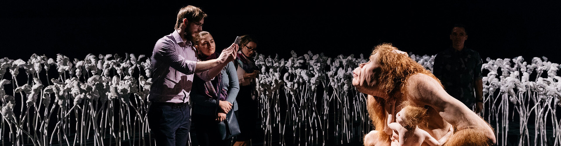
Melbourne artist Patricia Piccinini’s Curious Imaginings is expected to be one of the most talked about installations of the exhibit. Her style of “oddly captivating, somewhat grotesque, human-animal hybrid creature” is meant to be shocking and thought-provoking.

Piccinini’s interactive [emphasis mine] experience will “challenge us to explore the social impacts of emerging biotechnology and our ethical limits in an age where genetic engineering and digital technologies are already pushing the boundaries of humanity.”

Piccinini’s work will be displayed in the 105-year-old Patricia Hotel in Vancouver’s Strathcona neighbourhood. The 90-day ticketed exhibition [emphasis mine] is scheduled to open this September [2018].

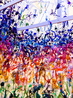
Given that this blog is focused on nanotechnology and other emerging technologies such as CRISPR, I’m focusing on Piccinini’s work and its art/science or sci-art status. This image from the GOMA Gallery where Piccinini’s ‘Curious Affection‘ installation is being shown from March 24 – Aug. 5, 2018 in Brisbane, Queensland, Australia may give you some sense of what one of her installations is like,

Courtesy: Queensland Art Gallery | Gallery of Modern Art (QAGOMA)

I spoke with Serena at the Vancouver Biennale office and asked about the ‘interactive’ aspect of Piccinini’s installation. She suggested the term ‘immersive’ as an alternative. In other words, you won’t be playing with the sculptures or pressing buttons and interacting with computer screens or robots. She also noted that the ticket prices have not been set yet and they are currently developing events focused on the issues raised by the installation. She knew that 2018 is the 200th anniversary of the publication of Mary Shelley’s Frankenstein but I’m not sure how the Biennale folks plan (or don’t plan)  to integrate any recognition of the novle’s impact on the discussions about ‘new’ technologies .They expect Piccinini will visit Vancouver. (Note 1: Piccinini’s work can  also be seen in a group exhibition titled: Frankenstein’s Birthday Party at the Hosfselt Gallery in San Francisco (California, US) from June 23 – August 11, 2018.  Note 2: I featured a number of international events commemorating the 200th anniversary of the publication of Mary Shelley’s novel, Frankenstein, in my Feb. 26, 2018 posting. Note 3: The term ‘Frankenfoods’ helped to shape the discussion of genetically modified organisms and food supply on this planet. It was a wildly successful campaign for activists affecting legislation in some areas of research. Scientists have not been as enthusiastic about the effects. My January 15, 2009 posting briefly traces a history of the term.)

The 2018 – 2020 Vancouver Biennale and science

A June 7, 2018 Vancouver Biennale news release provides more detail about the current series of exhibitions,

The Biennale is also committed to presenting artwork at the cutting edge of discussion and in keeping with the STEAM (science, technology, engineering, arts, math[ematics]) approach to integrating the arts and sciences. In August [2018], Colombian/American visual artist Jessica Angel will present her monumental installation Dogethereum Bridge at Hinge Park in Olympic Village. Inspired by blockchain technology, the artwork’s design was created through the integration of scientific algorithms, new developments in technology, and the arts. This installation, which will serve as an immersive space and collaborative hub for artists and technologists, will host a series of activations with blockchain as the inspirational jumping-off point.

In what is expected to become one of North America’s most talked-about exhibitions of the year, Melbourne artist Patricia Piccinini’s Curious Imaginings will see the intersection of art, science, and ethics. For the first time in the Biennale’s fifteen years of creating transformative experiences, and in keeping with the 2018-2020 theme of “re-IMAGE-n,” the Biennale will explore art in unexpected places by exhibiting in unconventional interior spaces.  The hyperrealist “world of oddly captivating, somewhat grotesque, human-animal hybrid creatures” will be the artist’s first exhibit in a non-museum setting, transforming a wing of the 105-year-old Patricia Hotel. Situated in Vancouver’s oldest neighbourbood of Strathcona, Piccinini’s interactive experience will “challenge us to explore the social impacts of emerging bio-technology and our ethical limits in an age where genetic engineering and digital technologies are already pushing the boundaries of humanity.” In this intimate hotel setting located in a neighborhood continually undergoing its own change, Curious Imaginings will empower visitors to personally consider questions posed by the exhibition, including the promises and consequences of genetic research and human interference. …

There are other pieces being presented at the Biennale but my special interest is in the art/sci pieces and, at this point, CRISPR.

Piccinini in more depth

You can find out more about Patricia Piccinini in her biography on the Vancouver Biennale website but I found this Char Larsson April 7, 2018 article for the Independent (UK) more informative (Note: A link has been removed),

Patricia Piccinini’s sculptures are deeply disquieting. Walking through Curious Affection, her new solo exhibition at Brisbane’s Gallery of Modern Art, is akin to entering a science laboratory full of DNA experiments. Made from silicone, fibreglass and even human hair, her sculptures are breathtakingly lifelike, however, we can’t be sure what life they are like. The artist creates an exuberant parallel universe where transgenic experiments flourish and human evolution has given way to genetic engineering and DNA splicing.

Curious Affection is a timely and welcome recognition of Piccinini’s enormous contribution to reaching back to the mid-1990s. Working across a variety of mediums including photography, video and drawing, she is perhaps best known for her hyperreal creations.

As a genre, hyperrealism depends on the skill of the artist to create the illusion of reality. To be truly successful, it must convince the spectator of its realness. Piccinini acknowledges this demand, but with a delightful twist. The excruciating attention to detail deliberately solicits our desire to look, only to generate unease, as her sculptures are imbued with a fascinating otherness. Part human, part animal, the works are uncannily familiar, but also alarmingly “other”.

Inspired by advances in genetically modified pigs to generate replacement organs for humans [also known as xenotransplantation], we are reminded that Piccinini has always been at the forefront of debates concerning the possibilities of science, technology and DNA cloning. She does so, however, with a warm affection and sense of humour, eschewing the hysterical anxiety frequently accompanying these scientific developments.

Beyond the astonishing level of detail achieved by working with silicon and fibreglass, there is an ethics at work here. Piccinini is asking us not to avert our gaze from the other, and in doing so, to develop empathy and understanding through the encounter.

I encourage anyone who’s interested to read Larsson’s entire piece (April 7, 2018 article).

According to her Wikipedia entry, Piccinini works in a variety of media including video, sound, sculpture, and more. She also has her own website.

Gene editing and xenotransplantation

Sarah Zhang’s June 8, 2018 article for The Atlantic provides a peek at the extraordinary degree of interest and competition in the field of gene editing and CRISPR ((clustered regularly interspaced short palindromic repeats))/Cas9 research (Note: A link has been removed),

China Is Genetically Engineering Monkeys With Brain Disorders

Guoping Feng applied to college the first year that Chinese universities reopened after the Cultural Revolution. It was 1977, and more than a decade’s worth of students—5.7 million—sat for the entrance exams. Feng was the only one in his high school to get in. He was assigned—by chance, essentially—to medical school. Like most of his contemporaries with scientific ambitions, he soon set his sights on graduate studies in the United States. “China was really like 30 to 50 years behind,” he says. “There was no way to do cutting-edge research.” So in 1989, he left for Buffalo, New York, where for the first time he saw snow piled several feet high. He completed his Ph.D. in genetics at the State University of New York at Buffalo.

Feng is short and slim, with a monk-like placidity and a quick smile, and he now holds an endowed chair in neuroscience at MIT, where he focuses on the genetics of brain disorders. His 45-person lab is part of the McGovern Institute for Brain Research, which was established in 2000 with the promise of a $350 million donation, the largest ever received by the university. In short, his lab does not lack for much.

Yet Feng now travels to China several times a year, because there, he can pursue research he has not yet been able to carry out in the United States. [emphasis mine] …

Feng had organized a symposium at SIAT [Shenzhen Institutes of Advanced Technology], and he was not the only scientist who traveled all the way from the United States to attend: He invited several colleagues as symposium speakers, including a fellow MIT neuroscientist interested in tree shrews, a tiny mammal related to primates and native to southern China, and Chinese-born neuroscientists who study addiction at the University of Pittsburgh and SUNY Upstate Medical University. Like Feng, they had left China in the ’80s and ’90s, part of a wave of young scientists in search of better opportunities abroad. Also like Feng, they were back in China to pursue a type of cutting-edge research too expensive and too impractical—and maybe too ethically sensitive—in the United States.

Here’s what precipitated Feng’s work in China, (from Zhang’s article; Note: Links have been removed)

At MIT, Feng’s lab worked on genetically engineering a monkey species called marmosets, which are very small and genuinely bizarre-looking. They are cheaper to keep due to their size, but they are a relatively new lab animal, and they can be difficult to train on lab tasks. For this reason, Feng also wanted to study Shank3 on macaques in China. Scientists have been cataloging the social behavior of macaques for decades, making it an obvious model for studies of disorders like autism that have a strong social component. Macaques are also more closely related to humans than marmosets, making their brains a better stand-in for those of humans.

The process of genetically engineering a macaque is not trivial, even with the advanced tools of CRISPR. Researchers begin by dosing female monkeys with the same hormones used in human in vitro fertilization. They then collect and fertilize the eggs, and inject the resulting embryos with CRISPR proteins using a long, thin glass needle. Monkey embryos are far more sensitive than mice embryos, and can be affected by small changes in the pH of the injection or the concentration of CRISPR proteins. Only some of the embryos will have the desired mutation, and only some will survive once implanted in surrogate mothers. It takes dozens of eggs to get to just one live monkey, so making even a few knockout monkeys required the support of a large breeding colony.

The first Shank3 macaque was born in 2015. Four more soon followed, bringing the total to five.

To visit his research animals, Feng now has to fly 8,000 miles across 12 time zones. It would be a lot more convenient to carry out his macaque research in the United States, of course, but so far, he has not been able to.

He originally inquired about making Shank3 macaques at the New England Primate Research Center, one of eight national primate research centers then funded by the National Institutes of Health in partnership with a local institution (Harvard Medical School, in this case). The center was conveniently located in Southborough, Massachusetts, just 20 miles west of the MIT campus. But in 2013, Harvard decided to shutter the center.

The decision came as a shock to the research community, and it was widely interpreted as a sign of waning interest in primate research in the United States. While the national primate centers have been important hubs of research on HIV, Zika, Ebola, and other diseases, they have also come under intense public scrutiny. Animal-rights groups like the Humane Society of the United States have sent investigators to work undercover in the labs, and the media has reported on monkey deaths in grisly detail. Harvard officially made its decision to close for “financial” reasons. But the announcement also came after the high-profile deaths of four monkeys from improper handling between 2010 and 2012. The deaths sparked a backlash; demonstrators showed up at the gates. The university gave itself two years to wind down their primate work, officially closing the center in 2015.

“They screwed themselves,” Michael Halassa, the MIT neuroscientist who spoke at Feng’s symposium, told me in Shenzhen. Wei-Dong Yao, another one of the speakers, chimed in, noting that just two years later CRISPR has created a new wave of interest in primate research. Yao was one of the researchers at Harvard’s primate center before it closed; he now runs a lab at SUNY Upstate Medical University that uses genetically engineered mouse and human stem cells, and he had come to Shenzhen to talk about restarting his addiction research on primates.

Here’s comes the competition (from Zhang’s article; Note: Links have been removed),

While the U.S. government’s biomedical research budget has been largely flat, both national and local governments in China are eager to raise their international scientific profiles, and they are shoveling money into research. A long-rumored, government-sponsored China Brain Project is supposed to give neuroscience research, and primate models in particular, a big funding boost. Chinese scientists may command larger salaries, too: Thanks to funding from the Shenzhen local government, a new principal investigator returning from overseas can get 3 million yuan—almost half a million U.S. dollars—over his or her first five years. China is even finding success in attracting foreign researchers from top U.S. institutions like Yale.

In the past few years, China has seen a miniature explosion of genetic engineering in monkeys. In Kunming, Shanghai, and Guangzhou, scientists have created monkeys engineered to show signs of Parkinson’s, Duchenne muscular dystrophy, autism, and more. And Feng’s group is not even the only one in China to have created Shank3 monkeys. Another group—a collaboration primarily between researchers at Emory University and scientists in China—has done the same.

Chinese scientists’ enthusiasm for CRISPR also extends to studies of humans, which are moving much more quickly, and in some cases under less oversight, than in the West. The first studies to edit human embryos and first clinical trials for cancer therapies using CRISPR have all happened in China. [emphases mine]

Some ethical issues are also covered (from Zhang’s article),

Parents with severely epileptic children had asked him if it would be possible to study the condition in a monkey. Feng told them what he thought would be technically possible. “But I also said, ‘I’m not sure I want to generate a model like this,’” he recalled. Maybe if there were a drug to control the monkeys’ seizures, he said: “I cannot see them seizure all the time.”

But is it ethical, he continued, to let these babies die without doing anything? Is it ethical to generate thousands or millions of mutant mice for studies of brain disorders, even when you know they will not elucidate much about human conditions?

Primates should only be used if other models do not work, says Feng, and only if a clear path forward is identified. The first step in his work, he says, is to use the Shank3 monkeys to identify the changes the mutations cause in the brain. Then, researchers might use that information to find targets for drugs, which could be tested in the same monkeys. He’s talking with the Oregon National Primate Research Center about carrying out similar work in the United States. ….[Note: I have a three-part series about CRISPR and germline editing* in the US, precipitated by research coming out of Oregon, Part 1, which links to the other parts, is here.]

Zhang’s June 8, 2018 article is excellent and I highly recommend reading it.

I touched on the topic of xenotransplanttaion in a commentary on a book about the science  of the television series, Orphan Black in a January 31,2018 posting (Note: A chimera is what you use to incubate a ‘human’ organ for transplantation or, more accurately, xenotransplantation),

On the subject of chimeras, the Canadian Broadcasting Corporation (CBC) featured a January 26, 2017 article about the pig-human chimeras on its website along with a video,

The end

I am very excited to see Piccinini’s work come to Vancouver. There have been a number of wonderful art and art/science installations and discussions here but this is the first one (I believe) to tackle the emerging gene editing technologies and the issues they raise. (It also fits in rather nicely with the 200th anniversary of the publication of Mary Shelley’s Frankenstein which continues to raise issues and stimulate discussion.)

In addition to the ethical issues raised in Zhang’s article, there are some other philosophical questions:

  • what does it mean to be human
  • if we are going to edit genes to create hybrid human/animals, what are they and how do they fit into our current animal/human schema
  • are you still human if you’ve had an organ transplant where the organ was incubated in a pig

There are also going to be legal issues. In addition to any questions about legal status, there are also fights about intellectual property such as the one involving Harvard & MIT’s [Massachusetts Institute of Technology] Broad Institute vs the University of California at Berkeley (March 15, 2017 posting)..

While I’m thrilled about the Piccinini installation, it should be noted the issues raised by other artworks hosted in this version of the Biennale are important. Happily, they have been broached here in Vancouver before and I suspect this will result in more nuanced  ‘conversations’ than are possible when a ‘new’ issue is introduced.

Bravo 2018 – 2020 Vancouver Biennale!

* Germline editing is when your gene editing will affect subsequent generations as opposed to editing out a mutated gene for the lifetime of a single individual.

Art/sci and CRISPR links

This art/science posting may prove of some interest:

The connectedness of living things: an art/sci project in Saskatchewan: evolutionary biology (February 16, 2018)

A selection of my CRISPR posts:

CRISPR and editing the germline in the US (part 1 of 3): In the beginning (August 15, 2017)

NOTE: An introductory CRISPR video describing how CRISPR/Cas9 works was embedded in part1.

Why don’t you CRISPR yourself? (January 25, 2018)

Editing the genome with CRISPR ((clustered regularly interspaced short palindromic repeats)-carrying nanoparticles (January 26, 2018)

Immune to CRISPR? (April 10, 2018)

Santiago Ramón y Cajal and the butterflies of the soul

The Cajal exhibit of drawings was here in Vancouver (Canada) this last fall (2017) and I still carry the memory of that glorious experience (see my Sept. 11, 2017 posting for more about the show and associated events). It seems Cajal’s drawings had a similar response in New York city, from a January 18, 2018 article by Roberta Smith for the New York Times,

It’s not often that you look at an exhibition with the help of the very apparatus that is its subject. But so it is with “The Beautiful Brain: The Drawings of Santiago Ramón y Cajal” at the Grey Art Gallery at New York University, one of the most unusual, ravishing exhibitions of the season.

The show finished its run on March 31, 2018 and is now on its way to the Massachusetts Institute of Technology (MIT) in Boston, Massachusetts for its opening on May 3, 2018. It looks like they have an exciting lineup of events to go along with the exhibit (from MIT’s The Beautiful Brain: The Drawings of Santiago Ramón y Cajal exhibit and event page),

SUMMER PROGRAMS

ONGOING

Spotlight Tours
Explorations led by local and Spanish scientists, artists, and entrepreneurs who will share their unique perspectives on particular aspects of the exhibition. (2:00 pm on select Tuesdays and Saturdays)

Tue, May 8 – Mark Harnett, Fred and Carole Middleton Career Development Professor at MIT and McGovern Institute Investigator Sat, May 26 – Marion Boulicault, MIT Graduate Student and Neuroethics Fellow in the Center for Sensorimotor Neural Engineering Tue, June 5 – Kelsey Allen, Graduate researcher, MIT Center for Brains, Minds, and Machines Sat, Jun 23 – Francisco Martin-Martinez, Research Scientist in MIT’s Laboratory for Atomistic & Molecular Mechanics and President of the Spanish Foundation for Science and Technology Jul 21 – Alex Gomez-Marin, Principal Investigator of the Behavior of Organisms Laboratory in the Instituto de Neurociencias, Spain Tue, Jul 31– Julie Pryor, Director of Communications at the McGovern Institute for Brain Research at MIT Tue, Aug 28 – Satrajit Ghosh, Principal Research Scientist at the McGovern Institute for Brain Research at MIT, Assistant Professor in the Department of Otolaryngology at Harvard Medical School, and faculty member in the Speech and Hearing Biosciences and Technology program in the Harvard Division of Medical Sciences

Idea Hub
Drop in and explore expansion microscopy in our maker-space.

Visualizing Science Workshop
Experiential learning with micro-scale biological images. (pre-registration required)

Gallery Demonstrations
Researchers share the latest on neural anatomy, signal transmission, and modern imaging techniques.

EVENTS

Teen Science Café: Mindful Matters
MIT researchers studying the brain share their mind-blowing findings.

Neuron Paint Night
Create a painting of cerebral cortex neurons and learn about the EyeWire citizen science game.

Cerebral Cinema Series
Hear from researchers and then compare real science to depictions on the big screen.

Brainy Trivia
Test your brain power in a night of science trivia and short, snappy research talks.

Come back to see our exciting lineup for the fall!

If you don’t have a chance to see the show or if you’d like a preview, I encourage you to read Smith’s article as it has embedded several Cajal drawings and rendered them exceptionally well.

For those who like a little contemporary (and related) science with their art, there’s a March 30, 2018 Harvard Medical Schoo (HMS)l news release by Kevin Jang (also on EurekAlert), Note: All links save one have been removed,

Drawing of the cells of the chick cerebellum by Santiago Ramón y Cajal, from “Estructura de los centros nerviosos de las aves,” Madrid, circa 1905


Modern neuroscience, for all its complexity, can trace its roots directly to a series of pen-and-paper sketches rendered by Nobel laureate Santiago Ramón y Cajal in the late 19th and early 20th centuries.

His observations and drawings exposed the previously hidden composition of the brain, revealing neuronal cell bodies and delicate projections that connect individual neurons together into intricate networks.

As he explored the nervous systems of various organisms under his microscope, a natural question arose: What makes a human brain different from the brain of any other species?

At least part of the answer, Ramón y Cajal hypothesized, lay in a specific class of neuron—one found in a dazzling variety of shapes and patterns of connectivity, and present in higher proportions in the human brain than in the brains of other species. He dubbed them the “butterflies of the soul.”

Known as interneurons, these cells play critical roles in transmitting information between sensory and motor neurons, and, when defective, have been linked to diseases such as schizophrenia, autism and intellectual disability.

Despite more than a century of study, however, it remains unclear why interneurons are so diverse and what specific functions the different subtypes carry out.

Now, in a study published in the March 22 [2018] issue of Nature, researchers from Harvard Medical School, New York Genome Center, New York University and the Broad Institute of MIT and Harvard have detailed for the first time how interneurons emerge and diversify in the brain.

Using single-cell analysis—a technology that allows scientists to track cellular behavior one cell at a time—the team traced the lineage of interneurons from their earliest precursor states to their mature forms in mice. The researchers identified key genetic programs that determine the fate of developing interneurons, as well as when these programs are switched on or off.

The findings serve as a guide for efforts to shed light on interneuron function and may help inform new treatment strategies for disorders involving their dysfunction, the authors said.

“We knew more than 100 years ago that this huge diversity of morphologically interesting cells existed in the brain, but their specific individual roles in brain function are still largely unclear,” said co-senior author Gordon Fishell, HMS professor of neurobiology and a faculty member at the Stanley Center for Psychiatric Research at the Broad.

“Our study provides a road map for understanding how and when distinct interneuron subtypes develop, giving us unprecedented insight into the biology of these cells,” he said. “We can now investigate interneuron properties as they emerge, unlock how these important cells function and perhaps even intervene when they fail to develop correctly in neuropsychiatric disease.”

A hippocampal interneuron. Image: Biosciences Imaging Gp, Soton, Wellcome Trust via Creative CommonsA hippocampal interneuron. Image: Biosciences Imaging Gp, Soton, Wellcome Trust via Creative Commons

Origins and Fates

In collaboration with co-senior author Rahul Satija, core faculty member of the New York Genome Center, Fishell and colleagues analyzed brain regions in developing mice known to contain precursor cells that give rise to interneurons.

Using Drop-seq, a single-cell sequencing technique created by researchers at HMS and the Broad, the team profiled gene expression in thousands of individual cells at multiple time points.

This approach overcomes a major limitation in past research, which could analyze only the average activity of mixtures of many different cells.

In the current study, the team found that the precursor state of all interneurons had similar gene expression patterns despite originating in three separate brain regions and giving rise to 14 or more interneuron subtypes alone—a number still under debate as researchers learn more about these cells.

“Mature interneuron subtypes exhibit incredible diversity. Their morphology and patterns of connectivity and activity are so different from each other, but our results show that the first steps in their maturation are remarkably similar,” said Satija, who is also an assistant professor of biology at New York University.

“They share a common developmental trajectory at the earliest stages, but the seeds of what will cause them to diverge later—a handful of genes—are present from the beginning,” Satija said.

As they profiled cells at later stages in development, the team observed the initial emergence of four interneuron “cardinal” classes, which give rise to distinct fates. Cells were committed to these fates even in the early embryo. By developing a novel computational strategy to link precursors with adult subtypes, the researchers identified individual genes that were switched on and off when cells began to diversify.

For example, they found that the gene Mef2c—mutations of which are linked to Alzheimer’s disease, schizophrenia and neurodevelopmental disorders in humans—is an early embryonic marker for a specific interneuron subtype known as Pvalb neurons. When they deleted Mef2c in animal models, Pvalb neurons failed to develop.

These early genes likely orchestrate the execution of subsequent genetic subroutines, such as ones that guide interneuron subtypes as they migrate to different locations in the brain and ones that help form unique connection patterns with other neural cell types, the authors said.

The identification of these genes and their temporal activity now provide researchers with specific targets to investigate the precise functions of interneurons, as well as how neurons diversify in general, according to the authors.

“One of the goals of this project was to address an incredibly fascinating developmental biology question, which is how individual progenitor cells decide between different neuronal fates,” Satija said. “In addition to these early markers of interneuron divergence, we found numerous additional genes that increase in expression, many dramatically, at later time points.”

The association of some of these genes with neuropsychiatric diseases promises to provide a better understanding of these disorders and the development of therapeutic strategies to treat them, a particularly important notion given the paucity of new treatments, the authors said.

Over the past 50 years, there have been no fundamentally new classes of neuropsychiatric drugs, only newer versions of old drugs, the researchers pointed out.

“Our repertoire is no better than it was in the 1970s,” Fishell said.

“Neuropsychiatric diseases likely reflect the dysfunction of very specific cell types. Our study puts forward a clear picture of what cells to look at as we work to shed light on the mechanisms that underlie these disorders,” Fishell said. “What we will find remains to be seen, but we have new, strong hypotheses that we can now test.”

As a resource for the research community, the study data and software are open-source and freely accessible online.

A gallery of the drawings of Santiago Ramón y Cajal is currently on display in New York City, and will open at the MIT Museum in Boston in May 2018.

Christian Mayer, Christoph Hafemeister and Rachel Bandler served as co-lead authors on the study.

This work was supported by the National Institutes of Health (R01 NS074972, R01 NS081297, MH071679-12, DP2-HG-009623, F30MH114462, T32GM007308, F31NS103398), the European Molecular Biology Organization, the National Science Foundation and the Simons Foundation.

Here’s link to and a citation for the paper,

Developmental diversification of cortical inhibitory interneurons by Christian Mayer, Christoph Hafemeister, Rachel C. Bandler, Robert Machold, Renata Batista Brito, Xavier Jaglin, Kathryn Allaway, Andrew Butler, Gord Fishell, & Rahul Satija. Nature volume 555, pages 457–462 (22 March 2018) doi:10.1038/nature25999 Published: 05 March 2018

This paper is behind a paywall.

Mott memristor

Mott memristors (mentioned in my Aug. 24, 2017 posting about neuristors and brainlike computing) gets more fulsome treatment in an Oct. 9, 2017 posting by Samuel K. Moore on the Nanoclast blog (found on the IEEE [Institute of Electrical and Electronics Engineers] website) Note: 1: Links have been removed; Note 2 : I quite like Moore’s writing style but he’s not for the impatient reader,

When you’re really harried, you probably feel like your head is brimful of chaos. You’re pretty close. Neuroscientists say your brain operates in a regime termed the “edge of chaos,” and it’s actually a good thing. It’s a state that allows for fast, efficient analog computation of the kind that can solve problems that grow vastly more difficult as they become bigger in size.

The trouble is, if you’re trying to replicate that kind of chaotic computation with electronics, you need an element that both acts chaotically—how and when you want it to—and could scale up to form a big system.

“No one had been able to show chaotic dynamics in a single scalable electronic device,” says Suhas Kumar, a researcher at Hewlett Packard Labs, in Palo Alto, Calif. Until now, that is.

He, John Paul Strachan, and R. Stanley Williams recently reported in the journal Nature that a particular configuration of a certain type of memristor contains that seed of controlled chaos. What’s more, when they simulated wiring these up into a type of circuit called a Hopfield neural network, the circuit was capable of solving a ridiculously difficult problem—1,000 instances of the traveling salesman problem—at a rate of 10 trillion operations per second per watt.

(It’s not an apples-to-apples comparison, but the world’s most powerful supercomputer as of June 2017 managed 93,015 trillion floating point operations per second but consumed 15 megawatts doing it. So about 6 billion operations per second per watt.)

The device in question is called a Mott memristor. Memristors generally are devices that hold a memory, in the form of resistance, of the current that has flowed through them. The most familiar type is called resistive RAM (or ReRAM or RRAM, depending on who’s asking). Mott memristors have an added ability in that they can also reflect a temperature-driven change in resistance.

The HP Labs team made their memristor from an 8-nanometer-thick layer of niobium dioxide (NbO2) sandwiched between two layers of titanium nitride. The bottom titanium nitride layer was in the form of a 70-nanometer wide pillar. “We showed that this type of memristor can generate chaotic and nonchaotic signals,” says Williams, who invented the memristor based on theory by Leon Chua.

(The traveling salesman problem is one of these. In it, the salesman must find the shortest route that lets him visit all of his customers’ cities, without going through any of them twice. It’s a difficult problem because it becomes exponentially more difficult to solve with each city you add.)

Here’s what the niobium dioxide-based Mott memristor looks like,

Photo: Suhas Kumar/Hewlett Packard Labs
A micrograph shows the construction of a Mott memristor composed of an 8-nanometer-thick layer of niobium dioxide between two layers of titanium nitride.

Here’s a link to and a citation for the paper,

Chaotic dynamics in nanoscale NbO2 Mott memristors for analogue computing by Suhas Kumar, John Paul Strachan & R. Stanley Williams. Nature 548, 318–321 (17 August 2017) doi:10.1038/nature23307 Published online: 09 August 2017

This paper is behind a paywall.

Nano- and neuro- together for nanoneuroscience

This is not the first time I’ve posted about nanotechnology and neuroscience (see this April 2, 2013 piece about then new brain science initiative in the US and Michael Berger’s  Nanowerk Spotlight article/review of an earlier paper covering the topic of nanotechnology and neuroscience).

Interestingly, the European Union (EU) had announced its two  $1B Euro research initiatives, the Human Brain Project and the Graphene Flagship (see my Jan. 28, 2013 posting about it),  months prior to the US brain research push. For those unfamiliar with the nanotechnology effort, graphene is a nanomaterial and there is high interest in its potential use in biomedical technology, thus partially connecting both EU projects.

In any event, Berger is highlighting a nanotechnology and neuroscience connection again in his Oct. 18, 2017 Nanowerk Spotlight article, or overview of, a new paper, which updates our understanding of the potential connections between the two fields (Note: A link has been removed),

Over the past several years, nanoscale analysis tools and in the design and synthesis of nanomaterials have generated optical, electrical, and chemical methods that can readily be adapted for use in neuroscience and brain activity mapping.

A review paper in Advanced Functional Materials (“Nanotechnology for Neuroscience: Promising Approaches for Diagnostics, Therapeutics and Brain Activity Mapping”) summarizes the basic concepts associated with neuroscience and the current journey of nanotechnology towards the study of neuron function by addressing various concerns on the significant role of nanomaterials in neuroscience and by describing the future applications of this emerging technology.

The collaboration between nanotechnology and neuroscience, though still at the early stages, utilizes broad concepts, such as drug delivery, cell protection, cell regeneration and differentiation, imaging and surgery, to give birth to novel clinical methods in neuroscience.

Ultimately, the clinical translation of nanoneuroscience implicates that central nervous system (CNS) diseases, including neurodevelopmental, neurodegenerative and psychiatric diseases, have the potential to be cured, while the industrial translation of nanoneuroscience indicates the need for advancement of brain-computer interface technologies.

Future Developing Arenas in Nanoneuroscience

The Brain Activity Map (BAM) Project aims to map the neural activity of every neuron across all neural circuits with the ultimate aim of curing diseases associated with the nervous system. The announcement of this collaborative, public-private research initiative in 2013 by President Obama has driven the surge in developing methods to elucidate neural circuitry. Three current developing arenas in the context of nanoneuroscience applications that will push such initiative forward are 1) optogenetics, 2) molecular/ion sensing and monitoring and 3) piezoelectric effects.

In their review, the authors discuss these aspects in detail.

Neurotoxicity of Nanomaterials

By engineering particles on the scale of molecular-level entities – proteins, lipid bilayers and nucleic acids – we can stereotactically interface with many of the components of cell systems, and at the cutting edge of this technology, we can begin to devise ways in which we can manipulate these components to our own ends. However, interfering with the internal environment of cells, especially neurons, is by no means simple.

“If we are to continue to make great strides in nanoneuroscience, functional investigations of nanomaterials must be complemented with robust toxicology studies,” the authors point out. “A database on the toxicity of materials that fully incorporates these findings for use in future schema must be developed. These databases should include information and data on 1) the chemical nature of the nanomaterials in complex aqueous environments; 2) the biological interactions of nanomaterials with chemical specificity; 3) the effects of various nanomaterial properties on living systems; and 4) a model for the simulation and computation of possible effects of nanomaterials in living systems across varying time and space. If we can establish such methods, it may be possible to design nanopharmaceuticals for improved research as well as quality of life.”

“However, challenges in nanoneuroscience are present in many forms, such as neurotoxicity; the inability to cross the blood-brain barrier [emphasis mine]; the need for greater specificity, bioavailability and short half-lives; and monitoring of disease treatment,” the authors conclude their review. “The nanoneurotoxicity surrounding these nanomaterials is a barrier that must be overcome for the translation of these applications from bench-to-bedside. While the challenges associated with nanoneuroscience seem unending, they represent opportunities for future work.”

I have a March 26, 2015 posting about Canadian researchers breaching the blood-brain barrier and an April 13, 2016 posting about US researchers at Cornell University also breaching the blood-brain barrier. Perhaps the “inability” mentioned in this Spotlight article means that it can’t be done consistently or that it hasn’t been achieved on humans.

Here’s a link to and a citation for the paper,

Nanotechnology for Neuroscience: Promising Approaches for Diagnostics, Therapeutics and Brain Activity Mapping by Anil Kumar, Aaron Tan, Joanna Wong, Jonathan Clayton Spagnoli, James Lam, Brianna Diane Blevins, Natasha G, Lewis Thorne, Keyoumars Ashkan, Jin Xie, and Hong Liu. Advanced Functional Materials Volume 27, Issue 39, October 19, 2017 DOI: 10.1002/adfm.201700489 Version of Record online: 14 AUG 2017

© 2017 WILEY-VCH Verlag GmbH & Co. KGaA, Weinheim

I took a look at the authors’ information and found that most of these researchers are based in  China and in the UK, with a sole researcher based in the US.

Narrating neuroscience in Toronto (Canada) on Oct. 20, 2017 and knitting a neuron

What is it with the Canadian neuroscience community? First, there’s The Beautiful Brain an exhibition of the extraordinary drawings of Santiago Ramón y Cajal (1852–1934) at the Belkin Gallery on the University of British Columbia (UBC) campus in Vancouver and a series of events marking the exhibition (for more see my Sept. 11, 2017 posting ; scroll down about 30% for information about the drawings and the events still to come).

I guess there must be some money floating around for raising public awareness because now there’s a neuroscience and ‘storytelling’ event (Narrating Neuroscience) in Toronto, Canada. From a Sept. 25, 2017 ArtSci Salon announcement (received via email),

With NARRATING NEUROSCIENCE we plan to initiate a discussion on the  role and the use of storytelling and art (both in verbal and visual  forms) to communicate abstract and complex concepts in neuroscience to  very different audiences, ranging from fellow scientists, clinicians and patients, to social scientists and the general public. We invited four guests to share their research through case studies and experiences stemming directly from their research or from other practices they have adopted and incorporated into their research, where storytelling and the arts have played a crucial role not only in communicating cutting edge research in neuroscience, but also in developing and advancing it.

OUR GUESTS

MATTEO FARINELLA, PhD, Presidential Scholar in Society and Neuroscience – Columbia University

SHELLEY WALL , AOCAD, MSc, PhD – Assistant professor, Biomedical Communications Graduate Program and Department of Biology, UTM

ALFONSO FASANO, MD, PhD, Associate Professor – University of Toronto Clinician Investigator – Krembil Research Institute Movement Disorders Centre – Toronto Western Hospital

TAHANI BAAKDHAH, MD, MSc, PhD candidate – University of Toronto

DATE: October 20, 2017
TIME: 6:00-8:00 pm
LOCATION: The Fields Institute for Research in Mathematical Sciences
222 College Street, Toronto, ON

Events Facilitators: Roberta Buiani and Stephen Morris (ArtSci Salon) and Nina Czegledy (Leonardo Network)

TAHANI BAAKDHAH is a PhD student at the University of Toronto studying how the stem cells built our retina during development, the mechanism by which the light sensing cells inside the eye enable us to see this beautiful world and how we can regenerate these cells in case of disease or injury.

MATTEO FARINELLA combines a background in neuroscience with a lifelong passion for drawing, making comics and illustrations about the brain. He is the author of _Neurocomic_ (Nobrow 2013) published with the support of the Wellcome Trust, _Cervellopoli_ (Editoriale Scienza 2017) and he has collaborated with universities and educational institutions around
the world to make science more clear and accessible. In 2016 Matteo joined Columbia University as a Presidential Scholar in Society and Neuroscience, where he investigates the role of visual narratives in science communication. Working with science journalists, educators and cognitive neuroscientists he aims to understand how these tools may
affect the public perception of science and increase scientific literacy (cartoonscience.org [2]).

ALFONSO FASANO graduated from the Catholic University of Rome, Italy, in 2002 and became a neurologist in 2007. After a 2-year fellowship at the University of Kiel, Germany, he completed a PhD in neuroscience at the Catholic University of Rome. In 2013 he joined the Movement Disorder Centre at Toronto Western Hospital, where he is the co-director of the
surgical program for movement disorders. He is also an associate professor of medicine in the Division of Neurology at the University of Toronto and clinician investigator at the Krembil Research Institute. Dr. Fasano’s main areas of interest are the treatment of movement  disorders with advanced technology (infusion pumps and neuromodulation), pathophysiology and treatment of tremor and gait disorders. He is author of more than 170 papers and book chapters. He is principal investigator of several clinical trials.

SHELLEY WALL is an assistant professor in the University of Toronto’s Biomedical Communications graduate program, a certified medical illustrator, and inaugural Illustrator-in-Residence in the Faculty of Medicine, University of Toronto. One of her primary areas of research, teaching, and creation is graphic medicine—the intersection of comics with illness, medicine, and caregiving—and one of her ongoing projects is a series of comics about caregiving and young onset Parkinson’s disease.

You can register for this free Toronto event here.

One brief observation, there aren’t any writers (other than academics) or storytellers included in this ‘storytelling’ event. The ‘storytelling’ being featured is visual. To be blunt I’m not of the ‘one picture is worth a thousand words’ school of thinking (see my Feb. 22, 2011 posting). Yes, sometimes pictures are all you need but that tiresome aphorism which suggests  communication can be reduced to one means of communication really needs to be retired. As for academic writing, it’s not noted for its storytelling qualities or experimentation. Academics are not judged on their writing or storytelling skills although there are some who are very good.

Getting back to the Toronto event, they seem to have the visual part of their focus  ” … discussion on the  role and the use of storytelling and art (both in verbal and visual  forms) … ” covered. Having recently attended a somewhat similar event in Vancouver, which was announced n my Sept. 11, 2017 posting, there were some exciting images and ideas presented.

The ArtSci Salon folks also announced this (from the Sept. 25, 2017 ArtSci Salon announcement; received via email),

ATTENTION ARTSCI SALONISTAS AND FANS OF ART AND SCIENCE!!
CALL FOR KNITTING AND CROCHET LOVERS!

In addition to being a PhD student at the University of Toronto, Tahani Baakdhah is a prolific knitter and crocheter and has been the motor behind two successful Knit-a-Neuron Toronto initiatives. We invite all Knitters and Crocheters among our ArtSci Salonistas to pick a pattern
(link below) and knit a neuron (or 2! Or as many as you want!!)

http://bit.ly/2y05hRR

BRING THEM TO OUR OCTOBER 20 ARTSCI SALON!
Come to the ArtSci Salon and knit there!
You can’t come?
Share a picture with @ArtSci_Salon @SciCommTO #KnitANeuronTO [3] on
social media
Or…Drop us a line at artscisalon@gmail.com !

I think it’s been a few years since my last science knitting post. No, it was Oct. 18, 2016. Moving on, I found more neuron knitting while researching this piece. Here’s the Neural Knitworks group, which is part of Australia’s National Science Week (11-19 August 2018) initiative (from the Neural Knitworks webpage),

Neural Knitworks is a collaborative project about mind and brain health.

Whether you’re a whiz with yarn, or just discovering the joy of craft, now you can crochet wrap, knit or knot—and find out about neuroscience.

During 2014 an enormous number of handmade neurons were donated (1665 in total!) and used to build a giant walk-in brain, as seen here at Hazelhurst Gallery [scroll to end of this post]. Since then Neural Knitworks have been held in dozens of communities across Australia, with installations created in Queensland, the ACT, Singapore, as part of the Cambridge Science Festival in the UK and in Philadelphia, USA.

In 2017, the Neural Knitworks team again invites you to host your own home-grown Neural Knitwork for National Science Week*. Together we’ll create a giant ‘virtual’ neural network by linking your displays visually online.

* If you wish to host a Neural Knitwork event outside of National Science Week or internationally we ask that you contact us to seek permission to use the material, particularly if you intend to create derivative works or would like to exhibit the giant brain. Please outline your plans in an email.

Your creation can be big or small, part of a formal display, or simply consist of neighbourhood neuron ‘yarn-bombings’. Knitworks can be created at home, at work or at school. No knitting experience is required and all ages can participate.

See below for how to register your event and download our scientifically informed patterns.

What is a neuron?

Neurons are electrically excitable cells of the brain, spinal cord and peripheral nerves. The billions of neurons in your body connect to each other in neural networks. They receive signals from every sense, control movement, create memories, and form the neural basis of every thought.

Check out the neuron microscopy gallery for some real-world inspiration.

What happens at a Neural Knitwork?

Neural Knitworks are based on the principle that yarn craft, with its mental challenges, social connection and mindfulness, helps keep our brains and minds sharp, engaged and healthy.

Have fun as you

  • design your own woolly neurons, or get inspired by our scientifically-informed knitting, crochet or knot patterns;
  • natter with neuroscientists and teach them a few of your crafty tricks;
  • contribute to a travelling textile brain exhibition;
  • increase your attention span and test your memory.

Calm your mind and craft your own brain health as you

  • forge friendships;
  • solve creative and mental challenges;
  • practice mindfulness and relaxation;
  • teach and learn;
  • develop eye-hand coordination and fine motor dexterity.

Interested in hosting a Neural Knitwork?

  1. Log your event on the National Science Week calendar to take advantage of multi-channel promotion.
  2. Share the link^ for this Neural Knitwork page on your own website or online newsletter and add information your own event details.
  3. Use this flyer template (2.5 MB .docx) to promote your event in local shop windows and on noticeboards.
  4. Read our event organisers toolbox for tips on hosting a successful event.
  5. You’ll need plenty of yarn, needles, copies of our scientifically-based neuron crafting pattern books (3.4 MB PDF) and a comfy spot in which to create.
  6. Gather together a group of friends who knit, crochet, design, spin, weave and anyone keen to give it a go. Those who know how to knit can teach others how to do it, and there’s even an easy no knit pattern that you can knot.
  7. Download a neuroscience podcast to listen to, and you’ve got a Neural Knitwork!
  8. Join the Neural Knitworks community on Facebook  to share and find information about events including public talks featuring neuroscientists.
  9. Tweet #neuralknitworks to show us your creations.
  10. Find display ideas in the pattern book and on our Facebook page.

Finally,, the knitted neurons from Australia’s 2014 National Science Week brain exhibit,

[downloaded from https://www.scienceweek.net.au/neural-knitworks/]

ETA Oct. 24, 2017: If you’re interested on how the talk was received, there’s an Oct. 24, 2017 posting by Magosia Pakulska for the Research2Reality blog.

Oops—Greg Gage does it again! With a ‘neuroscience’ talk for TED and launch for the Plant SpikerBox

I’ve written a couple times about Greg Gage and his Backyard Brains,  first, in a March 28, 2012 posting (scroll down about 40% of the way for the mention of the first [?] ‘SpikerBox’) and, most recently, in a June 26, 2013 posting (scroll down about 25% of the way for the mention of a RoboRoach Kickstater project from Backyard Brains) which also featured the launch of a new educational product and a TED [technology education design] talk.

Here’s the latest from an Oct. 10, 2017 news release (received via email),

Backyard Brains Releases Plant SpikerBox, unlocking the Secret Electrical Language used in Plants

The first consumer device to investigate how plants create behaviors through electrophysiology and to enable interspecies plant to plant communication.

ANN ARBOR, MI, OCTOBER 10, 2017–Today Backyard Brains launched the Plant SpikerBox, the first ever science kit designed to reveal the wonderful nature behind plant behavior through electrophysiology experiments done at home or in the classroom. The new SpikerBox launched alongside three new experiments, enabling users to explore Venus Flytrap and Sensitive Mimosa signals and to perform a jaw-dropping Interspecies Plant-Plant-Communicator experiment. The Plant SpikerBox and all three experiments are featured in a live talk from TED2017 given by Backyard Brains CEO and cofounder Dr. Greg Gage which was released today on ​​https://ted.com.

Backyard Brains received viral attention for their previous videos, TED talks, and for their mission to create hands-on neuroscience experiments for everyone. The company (run by professional neuroscientists) produces consumer-friendly versions of expensive graduate lab equipment used at top research universities around the world. The new plant experiments and device facilitate the growing movement of DIY [do it yourself] scientists, made up of passionate amateurs, students, parents, and teachers.

Like previous inventions, the Plant SpikerBox is extremely easy to use, making it accessible for students as young as middle school. The device works by recording the electrical activity responsible for different plant behaviors. For example, the Venus Flytrap uses an electrical signal to determine if prey has landed in its trap; the SpikerBox reveals these invisible messages and allows you to visualize them on your mobile device. For the first time ever, you can peer into the fascinating world of plant signaling and plant behaviors.

The new SpikerBox features an “Interspecies Plant-Plant-Communicator” which demonstrates the ubiquitous nature of electrical signaling seen in humans, insects, and plants. With this device, one can capture the electrical message (called an action potential) from one plant’s behavior, and send it to a different plant to activate another behavior.

Co-founder and CEO Greg Gage explains, “Itis surprising to many people that plants use electrical messages similar to those used by the neurons in our brains. I was shocked to hear that. Many neuroscientists are. But if you think about it, it [sic] does make sense. Our nervous system evolved to react quickly. Electricity is fast. The plants we are studying also need to react quickly, so it makes sense they would develop a similar system. To be clear: No, plants don’t have brains, but they do exhibit behaviors and they do use electric messages called ‘Action Potentials’ like we do to send information. The benefit of these plant experiments then is twofold: First, we can simply demonstrate fundamental neuroscience principles, and second, we can spread the wonder of understanding how living creatures work and hopefully encourage others to make a career in life sciences!”

The Plant SpikerBox is a trailblazer, bringing plant electrophysiology to the public for the first time ever. It is designed to work with the Backyard Brains SpikeRecorder software which is available to download for free on their website or in mobile app stores. The three plant experiments are just a few of the dozens of free experiments available on the Backyard Brains website. The Plant SpikerBox is available now for $149.99.

About Backyard Brains

A staggering 1 in 5 people will develop a neurological disorder in their lifetime, making the need for neuroscience studies urgent. Backyard Brains passionately responds with their motto “Neuroscience for Everyone,” providing exposure, education, and experiment kits to students of all ages. Founded in 2010 in Ann Arbor, MI by University of Michigan Neuroscience graduate students Greg Gage and Tim Marzullo, Backyard Brains have been dubbed Champions of Change at an Obama White House ceremony and have won prestigious awards from the National Institutes of Health and the Society for Neuroscience. To learn more, visit BackyardBrains.com

You can find an embedded video of Greg Gage’s TED talk and Plant SpikerBox launch along with links to experiments you could run with it on Backyard Brains’ Plant SpikerBox product page.

For a sample of what they have on offer, here’s an excerpt from the Venus Flytrap Electrophysiology experiment webpage (Note: Links have been removed),

Background

Your nervous system allows you to sense and respond quickly to the environment around you. You have a nervous system, animals have nervous systems, but plants do not. But not having a nervous system does not mean you cannot sense and respond to the world. Plants can certainly sense the environment around them and move. You have seen your plants slowly turn their leaves towards sunlight by the window over a week, open their flowers in the day, and close their flowers during the night. Some plants can move in much more dramatic fashion, such as the Venus Flytrap and the Sensitive Mimosa.

The Venus Flytrap comes from the swamps of North Carolina, USA, and lives in very nutrient-poor, water-logged soil. It photosynthesizes like other plants, but it can’t always rely on the sunlight for food. To supplement its food supply it traps and eats insects, extracting from them the nitrogen and phosphorous needed to form plant food (amino acids, nucleic acids, and other molecules).

If you look closely at the Venus Flytrap, you will notice it has very tiny “Trigger Hairs” inside its trap leaves.

If a wayward, unsuspecting insect touches a trigger hair, an Action Potential occurs in the leaves. This is a different Action Potential than what we are used to seeing in neurons, as it’s based on the movement of calcium, potassium, and chloride ions (vs. movement of potassium and sodium as in the Action Potentials of neurons and muscles), and it is muuuuuuuuucccchhhhhh longer than anything we’ve seen before.

If the trigger hair is touched twice within 20 seconds (firing two Action Potentials within 20 seconds), the trap closes. The trap is not closing due to muscular action (plants do not have muscles), but rather due to an osmotic, rapid change in the shape of curvature of the trap leaves. Interestingly, the firing of Action Potentials is not always reliable, depending on time of year, temperature, health of plant, and/or other factors. Quite different from we humans, Action Potential failure is not devastating to a Venus Flytrap.

We can observe this plant Action Potential using our Plant SpikerBox. Welcome to the Brave New World of Plant Electrophysiology.

Downloads

Before you begin, make sure you have the Backyard Brains SpikeRecorder. The Backyard Brains SpikeRecorder program allows you to visualize and save data on your computer when doing experiments.

….

I did feel a bit sorry for the Venus Flytrap in Greg Gage’s TED talk which was fooled into closing its trap. According to Gage, the Venus Flytrap has limited number of times it can close its trap and after the last time, it dies. On the other hand, I eat meat and use leather goods so there is not pedestal for me to perch on.

For anyone who caught the Brittany Spears reference in the headline in this posting,

From exploring outer space with Brittany Spears to exploring plant communication and neuroscience in your back yard, science can be found in many different places.

Art in the details: A look at the role of art in science—a Sept. 19, 2017 Café Scientifique event in Vancouver, Canada

The Sept. 19, 2017 Café Scientifique event, “Art in the Details A look at the role of art in science,” in Vancouver seems to be part of a larger neuroscience and the arts program at the University of British Columbia. First, the details about the Sept. 13, 2017 event from the eventful Vancouver webpage,

Café Scientifique – Art in the Details: A look at the role of art in science

Art in the Details: A look at the role of art in science With so much beauty in the natural world, why does the misconception that art and science are vastly different persist? Join us for discussion and dessert as we hear from artists, researchers and academic professionals about the role art has played in scientific research – from the formative work of Santiago Ramon Y Cajal to modern imaging, and beyond – and how it might help shape scientific understanding in the future. September 19th, 2017  7:00 – 9:00 pm (doors open at 6:45pm)  TELUS World of Science [also known as Science World], 1455 Quebec St., Vancouver, BC V6A 3Z7 Free Admission [emphasis mine] Experts Dr Carol-Ann Courneya Associate Professor in the Department of Cellular and Physiological Science and Assistant Dean of Student Affairs, Faculty of Medicine, University of British Columbia   Dr Jason Snyder  Assistant Professor, Department of Psychology, University of British Columbia http://snyderlab.com/   Dr Steven Barnes Instructor and Assistant Head—Undergraduate Affairs, Department of Psychology, University of British Columbia http://stevenjbarnes.com/   Moderated By   Bruce Claggett Senior Managing Editor, NEWS 1130   This evening event is presented in collaboration with the Djavad Mowafaghian Centre for Brain Health. Please note: this is a private, adult-oriented event and TELUS World of Science will be closed during this discussion.

The Art in the Details event page on the Science World website provides a bit more information about the speakers (mostly in the form of links to their webpage),,

Experts

Dr Carol-Ann Courneya
Associate Professor in the Department of Cellular and Physiological Science and Assistant Dean of Student Affairs, Faculty of Medicine, University of British Columbia

Dr Jason Snyder 

Assistant Professor, Department of Psychology, University of British Columbi

Dr Steven Barnes

Instructor, Department of Psychology, University of British Columbia

Moderated By  

Bruce Claggett

Senior Managing Editor, NEWS 1130

Should you click though to obtain tickets from either the eventful Vancouver or Science World websites, you’ll find the event is sold out but perhaps the organizers will include a waitlist.

Even if you can’t get a ticket, there’s an exhibition of Santiago Ramon Y Cajal’s work (from the Djavad Mowafaghian Centre for Brain Health’s Beautiful brain’s webpage),

Drawings of Santiago Ramón y Cajal to be shown at UBC

Santiago Ramón y Cajal, injured Purkinje neurons, 1914, ink and pencil on paper. Courtesy of Instituto Cajal (CSIC).

Pictured: Santiago Ramón y Cajal, injured Purkinje neurons, 1914, ink and pencil on paper. Courtesy of Instituto Cajal (CSIC).

The Beautiful Brain is the first North American museum exhibition to present the extraordinary drawings of Santiago Ramón y Cajal (1852–1934), a Spanish pathologist, histologist and neuroscientist renowned for his discovery of neuron cells and their structure, for which he was awarded the Nobel Prize in Physiology and Medicine in 1906. Known as the father of modern neuroscience, Cajal was also an exceptional artist. He combined scientific and artistic skills to produce arresting drawings with extraordinary scientific and aesthetic qualities.

A century after their completion, Cajal’s drawings are still used in contemporary medical publications to illustrate important neuroscience principles, and continue to fascinate artists and visual art audiences. Eighty of Cajal’s drawings will be accompanied by a selection of contemporary neuroscience visualizations by international scientists. The Morris and Helen Belkin Art Gallery exhibition will also include early 20th century works that imaged consciousness, including drawings from Annie Besant’s Thought Forms (1901) and Charles Leadbeater’s The Chakras (1927), as well as abstract works by Lawren Harris that explored his interest in spirituality and mysticism.

After countless hours at the microscope, Cajal was able to perceive that the brain was made up of individual nerve cells or neurons rather than a tangled single web, which was only decisively proven by electron microscopy in the 1950s and is the basis of neuroscience today. His speculative drawings stemmed from an understanding of aesthetics in their compressed detail and lucid composition, as he laboured to clearly represent matter and processes that could not be seen.

In a special collaboration with the Morris and Helen Belkin Art Gallery and the VGH & UBC Hospital Foundation this project will encourage meaningful dialogue amongst artists, curators, scientists and scholars on concepts of neuroplasticity and perception. Public and Academic programs will address the emerging field of art and neuroscience and engage interdisciplinary research of scholars from the sciences and humanities alike.

“This is an incredible opportunity for the neuroscience and visual arts communities at the University and Vancouver,” says Dr. Brian MacVicar, who has been working diligently with Director Scott Watson at the Morris and Helen Belkin Art Gallery and with his colleagues at the University of Minnesota for the past few years to bring this exhibition to campus. “Without Cajal’s impressive body of work, our understanding of the anatomy of the brain would not be so well-formed; Cajal’s legacy has been of critical importance to neuroscience teaching and research over the past century.”

A book published by Abrams accompanies the exhibition, containing full colour reproductions of all 80 of the exhibition drawings, commentary on each of the works and essays on Cajal’s life and scientific contributions, artistic roots and achievements and contemporary neuroscience imaging techniques.

Cajal’s work will be on display at the Morris and Helen Belkin Art Gallery from September 5 to December 3, 2017.

Join the UBC arts and neuroscience communities for a free symposium and dance performance celebrating The Beautiful Brain at UBC on September 7. [link removed]

The Beautiful Brain: The Drawings of Santiago Ramón y Cajal was developed by the Frederick R. Weisman Art Museum, University of Minnesota with the Instituto Cajal. The exhibition at the Morris and Helen Belkin Art Gallery, University British Columbia is presented in partnership with the Djavad Mowafaghian Centre for Brain Health with support from the VGH & UBC Hospital Foundation. We gratefully acknowledge the generous support of the Canada Council for the Arts, the British Columbia Arts Council and Belkin Curator’s Forum members.

The Morris and Helen Belkin Art Gallery’s Beautiful Brain webpage has a listing of upcoming events associated with the exhibition as well as instructions on how to get there (if you click on About),

SEMINAR & READING GROUP: Plasticity at SFU Vancouver and 221A: Wednesdays, October 4, 18, November 1, 15 and 21 at 7 pm

CONVERSATION with Anthony Phillips and Timothy Taylor: Wednesday, October 11, 2017 at 7 pm

LECTURE with Catherine Malabou at the Liu Institute: Thursday, November 23 at 6 pm

CONCERT with UBC Contemporary Players: Friday, December 1 at 2 pm

Cajal was also an exceptional artist and studied as a teenager at the Academy of Arts in Huesca, Spain. He combined scientific and artistic skills to produce arresting drawings with extraordinary scientific and aesthetic qualities. A century after their completion, his drawings are still used in contemporary medical publications to illustrate important neuroscience principles, and continue to fascinate artists and visual art audiences. Eighty of Cajal’s drawings are accompanied by a selection of contemporary neuroscience visualizations by international scientists.

Organizationally, this seems a little higgledy piggledy with the Cafe Scientifique event found on some sites, the Belkin Gallery events found on one site, and no single listing of everything on any one site for the Beautiful Brain. Please let me know if you find something I’ve missed.

Multi-level thinking in science—the art of seeing systems

I’ve quickly read Michael Edgeworth McIntyre’s paper on multi-level thinking and find it provides fascinating insight and some good writing style (I’ve provided a few excerpts from the paper further down in the posting).

Here’s more about the paper from an Aug. 17, 2017 Institute of Atmospheric Physics, Chinese Academy of Sciences press release on EurekAlert,

An unusual paper “On multi-level thinking and scientific understanding” appears in the October issue of Advances in Atmospheric Sciences. The author is Professor Michael Edgeworth McIntyre from University of Cambridge, whose work in atmospheric dynamics is well known. He has also had longstanding interests in astrophysics, music, perception psychology, and biological evolution.

The paper touches on a range of deep questions within and outside the atmospheric sciences. They include insights into the nature of science itself, and of scientific understanding — what it means to understand a scientific problem in depth — and into the communication skills necessary to convey that understanding and to mediate collaboration across specialist disciplines.

The paper appears in a Special Issue arising from last year’s Symposium held in Nanjing to commemorate the life of Professor Duzheng YE, who was well known as a national and international scientific leader and for his own wide range of interests, within and outside the atmospheric sciences. The symposium was organized by the Institute of Atmospheric Physics (IAP), Chinese Academy of Sciences, where Prof. YE had worked nearly 70 years before he passed away. Upon the invitation of Prof. Jiang ZHU, the Director General of IAP, also the Editor-in-Chief of Advances in Atmospheric Sciences (AAS), Prof. McIntyre agreed to contribute a review paper to an AAS special issue commemorating the centenary of Duzheng YE’s birth. Prof. YE was also the founding Editor-in-Chief of this journal.

One of Professor McIntyre’s themes is that we all have unconscious mathematics, including Euclidean geometry and the calculus of variations. This is easy to demonstrate and is key to understanding not only how science works but also, for instance, how music works. Indeed, it reveals some of the deepest connections between music and mathematics, going beyond the usual remarks about number-patterns. All this revolves around the biological significance of what Professor McIntyre calls the “organic-change principle”.

Further themes include the scientific value of looking at a problem from more than one viewpoint, and the need to use more than one level of description. Many scientific and philosophical controversies stem from confusing one level of description with another, for instance applying arguments to one level that belong on another. This confusion can be especially troublesome when it comes to questions about human biology and human nature, and about what Professor YE called multi-level “orderly human activities”.

Related to all these points are the contrasting modes of perception and understanding offered by the brain’s left and right hemispheres. Our knowledge of their functioning has progressed far beyond the narrow clichés of popular culture, thanks to recent work in the neurosciences. The two hemispheres automatically give us different levels of description, and complementary views of a problem. Good science takes advantage of this. When the two hemispheres cooperate, with each playing to its own strengths, our problem-solving is at its most powerful.

The paper ends with three examples of unconscious assumptions that have impeded scientific progress in the past. Two of them are taken from Professor McIntyre’s main areas of research. A third is from biology.

Here’s a link to and a citation for the paper,

On multi-level thinking and scientific understanding by Michael Edgeworth McIntyre. Advances in Atmospheric Sciences October 2017, Volume 34, Issue 10, pp 1150–1158 DOI: https://doi.org/10.1007/s00376-017-6283-3

This paper is open access.

To give you a sense of his writing and imagination, I’ve excerpted a few paragraphs from p. 1153 but first you need to see this .gif (he provides a number of ways to watch the .gif in his text but I think it’s easier to watch the copy of the one he has on his website),

Now for the excerpt,

Here is an example to show what I mean. It is a classic in experimental psychology, from the work of Professor Gunnar JOHANSSON in the 1970s. …

As soon as the twelve dots start moving, everyone with normal vision sees a person walking. This immediately illustrates several things. First, it illustrates that we all make unconscious assumptions. Here, we unconsciously assume a particular kind of three-dimensional motion. In this case the unconscious assumption is completely involuntary. We cannot help seeing a person walking, despite knowing that it is only twelve moving dots.

The animation also shows that we have unconscious mathematics, Euclidean geometry in this case. In order to generate the percept of a person walking, your brain has to fit a mathematical model to the incoming visual data, in this case a mathematical model based on Euclidean geometry. (And the model-fitting process is an active, and highly complex, predictive process most of which is inaccessible to conscious introspection.)

This brings me to the most central point in our discussion. Science does essentially the same thing. It fits models to data. So science is, in the most fundamental possible sense, an extension of ordinary perception. That is a simple way of saying what was said many decades ago by great thinkers such as Professor Sir Karl POPPER….

I love that phase “unconscious mathematics” for the way it includes even those of us who would never dream of thinking we had any kind of mathematics. I encourage you to read his paper in its entirety, which does include a little technical language in a few spots but the overall thesis is clear and easily understood.

Brain stuff: quantum entanglement and a multi-dimensional universe

I have two brain news bits, one about neural networks and quantum entanglement and another about how the brain operates in* more than three dimensions.

Quantum entanglement and neural networks

A June 13, 2017 news item on phys.org describes how machine learning can be used to solve problems in physics (Note: Links have been removed),

Machine learning, the field that’s driving a revolution in artificial intelligence, has cemented its role in modern technology. Its tools and techniques have led to rapid improvements in everything from self-driving cars and speech recognition to the digital mastery of an ancient board game.

Now, physicists are beginning to use machine learning tools to tackle a different kind of problem, one at the heart of quantum physics. In a paper published recently in Physical Review X, researchers from JQI [Joint Quantum Institute] and the Condensed Matter Theory Center (CMTC) at the University of Maryland showed that certain neural networks—abstract webs that pass information from node to node like neurons in the brain—can succinctly describe wide swathes of quantum systems.

An artist’s rendering of a neural network with two layers. At the top is a real quantum system, like atoms in an optical lattice. Below is a network of hidden neurons that capture their interactions (Credit: E. Edwards/JQI)

A June 12, 2017 JQI news release by Chris Cesare, which originated the news item, describes how neural networks can represent quantum entanglement,

Dongling Deng, a JQI Postdoctoral Fellow who is a member of CMTC and the paper’s first author, says that researchers who use computers to study quantum systems might benefit from the simple descriptions that neural networks provide. “If we want to numerically tackle some quantum problem,” Deng says, “we first need to find an efficient representation.”

On paper and, more importantly, on computers, physicists have many ways of representing quantum systems. Typically these representations comprise lists of numbers describing the likelihood that a system will be found in different quantum states. But it becomes difficult to extract properties or predictions from a digital description as the number of quantum particles grows, and the prevailing wisdom has been that entanglement—an exotic quantum connection between particles—plays a key role in thwarting simple representations.

The neural networks used by Deng and his collaborators—CMTC Director and JQI Fellow Sankar Das Sarma and Fudan University physicist and former JQI Postdoctoral Fellow Xiaopeng Li—can efficiently represent quantum systems that harbor lots of entanglement, a surprising improvement over prior methods.

What’s more, the new results go beyond mere representation. “This research is unique in that it does not just provide an efficient representation of highly entangled quantum states,” Das Sarma says. “It is a new way of solving intractable, interacting quantum many-body problems that uses machine learning tools to find exact solutions.”

Neural networks and their accompanying learning techniques powered AlphaGo, the computer program that beat some of the world’s best Go players last year (link is external) (and the top player this year (link is external)). The news excited Deng, an avid fan of the board game. Last year, around the same time as AlphaGo’s triumphs, a paper appeared that introduced the idea of using neural networks to represent quantum states (link is external), although it gave no indication of exactly how wide the tool’s reach might be. “We immediately recognized that this should be a very important paper,” Deng says, “so we put all our energy and time into studying the problem more.”

The result was a more complete account of the capabilities of certain neural networks to represent quantum states. In particular, the team studied neural networks that use two distinct groups of neurons. The first group, called the visible neurons, represents real quantum particles, like atoms in an optical lattice or ions in a chain. To account for interactions between particles, the researchers employed a second group of neurons—the hidden neurons—which link up with visible neurons. These links capture the physical interactions between real particles, and as long as the number of connections stays relatively small, the neural network description remains simple.

Specifying a number for each connection and mathematically forgetting the hidden neurons can produce a compact representation of many interesting quantum states, including states with topological characteristics and some with surprising amounts of entanglement.

Beyond its potential as a tool in numerical simulations, the new framework allowed Deng and collaborators to prove some mathematical facts about the families of quantum states represented by neural networks. For instance, neural networks with only short-range interactions—those in which each hidden neuron is only connected to a small cluster of visible neurons—have a strict limit on their total entanglement. This technical result, known as an area law, is a research pursuit of many condensed matter physicists.

These neural networks can’t capture everything, though. “They are a very restricted regime,” Deng says, adding that they don’t offer an efficient universal representation. If they did, they could be used to simulate a quantum computer with an ordinary computer, something physicists and computer scientists think is very unlikely. Still, the collection of states that they do represent efficiently, and the overlap of that collection with other representation methods, is an open problem that Deng says is ripe for further exploration.

Here’s a link to and a citation for the paper,

Quantum Entanglement in Neural Network States by Dong-Ling Deng, Xiaopeng Li, and S. Das Sarma. Phys. Rev. X 7, 021021 – Published 11 May 2017

This paper is open access.

Blue Brain and the multidimensional universe

Blue Brain is a Swiss government brain research initiative which officially came to life in 2006 although the initial agreement between the École Politechnique Fédérale de Lausanne (EPFL) and IBM was signed in 2005 (according to the project’s Timeline page). Moving on, the project’s latest research reveals something astounding (from a June 12, 2017 Frontiers Publishing press release on EurekAlert),

For most people, it is a stretch of the imagination to understand the world in four dimensions but a new study has discovered structures in the brain with up to eleven dimensions – ground-breaking work that is beginning to reveal the brain’s deepest architectural secrets.

Using algebraic topology in a way that it has never been used before in neuroscience, a team from the Blue Brain Project has uncovered a universe of multi-dimensional geometrical structures and spaces within the networks of the brain.

The research, published today in Frontiers in Computational Neuroscience, shows that these structures arise when a group of neurons forms a clique: each neuron connects to every other neuron in the group in a very specific way that generates a precise geometric object. The more neurons there are in a clique, the higher the dimension of the geometric object.

“We found a world that we had never imagined,” says neuroscientist Henry Markram, director of Blue Brain Project and professor at the EPFL in Lausanne, Switzerland, “there are tens of millions of these objects even in a small speck of the brain, up through seven dimensions. In some networks, we even found structures with up to eleven dimensions.”

Markram suggests this may explain why it has been so hard to understand the brain. “The mathematics usually applied to study networks cannot detect the high-dimensional structures and spaces that we now see clearly.”

If 4D worlds stretch our imagination, worlds with 5, 6 or more dimensions are too complex for most of us to comprehend. This is where algebraic topology comes in: a branch of mathematics that can describe systems with any number of dimensions. The mathematicians who brought algebraic topology to the study of brain networks in the Blue Brain Project were Kathryn Hess from EPFL and Ran Levi from Aberdeen University.

“Algebraic topology is like a telescope and microscope at the same time. It can zoom into networks to find hidden structures – the trees in the forest – and see the empty spaces – the clearings – all at the same time,” explains Hess.

In 2015, Blue Brain published the first digital copy of a piece of the neocortex – the most evolved part of the brain and the seat of our sensations, actions, and consciousness. In this latest research, using algebraic topology, multiple tests were performed on the virtual brain tissue to show that the multi-dimensional brain structures discovered could never be produced by chance. Experiments were then performed on real brain tissue in the Blue Brain’s wet lab in Lausanne confirming that the earlier discoveries in the virtual tissue are biologically relevant and also suggesting that the brain constantly rewires during development to build a network with as many high-dimensional structures as possible.

When the researchers presented the virtual brain tissue with a stimulus, cliques of progressively higher dimensions assembled momentarily to enclose high-dimensional holes, that the researchers refer to as cavities. “The appearance of high-dimensional cavities when the brain is processing information means that the neurons in the network react to stimuli in an extremely organized manner,” says Levi. “It is as if the brain reacts to a stimulus by building then razing a tower of multi-dimensional blocks, starting with rods (1D), then planks (2D), then cubes (3D), and then more complex geometries with 4D, 5D, etc. The progression of activity through the brain resembles a multi-dimensional sandcastle that materializes out of the sand and then disintegrates.”

The big question these researchers are asking now is whether the intricacy of tasks we can perform depends on the complexity of the multi-dimensional “sandcastles” the brain can build. Neuroscience has also been struggling to find where the brain stores its memories. “They may be ‘hiding’ in high-dimensional cavities,” Markram speculates.

###

About Blue Brain

The aim of the Blue Brain Project, a Swiss brain initiative founded and directed by Professor Henry Markram, is to build accurate, biologically detailed digital reconstructions and simulations of the rodent brain, and ultimately, the human brain. The supercomputer-based reconstructions and simulations built by Blue Brain offer a radically new approach for understanding the multilevel structure and function of the brain. http://bluebrain.epfl.ch

About Frontiers

Frontiers is a leading community-driven open-access publisher. By taking publishing entirely online, we drive innovation with new technologies to make peer review more efficient and transparent. We provide impact metrics for articles and researchers, and merge open access publishing with a research network platform – Loop – to catalyse research dissemination, and popularize research to the public, including children. Our goal is to increase the reach and impact of research articles and their authors. Frontiers has received the ALPSP Gold Award for Innovation in Publishing in 2014. http://www.frontiersin.org.

Here’s a link to and a citation for the paper,

Cliques of Neurons Bound into Cavities Provide a Missing Link between Structure and Function by Michael W. Reimann, Max Nolte, Martina Scolamiero, Katharine Turner, Rodrigo Perin, Giuseppe Chindemi, Paweł Dłotko, Ran Levi, Kathryn Hess, and Henry Markram. Front. Comput. Neurosci., 12 June 2017 | https://doi.org/10.3389/fncom.2017.00048

This paper is open access.

*Feb. 3, 2021: ‘on’ changed to ‘in’