Tag Archives: osteoarthritis

Lab-made cartilage gel for stiff, achy knees

Researchers claim their lab-made cartilage is better than the real thing in an August 11, 2022 news item on phys.org, Note: Links have been removed,

Over-the-counter pain relievers, physical therapy, steroid injections—some people have tried it all and are still dealing with knee pain.

Often knee pain comes from the progressive wear and tear of cartilage known as osteoarthritis, which affects nearly one in six adults—867 million people—worldwide. For those who want to avoid replacing the entire knee joint, there may soon be another option that could help patients get back on their feet fast, pain-free, and stay that way.

Writing in the journal Advanced Functional Materials, a Duke University-led team says they have created the first gel-based cartilage substitute that is even stronger and more durable than the real thing.

Caption: Duke researchers have developed a gel-based cartilage substitute to relieve achy knees that’s even stronger and more durable than the real thing. Clinical trials to start next year. Credit: Canva Credit: Benjamin Wiley, Duke University

Here’s the August 11, 2022 Duke University news release (also on EurekAlert), which originated the news item, where you’ll find more details about the research, Note: Links have been removed,

Mechanical testing reveals that the Duke team’s hydrogel — a material made of water-absorbing polymers — can be pressed and pulled with more force than natural cartilage, and is three times more resistant to wear and tear.

Implants made of the material are currently being developed by Sparta Biomedical and tested in sheep. Researchers are gearing up to begin clinical trials in humans next year.

“If everything goes according to plan, the clinical trial should start as soon as April 2023,” said Duke chemistry professor Benjamin Wiley, who led the research along with Duke mechanical engineering and materials science professor Ken Gall.

To make this material, the Duke team took thin sheets of cellulose fibers and infused them with a polymer called polyvinyl alcohol — a viscous goo consisting of stringy chains of repeating molecules — to form a gel.

The cellulose fibers act like the collagen fibers in natural cartilage, Wiley said — they give the gel strength when stretched. The polyvinyl alcohol helps it return to its original shape. The result is a Jello-like material, 60% water, which is supple yet surprisingly strong.

Natural cartilage can withstand a whopping 5,800 to 8,500 pounds per inch of tugging and squishing, respectively, before reaching its breaking point. Their lab-made version is the first hydrogel that can handle even more. It is 26% stronger than natural cartilage in tension, something like suspending seven grand pianos from a key ring, and 66% stronger in compression — which would be like parking a car on a postage stamp.

“It’s really off the charts in terms of hydrogel strength,” Wiley said.

The team has already made hydrogels with remarkable properties. In 2020, they reported that they had created the first hydrogel strong enough for knees, which feel the force of two to three times body weight with each step.

Putting the gel to practical use as a cartilage replacement, however, presented additional design challenges. One was achieving the upper limits of cartilage’s strength. Activities like hopping, lunging, or climbing stairs put some 10 Megapascals of pressure on the cartilage in the knee, or about 1,400 pounds per square inch. But the tissue can take up to four times that before it breaks.

“We knew there was room for improvement,” Wiley said.

In the past, researchers attempting to create stronger hydrogels used a freeze-thaw process to produce crystals within the gel, which drive out water and help hold the polymer chains together. In the new study, instead of freezing and thawing the hydrogel, the researchers used a heat treatment called annealing to coax even more crystals to form within the polymer network.

By increasing the crystal content, the researchers were able to produce a gel that can withstand five times as much stress from pulling and nearly twice as much squeezing relative to freeze-thaw methods.

The improved strength of the annealed gel also helped solve a second design challenge: securing it to the joint and getting it to stay put.

Cartilage forms a thin layer that covers the ends of bones so they don’t grind against one another. Previous studies haven’t been able to attach hydrogels directly to bone or cartilage with sufficient strength to keep them from breaking loose or sliding off. So the Duke team came up with a different approach.

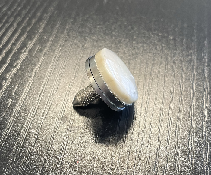
Their method of attachment involves cementing and clamping the hydrogel to a titanium base. This is then pressed and anchored into a hole where the damaged cartilage used to be. Tests show the design stays fastened 68% more firmly than natural cartilage on bone.

“Another concern for knee implants is wear over time, both of the implant itself and the opposing cartilage,” Wiley said.

Other researchers have tried replacing damaged cartilage with knee implants made of metal or polyethylene, but because these materials are stiffer than cartilage they can chafe against other parts of the knee.

In wear tests, the researchers took artificial cartilage and natural cartilage and spun them against each other a million times, with a pressure similar to what the knee experiences during walking. Using a high-resolution X-ray scanning technique called micro-computed tomography (micro-CT), the scientists found that the surface of their lab-made version held up three times better than the real thing. Yet because the hydrogel mimics the smooth, slippery, cushiony nature of real cartilage, it protects other joint surfaces from friction as they slide against the implant.

Natural cartilage is remarkably durable stuff. But once damaged, it has limited ability to heal because it doesn’t have any blood vessels, Wiley said.

In the United States, osteoarthritis is twice as common today than it was a century ago. Surgery is an option when conservative treatments fail. Over the decades surgeons have developed a number of minimally invasive approaches, such as removing loose cartilage, or making holes to stimulate new growth, or transplanting healthy cartilage from a donor. But all of these methods require months of rehab, and some percentage of them fail over time.

Generally considered a last resort, total knee replacement is a proven way to relieve pain. But artificial joints don’t last forever, either. Particularly for younger patients who want to avoid major surgery for a device that will only need to be replaced again down the line, Wiley said, “there’s just not very good options out there.”

“I think this will be a dramatic change in treatment for people at this stage,” Wiley said.

This work was supported in part by Sparta Biomedical and by the Shared Materials Instrumentation Facility at Duke University. Wiley and Gall are shareholders in Sparta Biomedical.

Here’s a link to and a citation for the paper,

A Synthetic Hydrogel Composite with a Strength and Wear Resistance Greater than Cartilage by Jiacheng Zhao, Huayu Tong, Alina Kirillova, William J. Koshut, Andrew Malek, Natasha C. Brigham, Matthew L. Becker, Ken Gall, Benjamin J. Wiley. Advanced Functional Materials DOI: https://doi.org/10.1002/adfm.202205662 First published: 04 August 2022

This paper is behind a paywall.

You can find Sparta Biomedical here.

Nanoscale measurements for osteoarthritis biomarker

There’s a new technique for measuring hyaluronic acid (HA), which appears to be associated with osteoarthritis. A March 12, 2018 news item on ScienceDaily makes the announcement,

For the first time, scientists at Wake Forest Baptist Medical Center have been able to measure a specific molecule indicative of osteoarthritis and a number of other inflammatory diseases using a newly developed technology.

This preclinical [emphasis mine] study used a solid-state nanopore sensor as a tool for the analysis of hyaluronic acid (HA).

I looked at the abstract for the paper (citation and link follow at end of this post) and found that it has been tested on ‘equine models’. Presumably they mean horses or, more accurately, members of the horse family. The next step is likely to be testing on humans, i.e., clinical trials.

A March 12, 2018 Wake Forest Baptist Medical Center news release (also on EurekAlert), which originated the news item, provides more details,

HA is a naturally occurring molecule that is involved in tissue hydration, inflammation and joint lubrication in the body. The abundance and size distribution of HA in biological fluids is recognized as an indicator of inflammation, leading to osteoarthritis and other chronic inflammatory diseases. It can also serve as an indicator of how far the disease has progressed.

“Our results established a new, quantitative method for the assessment of a significant molecular biomarker that bridges a gap in the conventional technology,” said lead author Adam R. Hall, Ph.D., assistant professor of biomedical engineering at Wake Forest School of Medicine, part of Wake Forest Baptist.

“The sensitivity, speed and small sample requirements of this approach make it attractive as the basis for a powerful analytic tool with distinct advantages over current assessment technologies.”

The most widely used method is gel electrophoresis, which is slow, messy, semi-quantitative, and requires a lot of starting material, Hall said. Other technologies include mass spectrometry and size-exclusion chromatography, which are expensive and limited in range, and multi-angle light scattering, which is non-quantitative and has limited precision.

The study, which is published in the current issue of Nature Communications, was led by Hall and Elaheh Rahbar, Ph.D., of Wake Forest Baptist, and conducted in collaboration with scientists at Cornell University and the University of Oklahoma.

In the study, Hall, Rahbar and their team first employed synthetic HA polymers to validate the measurement approach. They then used the platform to determine the size distribution of as little as 10 nanograms (one-billionth of a gram) of HA extracted from the synovial fluid of a horse model of osteoarthritis.

The measurement approach consists of a microchip with a single hole or pore in it that is a few nanometers wide – about 5,000 times smaller than a human hair. This is small enough that only individual molecules can pass through the opening, and as they do, each can be detected and analyzed. By applying the approach to HA molecules, the researchers were able to determine their size one-by-one. HA size distribution changes over time in osteoarthritis, so this technology could help better assess disease progression, Hall said.

“By using a minimally invasive procedure to extract a tiny amount of fluid – in this case synovial fluid from the knee – we may be able to identify the disease or determine how far it has progressed, which is valuable information for doctors in determining appropriate treatments,” he said.

Hall, Rahbar and their team hope to conduct their next study in humans, and then extend the technology with other diseases where HA and similar molecules play a role, including traumatic injuries and cancer.

Here’s a link to and a citation for the paper,

Label-free analysis of physiological hyaluronan size distribution with a solid-state nanopore sensor by Felipe Rivas, Osama K. Zahid, Heidi L. Reesink, Bridgette T. Peal, Alan J. Nixon, Paul L. DeAngelis, Aleksander Skardal, Elaheh Rahbar, & Adam R. Hall. Nature Communications volume 9, Article number: 1037 (2018) doi:10.1038/s41467-018-03439-x
Published online: 12 March 2018

This paper is open access.

Recycling apples to regenerate bone and cartilage tissue

A March 30, 2017 news item on phys.org announces research utilizing apple waste as a matrix for regenerating bones and cartilage,

Researchers from UPM and CSIC [both organizations are in Spain] have employed waste from the agri-food industry to develop biomaterials that act as matrices to regenerate bone and cartilage tissues, which is of great interest for the treatment of diseases related to aging.

The researchers have produced biocompatible materials from apple pomace resulting from juice production. These materials can be used as 3-D matrices for the regeneration of bone and cartilage tissues, useful in regenerative medicine for diseases such as osteoporosis, arthritis or osteoarthritis, all of them rising due to the increasing average age of the population.

A March 30, 2017 Universidad Politécnica de Madrid (UPM) press release, which originated the news item,, expands on the theme,

Apple pomace is an abundant raw material. The world production of apples was more than 70 million tons in 2015, of which the European Union contributed with more than 15%, while half a million tons of which came from Spain. About 75% of apples can be converted into juice and the rest, known as apple pomace, that contains approximately 20–30% dried matter, is used mainly as animal feed or for compost. Since apple pomace is generated in vast quantities and contains a large fraction of water, it poses storage problems and requires immediate treatments to prevent putrefaction. An alternative of great environmental interest is its transformation into value added commodities, thus reducing the volume of waste.

The procedure of the multivalorization of apple pomace carried out by the UPM and CSIC researchers are based on sequential extractions of different bioactive molecules, such as antioxidants or pectin, to finally obtain the waste from which they prepare a biomaterial with suitable porosity and texture to be used in tissue engineering.

The primary extraction of antioxidants and carbohydrates constitutes 2% of the dry weight of apple pomace and pectin extraction is 10%. The extracted chemical cells have a recognized value as nutraceuticals and pectin is a material of great utility in different medical applications, given its high biocompatibility and being part of antitumor drugs or in the treatment of coetaneous wounds.

Furthermore, it has been found that the materials remaining after antioxidant and pectin removal from apple pomace can still be designed with adequate structure, texture and composition to grow diverse types of cells. In this particularly case, the chosen cells were osteoblasts and chondrocytes, both of them related to the regeneration of bone and cartilage tissues because of their application in regenerative medicine in diseases such as osteoporosis, arthritis or osteoarthritis.

Today, there are products in the market with the same applications, however they have a high price reaching over €100 per gram, while waste used in this work hardly reaches €100 per ton. For this reason, there are consistent incentives to convert this waste into final products of great added value.

According to Milagro Ramos, a female researcher of the study, “with this approach we achieve a double goal, firstly using waste as a renewable raw material of high value and chemical diversity, and secondly, to reduce the impact of such waste accumulation on the environment”.

Thanks to the new materials obtained in this work, researchers are developing new technological applications that allow them to structure customized biomaterials through 3D printing techniques.

Here’s a link to and a citation for the paper,

Multivalorization of apple pomace towards materials and chemicals. Waste to wealth by Malcolm Yates, Milagros Ramos Gomez, Maria A. Martin-Luengo, Violeta Zurdo Ibañez, Ana Maria Martinez Serrano. Journal of Cleaner Production Volume 143, 1 February 2017, Pages 847–853  http://doi.org/10.1016/j.jclepro.2016.12.036

This paper is behind a paywall.

Replacement cartilage grown on laboratory chip

Most of us don’t think too much about cartilage (soft, flexible connective tissue found in the body) unless it’s damaged in which case it’s importance becomes immediately apparent. There is no substitute for cartilage although scientists are working on that problem and it seems that one team may have made a significant breakthrough according to an April 27, 2014 news item on ScienceDaily,

In a significant step toward reducing the heavy toll of osteoarthritis around the world, scientists have created the first example of living human cartilage grown on a laboratory chip. The researchers ultimately aim to use their innovative 3-D printing approach to create replacement cartilage for patients with osteoarthritis or soldiers with battlefield injuries.

“Osteoarthritis has a severe impact on quality of life, and there is an urgent need to understand the origin of the disease and develop effective treatments” said Rocky Tuan, Ph.D., director of the Center for Cellular and Molecular Engineering at the University of Pittsburgh School of Medicine, member of the American Association of Anatomists and the study’s senior investigator. “We hope that the methods we’re developing will really make a difference, both in the study of the disease and, ultimately, in treatments for people with cartilage degeneration or joint injuries.”

Osteoarthritis is marked by a gradual disintegration of cartilage, a flexible tissue that provides padding where bones come together in a joint. Causing severe pain and loss of mobility in joints such as knees and fingers, osteoarthritis is one of the leading causes of physical disability in the United States. It is estimated that up to 1 in 2 Americans will develop some form of the disease in their lifetime.

Although some treatments can help relieve arthritis symptoms, there is no cure. Many patients with severe arthritis ultimately require a joint replacement.

An April 27,2014 Experimental Biology (EB) 2014 news release provides more insight,

Tuan said artificial cartilage built using a patient’s own stem cells could offer enormous therapeutic potential. “Ideally we would like to be able to regenerate this tissue so people can avoid having to get a joint replacement, which is a pretty drastic procedure and is unfortunately something that some patients have to go through multiple times,” said Tuan.

In addition to offering relief for people with osteoarthritis, Tuan said replacement cartilage could also be a game-changer for people with debilitating joint injuries, such as soldiers with battlefield injuries. “We really want these technologies to help wounded warriors return to service or pursue a meaningful post-combat life,” said Tuan, who co-directs the Armed Forces Institute of Regenerative Medicine, a national consortium focused on developing regenerative therapies for injured soldiers. “We are on a mission.”

Creating artificial cartilage requires three main elements: stem cells, biological factors to make the cells grow into cartilage, and a scaffold to give the tissue its shape. Tuan’s 3-D printing approach achieves all three by extruding thin layers of stem cells embedded in a solution that retains its shape and provides growth factors. “We essentially speed up the development process by giving the cells everything they need, while creating a scaffold to give the tissue the exact shape and structure that we want,” said Tuan.

The ultimate vision is to give doctors a tool they can thread through a catheter to print new cartilage right where it’s needed in the patient’s body. Although other researchers have experimented with 3-D printing approaches for cartilage, Tuan’s method represents a significant step forward because it uses visible light, while others have required UV light, which can be harmful to living cells.

In another significant step, Tuan has successfully used the 3-D printing method to produce the first “tissue-on-a-chip” replica of the bone-cartilage interface. Housing 96 blocks of living human tissue 4 millimeters across by 8 millimeters deep, the chip could serve as a test-bed for researchers to learn about how osteoarthritis develops and develop new drugs. “With more testing, I think we’ll be able to use our platform to simulate osteoarthritis, which would be extremely useful since scientists really know very little about how the disease develops,” said Tuan.

As a next step, the team is working to combine their 3-D printing method with a nanofiber spinning technique they developed previously. They hope combining the two methods will provide a more robust scaffold and allow them to create artificial cartilage that even more closely resembles natural cartilage.

Rocky Tuan presented the research during the Experimental Biology 2014 meeting on Sunday, April 27 [2014].

I haven’t been able to find any papers published on this work but you can find Rocky Tuan’s faculty page (along with a list of publications) here and you may have more luck with the EB 2014 conference website than I did.