Tag Archives: osteoporosis

Canadian researchers develop bone implant material from cellulose nanocrystals (CNC) while Russian scientists restore internal structure of bone with polycaprolactone nanofibers

Two research groups are working to the same end where bone marrow is concerned, encourage bone cell growth, but they are using different strategies.

University of British Columbia and McMaster University (Canada)

Caption: Researchers treated nanocrystals derived from plant cellulose so that they can link up and form a strong but lightweight sponge (an aerogel) that can compress or expand as needed to completely fill out a bone cavity. Credit: Clare Kiernan, UBC

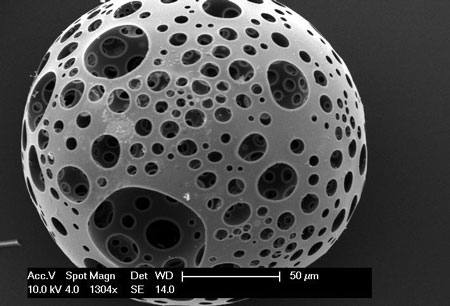
The samples look a little like teeth, don’t they?

Before diving into the research news, there’s a terminology issue that should be noted as you’ll see when you read the news/press releases. Nanocrystal cellulose/nanocrystalline cellulose (NCC) is a term coined by Canadian researchers. Since those early day, most researchers, internationally, have adopted the term cellulose nanocrystals (CNC) as the standard term. It fits better with the naming conventions for other nnanocellulose materials such as cellulose nanofibrils, etc. By the way, a Canadian company (CelluForce) that produces CNC retained the term nanocrystalline cellulose (NCC) as a trademark for the product, CelluForce NCC®.

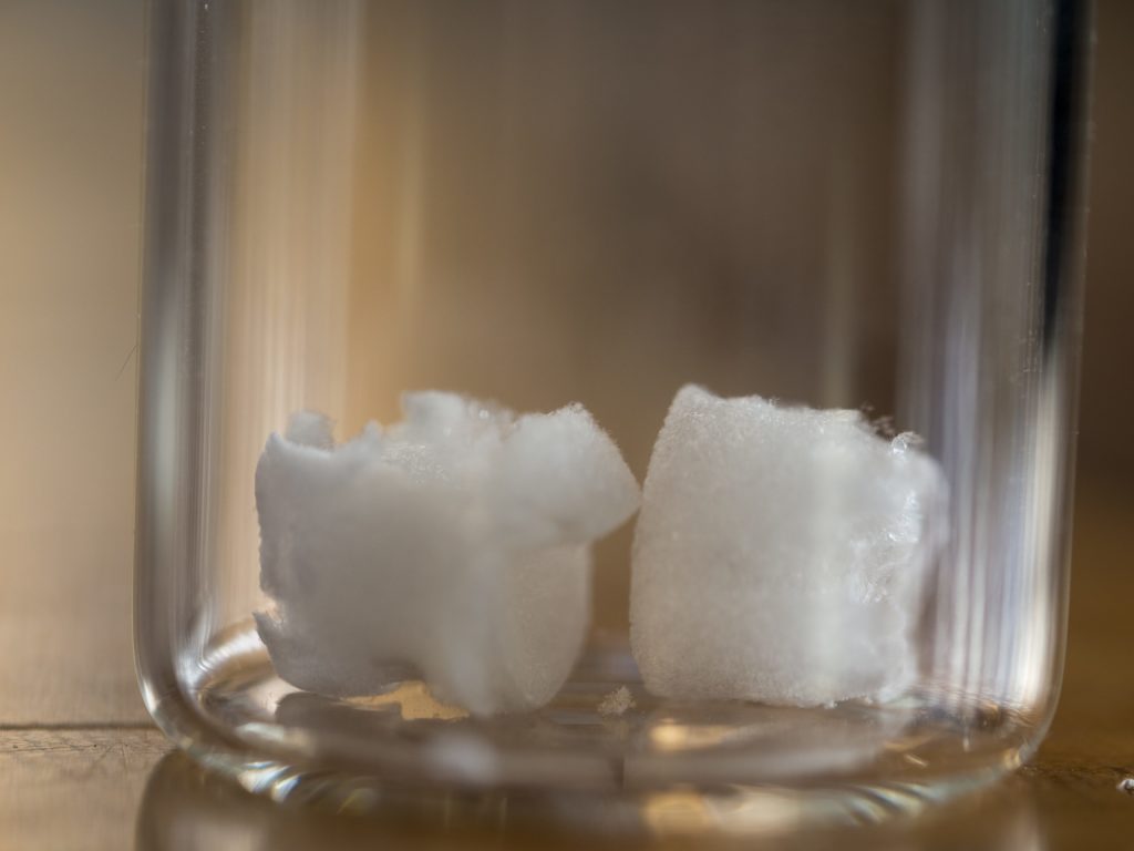
For anyone not familiar with aerogels, what the University of British Columbia (UBC) and McMaster University researchers are developing, are also popularly known known as ‘frozen smoke’ (see the Aerogel Wikipedia entry for more).

A March 19, 2019 news item on ScienceDaily announces the research,

Researchers from the University of British Columbia and McMaster University have developed what could be the bone implant material of the future: an airy, foamlike substance that can be injected into the body and provide scaffolding for the growth of new bone.

It’s made by treating nanocrystals derived from plant cellulose so that they link up and form a strong but lightweight sponge — technically speaking, an aerogel — that can compress or expand as needed to completely fill out a bone cavity.

A March 19, 2019 UBC news release (also on EurekAlert), which originated the news item, describes the research in more detail,

“Most bone graft or implants are made of hard, brittle ceramic that doesn’t always conform to the shape of the hole, and those gaps can lead to poor growth of the bone and implant failure,” said study author Daniel Osorio, a PhD student in chemical engineering at McMaster. “We created this cellulose nanocrystal aerogel as a more effective alternative to these synthetic materials.”

For their research, the team worked with two groups of rats, with the first group receiving the aerogel implants and the second group receiving none. Results showed that the group with implants saw 33 per cent more bone growth at the three-week mark and 50 per cent more bone growth at the 12-week mark, compared to the controls.

“These findings show, for the first time in a lab setting, that a cellulose nanocrystal aerogel can support new bone growth,” said study co-author Emily Cranston, a professor of wood science and chemical and biological engineering who holds the President’s Excellence Chair in Forest Bio-products at UBC. She added that the implant should break down into non-toxic components in the body as the bone starts to heal.

The innovation can potentially fill a niche in the $2-billion bone graft market in North America, said study co-author Kathryn Grandfield, a professor of materials science and engineering, and biomedical engineering at McMaster who supervised the work.

“We can see this aerogel being used for a number of applications including dental implants and spinal and joint replacement surgeries,” said Grandfield. “And it will be economical because the raw material, the nanocellulose, is already being produced in commercial quantities.”

The researchers say it will be some time before the aerogel makes it out of the lab and into the operating room.

“This summer, we will study the mechanisms between the bone and implant that lead to bone growth,” said Grandfield. “We’ll also look at how the implant degrades using advanced microscopes. After that, more biological testing will be required before it is ready for clinical trials.”

Here’s a link to and a citation for the paper,

Cross-linked cellulose nanocrystal aerogels as viable bone tissue scaffolds by Daniel A. Osorio, Bryan E. J. Lee, Jacek M. Kwiecien, Xiaoyue Wang, Iflah Shahid, Ariana L. Hurley, Emily D. Cranston and Kathryn Grandfield. Acta Biomaterialia Volume 87, 15 March 2019, Pages 152-165 DOI: https://doi.org/10.1016/j.actbio.2019.01.049

This paper is behind a paywall

Now for the Russian team.

National University of Science and Technology “MISIS” (formerly part of the Moscow Mining Academy)

These scientists have adopted a different strategy as you’ll see in the March 19, 2019 news item on Nanwerk, which, coincidentally, was published on the same day as the Canadian research,

Scientists from the National University of Science and Technology “MISIS” developed a nanomaterial, which will be able to rstore the internal structure of bones damaged due to osteoporosis and osteomyelitis. A special bioactive coating of the material helped to increase the rate of division of bone cells by 3 times. In the future, it can allow to abandon bone marrow transplantation and patients will no longer need to wait for suitable donor material.

A March 19, 2019 National University of Science and Technology (MISIS) press release (also on EurekAlert), which originated the news item, provides detail about the impetus for the research and the technique being developed,

Such diseases as osteoporosis and osteomyelitis cause irreversible degenerative changes in the bone structure. Such diseases require serious complex treatment and surgery and transplantation of the destroyed bone marrow in severe stages. Donor material should have a number of compatibility indicators and even close relationship with the donor cannot guarantee full compatibility.

Research group from the National University of Science and Technology “MISIS” (NUST MISIS), led by Anton Manakhov (Laboratory for Inorganic Nanomaterials) developed material that will allow to restore damaged internal bone structure without bone marrow transplantation.
It is based on nanofibers of polycaprolactone, which is biocompatible self-dissolvable material. Earlier, the same research group has already worked with this material: by adding antibiotics to the nanofibers, scientists have managed to create non-changeable healing bandages.

“If we want the implant to take, not only biocompatibility is needed, but also activation of the natural cell growth on the surface of the material. Polycaprolactone as such is a hydrophobic material, meaning, and cells feel uncomfortable on its surface. They gather on the smooth surface and divide extremely slow”, Elizaveta Permyakova, one of the co-authors and researcher at NUST MISIS Laboratory for Inorganic Nanomaterials, explains.

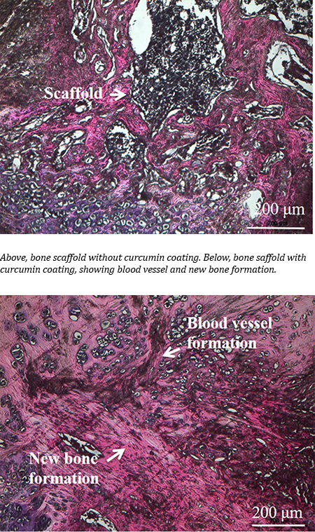
To increase the hydrophilicity of the material, a thin layer of bioactive film consisting of titanium, calcium, phosphorus, carbon, oxygen and nitrogen (TiCaPCON) was deposited on it. The structure of nanofibers identical to the cell surface was preserved. These films, when immersed in a special salt medium, which chemical composition is identical to human blood plasma, are able to form on its surface a special layer of calcium and phosphorus, which in natural conditions forms the main part of the bone. Due to the chemical similarity and the structure of nanofibers, new bone tissue begins to grow rapidly on this layer. Most importantly, polycaprolactone nanofibers dissolve, having fulfilled their functions. Only new “native” tissue remains in the bone.

In the experimental part of the study, the researchers compared the rate of division of osteoblastic bone cells on the surface of the modified and unmodified material. It was found that the modified material TiCaPCON has a high hydrophilicity. In contrast to the unmodified material, the cells on its surface felt clearly more comfortable, and divided three times faster.

According to scientists, such results open up great prospects for further work with modified polycaprolactone nanofibers as an alternative to bone marrow transplantation.

Here’s a link to and a citation for the paper,

Bioactive TiCaPCON-coated PCL nanofibers as a promising material for bone tissue engineering by Anton Manakhov, Elizaveta S. Permyakova, Sergey Ershov, Alexander Sheveyko, Andrey Kovalskii, Josef Polčák, Irina Y. Zhitnyak, Natalia A. Gloushankova, Lenka Zajíčková, Dmitry V. Shtansky. Applied Surface Science Volume 479, 15 June 2019, Pages 796-802 DOI: https://doi.org/10.1016/j.apsusc.2019.02.163

This paper is behind a paywall.

Bone regeneration with a mix of 21st century techniques and an age-old natural cure

Curry was how I was introduced to turmeric. My father who came from Mauritius loved curry and we had it at least once a week. Nobody mentioned healing properties, which I was to discover them only after I started this blog. Usually, turmeric is mentioned in cancer cures but not this time.

Turmeric Courtesy: Washington State University

From a May 2, 2018 Washington State University news release by Tina Hilding (also on EurekAlert but dated May 3, 2018),

A WSU research team is bringing together natural medical cures with modern biomedical devices in hopes of bringing about better health outcomes for people with bone diseases.

In this first-ever effort, the team improved bone-growing capabilities on 3D-printed, ceramic bone scaffolds by 30-45 percent when coated with curcumin, a compound found in the spice, turmeric. They have published their work in the journal, Materials Today Chemistry.

The work could be important for the millions of Americans who suffer from injuries or bone diseases like osteoporosis.

Human bone includes bone forming and resorbing cells that constantly remodel throughout our lives. As people age, the bone cell cycling process often doesn’t work as well. Bones become weaker and likely to fracture. Many of the medicines used for osteoporosis work by slowing down or stopping the destruction of old bone or by forming new bone. While they may increase bone density, they also create an imbalance in the natural bone remodeling cycle and may create poorer quality bone.

Turmeric has been used as medicine for centuries in Asian countries, and curcumin has been shown to have antioxidant, anti-inflammatory and bone-building capabilities. It can also prevent various forms of cancers. However, when taken orally as medicine, the compound can’t be absorbed well in the body. It is metabolized and eliminated too quickly.

Led by Susmita Bose, Herman and Brita Lindholm Endowed Chair Professor in the School of Mechanical and Materials Engineering, the researchers encased the curcumin within a water-loving polymer, a large molecule, so that it could be gradually released from their ceramic scaffolds. The curcumin increased the viability and proliferation of new bone cells and blood vessels in surrounding tissue as well as accelerated the healing process.

Bose hopes that the work will lead to medicines that naturally create healthier bone without affecting the bone remodeling cycle.

“In the end, it’s the bone quality that matters,” she said.

The researchers are continuing the studies, looking at the protein and cellular level to gain better understanding of exactly how the natural compound works. They are also working to improve the process’ efficiency and control. The challenge with the natural compounds, said Bose, is that they are often large organic molecules.

“You have to use the right vehicle for delivery,” she said. “We need to load and get it released in a controlled and sustained way. The chemistry of vehicle delivery is very important.”

In addition to curcumin, the researchers are studying other natural remedies, including compounds from aloe vera, saffron, Vitamin D, garlic, oregano and ginger. Bose is focused on compounds that might help with bone disorders, including those that encourage bone growth or that have anti-inflammatory, infection control, or anti-cancer properties.

Starting with her own health issues, Bose has had a longtime interest in bridging natural medicinal compounds with modern medicine. That interest increased after she had her children.

“As a mother and having a chemistry background, I realized I didn’t want my children to be exposed to so many chemicals for every illness,” Bose said. “I started looking at home remedies.”

To her students, she always emphasizes healthy living as the best way to guarantee the best health outcomes, including healthy eating, proper sleep, interesting hobbies, and exercise.

Courtesy Washington State University

Here’s a link to and a citation for the paper,

Effects of PCL, PEG and PLGA polymers on curcumin release from calcium phosphate matrix for in vitro and in vivo bone regeneration by Susmita Bose, Naboneeta Sarkar, Dishary Banerjee. Materials Today Chemistry Vol. 8 June 2018, pp. 110-130 [Published online May 2, 2018] https://doi.org/10.1016/j.mtchem.2018.03.005

This paper is behind a paywall.

Lighting the way to improvements for the bond between dental implants and bone

A July 3, 2018 Canadian Light Source news release by Colleen MacPherson describes an investigation into how dental implants and bones interact with the hope of making dental implantation safer and more certain,

Research carried out recently at the Canadian Light Source (CLS) [also known as a synchrotron] in Saskatoon [Saskatchewan, Canada] has revealed promising information about how to build a better dental implant, one that integrates more readily with bone to reduce the risk of failure.

“There are millions of dental and orthopedic implants placed every year in North America and a certain number of them always fail, even in healthy people with healthy bone,” said Kathryn Grandfield, assistant professor in the Department of Materials Science and Engineering at McMaster University in Hamilton [Ontario, Canada].

A dental implant restores function after a tooth is lost or removed. It is usually a screw shaped implant that is placed in the jaw bone and acts as the tooth roots, while an artificial tooth is placed on top. The implant portion is the artificial root that holds an artificial tooth in place.

Grandfield led a study that showed altering the surface of a titanium implant improved its connection to the surrounding bone. It is a finding that may well be applicable to other kinds of metal implants, including engineered knees and hips, and even plates used to secure bone fractures.

About three million people in North America receive dental implants annually. While the failure rate is only one to two percent, “one or two percent of three million is a lot,” she said. Orthopedic implants fail up to five per cent of the time within the first 10 years; the expected life of these devices is about 20 to 25 years, she added.

“What we’re trying to discover is why they fail, and why the implants that are successful work. Our goal is to understand the bone-implant interface in order to improve the design of implants.”

Grandfield’s research team, which included post-doctoral fellow Xiaoyue Wang and McMaster colleague Adam Hitchcock from the Department of Chemistry and Chemical Biology. The team members used the soft X-ray spectromicroscopy beamline at the CLS as well as facilities at the Canadian Centre for Electron Microscopy in Hamilton to examine a failed dental implant that had to be removed, along with a small amount of surrounding bone, from a patient. Prior to implantation, a laser beam was used to alter the implant, to roughen the surface, creating what looked like “little volcanoes” on the surface. After removal from the patient, the point of connection between bone and metal was then carefully studied to understand how the implant behaved.

“What we found was that the surface modification changed the chemistry of the implant. The modification created an oxide layer, but not a bad oxide layer like rust but a better, more beneficial layer that helps integrate with bone material.”

The research results were published in Advanced Materials Interfaces in May [2018], ensuring the findings are available “to implant companies interested in using nanotechnology to change the structure of the implants they produce,” said Grandfield.

The next steps in the research will be to apply the surface modification technique to other types of implants “to be able to understand fully how they function.” Grandfield added the research done at the CLS involved healthy bone “so I’d be really interested in seeing the response when bone is a bit more compromised by age or disease, like osteoporosis. We need to find the best surface modifications … because the technology we have today to treat patients with healthier bone may not be sufficient with compromised bone.”

Here’s a link to (even though it’s in the news release text) and a citation for the paper,

Biomineralization at Titanium Revealed by Correlative 4D Tomographic and Spectroscopic Methods by Xiaoyue Wang, Brian Langelier, Furqan A. Shah, Andreas Korinek, Matthieu Bugnet, Adam P. Hitchcock, Anders Palmquist, Kathryn Grandfield. Advnaced Materials Interfaces https://doi-org.proxy.lib.sfu.ca/10.1002/admi.201800262 First published: 16 May 2018

This paper is behind a paywall.

Recycling apples to regenerate bone and cartilage tissue

A March 30, 2017 news item on phys.org announces research utilizing apple waste as a matrix for regenerating bones and cartilage,

Researchers from UPM and CSIC [both organizations are in Spain] have employed waste from the agri-food industry to develop biomaterials that act as matrices to regenerate bone and cartilage tissues, which is of great interest for the treatment of diseases related to aging.

The researchers have produced biocompatible materials from apple pomace resulting from juice production. These materials can be used as 3-D matrices for the regeneration of bone and cartilage tissues, useful in regenerative medicine for diseases such as osteoporosis, arthritis or osteoarthritis, all of them rising due to the increasing average age of the population.

A March 30, 2017 Universidad Politécnica de Madrid (UPM) press release, which originated the news item,, expands on the theme,

Apple pomace is an abundant raw material. The world production of apples was more than 70 million tons in 2015, of which the European Union contributed with more than 15%, while half a million tons of which came from Spain. About 75% of apples can be converted into juice and the rest, known as apple pomace, that contains approximately 20–30% dried matter, is used mainly as animal feed or for compost. Since apple pomace is generated in vast quantities and contains a large fraction of water, it poses storage problems and requires immediate treatments to prevent putrefaction. An alternative of great environmental interest is its transformation into value added commodities, thus reducing the volume of waste.

The procedure of the multivalorization of apple pomace carried out by the UPM and CSIC researchers are based on sequential extractions of different bioactive molecules, such as antioxidants or pectin, to finally obtain the waste from which they prepare a biomaterial with suitable porosity and texture to be used in tissue engineering.

The primary extraction of antioxidants and carbohydrates constitutes 2% of the dry weight of apple pomace and pectin extraction is 10%. The extracted chemical cells have a recognized value as nutraceuticals and pectin is a material of great utility in different medical applications, given its high biocompatibility and being part of antitumor drugs or in the treatment of coetaneous wounds.

Furthermore, it has been found that the materials remaining after antioxidant and pectin removal from apple pomace can still be designed with adequate structure, texture and composition to grow diverse types of cells. In this particularly case, the chosen cells were osteoblasts and chondrocytes, both of them related to the regeneration of bone and cartilage tissues because of their application in regenerative medicine in diseases such as osteoporosis, arthritis or osteoarthritis.

Today, there are products in the market with the same applications, however they have a high price reaching over €100 per gram, while waste used in this work hardly reaches €100 per ton. For this reason, there are consistent incentives to convert this waste into final products of great added value.

According to Milagro Ramos, a female researcher of the study, “with this approach we achieve a double goal, firstly using waste as a renewable raw material of high value and chemical diversity, and secondly, to reduce the impact of such waste accumulation on the environment”.

Thanks to the new materials obtained in this work, researchers are developing new technological applications that allow them to structure customized biomaterials through 3D printing techniques.

Here’s a link to and a citation for the paper,

Multivalorization of apple pomace towards materials and chemicals. Waste to wealth by Malcolm Yates, Milagros Ramos Gomez, Maria A. Martin-Luengo, Violeta Zurdo Ibañez, Ana Maria Martinez Serrano. Journal of Cleaner Production Volume 143, 1 February 2017, Pages 847–853  http://doi.org/10.1016/j.jclepro.2016.12.036

This paper is behind a paywall.

A grant for regenerating bones with injectable stem cell microspheres

I have a longstanding interest in bones partly due to my introduction to a skeleton in a dance course and to US artist Georgia O’Keeffe’s paintings. In any event, it’s been too long since I’ve featured any research on bones here.

This news comes from the UK’s University of Nottingham. A July 25, 2016 news item on Nanowerk announced a grant for stem cell research,

The University of Nottingham has secured £1.2m to develop injectable stem cell-carrying materials to treat and prevent fractures caused by osteoporosis and other bone-thinning diseases.

A July 25, 2016 University of Nottingham press release, which originated the news item, offers more information about the proposed therapy and the research project (Note: Links have been removed),

The experimental materials consist of porous microspheres produced from calcium phosphates – a key component in bones – to be filled with stem cells extracted from the patient.

The targeted therapy could offer a quick, easy and minimally-invasive treatment that is injected into areas considered to be at high-risk of fracture to promote bone regeneration.

The funding grant, from the National Institute for Health Research (NIHR i4i Challenge Award), also supports the development of a prototype delivery device to inject these stem cell loaded microspheres to the sites of interest.

In addition, project partners will investigate how well the materials stay in place once they have been injected inside the body.

Research leads, Dr Ifty Ahmed and Professor Brigitte Scammell explained that the aim was to develop a preventive treatment option to address the growing issue of fractures occurring due to bone-thinning diseases, which is exacerbated due to the worldwide ageing population.

Osteoporosis-related conditions affect some three million Britons, and cost the NHS over £1.73bn each year, according to the National Osteoporotic Society.

Dr Ahmed, from the Faculty of Engineering at The University of Nottingham, said, “We would advocate a national screening program, using a DEXA scan, which measures bone mineral density, to identify people at high risk of fracture due to osteoporosis.

“If we could strengthen these peoples bone before they suffered from fractures, using a simple injection procedure, it would save people the pain and trauma of broken bones and associated consequences such as surgery and loss of independence.”

The NIHR grant will also fund a Patient and Public Involvement study on the suitability of the technology, gauging the opinions and personal experience of people affected by osteoporosis as sufferers or carers, for example.

The project has already undertaken proof-of-concept work to test the feasibility of manufacturing the microsphere materials and lab work to ensure that stem cells attach and reside within these novel microsphere carriers.

The research is still at an early stage and the project team are working towards next phase pre-clinical trials.

This work reminded me of an unfinished piece of science fiction where I developed a society that had the ability to grow bone to replace lost limbs, replace lost bone matter, and restructure faces. I should get back to it one of these days. In the meantime, here’s an image of a microsphere,

A close-up of a injectable stem-cell carrying microsphere made of calcium phosphate which are injected to prevent and treat fractures caused by bone-thinning diseases. (Image: Ifty Ahmed; University of Nottingham)

A close-up of a injectable stem-cell carrying microsphere made of calcium phosphate which are injected to prevent and treat fractures caused by bone-thinning diseases. (Image: Ifty Ahmed; University of Nottingham)

One final note, fragile bones are no joke but there does seem to be a movement to diagnose more and more people with osteoporosis. Alan Cassels, in his July/August 2016 article for Common Ground magazine, points out that the guidelines for diagnosis have changed and more healthy people are being targeted,

… Americans, the experts tell us, are suffering an epidemic of osteoporosis. A new US osteoporosis guideline says that 72% of women over 65 are considered ‘diseased’ – a number which rises to 93% for those over 75 years old – and hence in need of drug therapy.

What is going on here?

Clearly, the only real ‘epidemic’ is the growing phenomenon where risks for disease are being turned into diseases, in and of themselves. In this racket, ‘high’ blood pressure, elevated cholesterol, low bone density, fluctuating blood sugars, high eyeball pressure and low testosterone, among other things, become worrying signs of chronic, lifelong conditions that demand attention and medication. As I’ve said in the past, “If you want to know why pharma is increasingly targeting healthy people with ‘preventive medicine,’ it’s because that’s where the money is.”

One thing all these risks-as-disease models have in common is they are shaped and supported by clinical practice guidelines. In these guidelines, doctors are told to measure their patients’ parameters. If your measurements are outside some preset levels deemed ‘high risk’ by the expert guidelines, you know what that means: more frequent trips to the pharmacy. The main downside of guidelines is they slap labels on people who aren’t sick and instill in physicians the constant idea their healthy patients are really disease-ridden.

But this is a good news story and if you haven’t sensed it, there’s a rising backlash against medical guidelines, mostly led by doctors, researchers and even some patients outraged at what they see going on. …

I don’t wish to generalize from the situation in the US to the situation in the UK. The medical systems and models are quite different but since at least some of my readership is from the US, I thought this digression might prove helpful. Regardless of where you live, it never hurts to ask questions.

Remotely controlling bone regeneration with metallic nanoparticles

A Nov. 24, 2014 news item on ScienceDaily heralds some bone regeneration research which was published back in Sept. 2014,

Researchers in bone tissue regeneration believe they have made a significant breakthrough for sufferers of bone trauma, disease or defects such as osteoporosis.

Medical researchers from Keele University and Nottingham University have found that magnetic nanoparticles coated with targeting proteins can stimulate stem cells to regenerate bone. Researchers were also able to deliver the cells directly to the injured area, remotely controlling the nanoparticles to generate mechanical forces and maintain the regeneration process through staged releases of a protein growth stimulant.

A Nov. 17, 2014 Keele University (UK) press release, which originated the news item, describes the issues the researchers are addressing and their research approach,

The current method for repairing bone that can’t heal itself is through a graft taken from the patient. Unfortunately, this can be a painful, invasive procedure, and when the area that needs repair is too large or the patient has a skeletal disorder such as there can sometimes be a lack of healthy bone for grafting.

For this reason, spurring the growth of new bone through injected stem cells is an area of great interest to medical researchers. Much progress has been made, but a major hurdle remains – finding an appropriate means to stimulate the differentiation of the stem cells so they become the quality of bone tissue needed in a quantity large enough to treat patients effectively.

James Henstock, Ph.D. led the Biotechnology and Biological Sciences Research Council (BBSRC)-funded study, alongside Alicia El Haj, Ph.D., and colleagues at Keele University’s Institute for Science and Technology in Medicine, as well as Kevin Shakesheff, Ph.D., from the University of Nottingham’s School of Pharmacy.

James Henstock said: “Injectable therapies for regenerative medicine show great potential as a minimally invasive route for introducing therapeutic stem cells, drug delivery vehicles and biomaterials efficiently to wound sites.”

“In our investigation we coated magnetic nanoparticles with specific targeting proteins then controlled them remotely with an external magnetic field to simulate exercise. We wanted to learn how this might affect the injected stem cells and their ability to restore functional bone.”

The team of researchers conducted their test using two models: chicken foetal femurs and tissue-engineered collagen hydrogels. In both instances the results showed an increase in bone formation and density without causing any mechanical stress to the construct or surrounding tissue.

“This work demonstrates that providing the appropriate mechanical cues in conjunction with controlled release of growth factors to these injectable cell therapies can have a significant impact on improving bone growth. It also could potentially improve tissue engineering approaches for translational medicine” Dr. Henstock said.

Here’s a link to and a citation for the published paper,

Remotely Activated Mechanotransduction via Magnetic Nanoparticles Promotes Mineralization Synergistically With Bone Morphogenetic Protein 2: Applications for Injectable Cell Therapy by James R. Henstock, Michael Rotherham, Hassan Rashidi, Kevin M. Shakesheff, and Alicia J. El Haja. Stem Cells Trans Med September 2014 sctm.2014-0017  (First Published Online September 22, 2014 doi: 10.5966/sctm.2014-0017)

This paper is open access but you do need to sign up for a free registration for access to the website.