Tag Archives: Paul Rothemund

DNA origami as Van Gogh’s Starry Night

This glowing reproduction of "The Starry Night" contains 65,536 pixels and is the width of a dime across. Credit: Ashwin Gopinath/Caltech

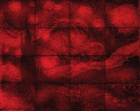
This glowing reproduction of “The Starry Night” contains 65,536 pixels and is the width of a dime across.
Credit: Ashwin Gopinath/Caltech

It may take you a few seconds (it did me) but it’s possible to see Van Gogh’s Starry Night in this image. A July 12, 2016 news item on ScienceDaily reveals more,

Using folded DNA [deoxyribonucleic acid] to precisely place glowing molecules within microscopic light resonators, researchers at Caltech have created one of the world’s smallest reproductions of Vincent van Gogh’s The Starry Night.

A July 12, 2016 Caltech news release (also on EurekAlert) by Richard Perkins, which originated the news item, provides more information about the image, DNA origami, and this latest research on coupling light emitters to photonic crystal cavities (Note: Links have been removed),

The monochrome image—just the width of a dime across—was a proof-of-concept project that demonstrated, for the first time, how the precision placement of DNA origami can be used to build chip-based devices like computer circuits at smaller scales than ever before.

DNA origami, developed 10 years ago by Caltech’s Paul Rothemund (BS ’94), is a technique that allows researchers to fold a long strand of DNA into any desired shape. The folded DNA then acts as a scaffold onto which researchers can attach and organize all kinds of nanometer-scale components, from fluorescent molecules to electrically conductive carbon nanotubes to drugs.

“Think of it a bit like the pegboards people use to organize tools in their garages, only in this case, the pegboard assembles itself from DNA strands and the tools likewise find their own positions,” says Rothemund, research professor of bioengineering, computing and mathematical sciences, and computation and neural systems. “It all happens in a test tube without human intervention, which is important because all of the parts are too small to manipulate efficiently, and we want to make billions of devices.”

The process has the potential to influence a variety of applications from drug delivery to the construction of nanoscale computers. But for many applications, organizing nanoscale components to create devices on DNA pegboards is not enough; the devices have to be wired together into larger circuits and need to have a way of communicating with larger-scale devices.

One early approach was to make electrodes first, and then scatter devices randomly on a surface, with the expectation that at least a few would land where desired, a method Rothemund describes as “spray and pray.”

In 2009, Rothemund and colleagues at IBM Research first described a technique through which DNA origami can be positioned at precise locations on surfaces using electron-beam lithography to etch sticky binding sites that have the same shape as the origami. For example, triangular sticky patches bind triangularly folded DNA.

Over the last seven years, Rothemund and Ashwin Gopinath, senior postdoctoral scholar in bioengineering at Caltech, have refined and extended this technique so that DNA shapes can be precisely positioned on almost any surface used in the manufacture of computer chips. In the Nature paper, they report the first application of the technique—using DNA origami to install fluorescent molecules into microscopic light sources.

“It’s like using DNA origami to screw molecular light bulbs into microscopic lamps,” Rothemund says.

In this case, the lamps are microfabricated structures called photonic crystal cavities (PCCs), which are tuned to resonate at a particular wavelength of light, much like a tuning fork vibrates with a particular pitch. Created within a thin glass-like membrane, a PCC takes the form of a bacterium-shaped defect within an otherwise perfect honeycomb of holes.

“Depending on the exact size and spacing of the holes, a particular wavelength of light reflects off the edge of the cavity and gets trapped inside,” says Gopinath, the lead author of the study. He built PCCs that are tuned to resonate at around 660 nanometers, the wavelength corresponding to a deep shade of the color red. Fluorescent molecules tuned to glow at a similar wavelength light up the lamps—provided they stick to exactly the right place within the PCC.

“A fluorescent molecule tuned to the same color as a PCC actually glows more brightly inside the cavity, but the strength of this coupling effect depends strongly on the molecule’s position within the cavity. A few tens of nanometers is the difference between the molecule glowing brightly, or not at all,” Gopinath says.

By moving DNA origami through the PCCs in 20-nanometer steps, the researchers found that they could map out a checkerboard pattern of hot and cold spots, where the molecular light bulbs either glowed weakly or strongly. As a result, they were able to use DNA origami to position fluorescent molecules to make lamps of varying intensity. Similar structures have been proposed to power quantum computers and for use in other optical applications that require many tiny light sources integrated together on a single chip.

“All previous work coupling light emitters to PCCs only successfully created a handful of working lamps, owing to the extraordinary difficulty of reproducibly controlling the number and position of emitters in a cavity,” Gopinath says. To prove their new technology, the researchers decided to scale-up and provide a visually compelling demonstration. By creating PCCs with different numbers of binding sites, Gopinath was able to reliably install any number from zero to seven DNA origami, allowing him to digitally control the brightness of each lamp. He treated each lamp as a pixel with one of eight different intensities, and produced an array of 65,536 of the PCC pixels (a 256 x 256 pixel grid) to create a reproduction of Van Gogh’s “The Starry Night.”

Now that the team can reliably combine molecules with PCCs, they are working to improve the light emitters. Currently, the fluorescent molecules last about 45 seconds before reacting with oxygen and “burning out,” and they emit a few shades of red rather than a single pure color. Solving both these problems will help with applications such as quantum computers.

“Aside from applications, there’s a lot of fundamental science to be done,” Gopinath says.

Here’s a link to and a citation for the paper,

Engineering and mapping nanocavity emission via precision placement of DNA origami by Ashwin Gopinath, Evan Miyazono, Andrei Faraon, & Paul W. K. Rothemund. Nature (2016) doi:10.1038/nature18287 Published online 11 July 2016

This paper is behind a paywall.

Nature celebrates some nanotechnology anniversaries

An April 5, 2016 editorial in Nature magazine celebrates some nanotechnology milestones (Note: Links have been removed),

In March 1986, the atomic force microscope (AFM) was introduced by Gerd Binnig, Calvin Quate and Christoph Gerber with a paper in the journal Physical Review Letters titled simply ‘Atomic force microscope’1. This was 5 years (to the month) after the precursor to the AFM, the scanning tunnelling microscope (STM), had first been successfully tested at IBM’s Zurich Research Laboratory by Binnig and the late Heinrich Rohrer, and 7 months before Binnig and Rohrer were awarded a share of the Nobel Prize in Physics for the design of the STM (the prize was shared with Ernst Ruska, the inventor of the electron microscope). Achieving atomic resolution with the AFM proved more difficult than with the STM. It was, for example, only two years after its invention that the STM provided atomic-resolution images of an icon of surface science, the 7 × 7 surface reconstruction of Si(111) (ref. 2), whereas it took 8 years to achieve a similar feat with the AFM3, 4.

The editorial also provides an explanation of how the AFM works,

The AFM works by scanning a sharp tip attached to a flexible cantilever across a sample while measuring the interaction between the tip and the sample surface. The technique can operate in a range of environments, including in liquid and in air, and unlike the STM, it can be used with insulating materials; in their original paper, Binnig and colleagues used the instrument to analyse an aluminium oxide sample.

Then, the editorial touches on DNA (deoxyribonucleic acid) nanotechnology (Note: Links have been removed),

The history of structural DNA nanotechnology can, like the AFM, be traced back to the early 1980s, when Nadrian Seeman suggested that the exquisite base-pairing rules of DNA could be exploited to build artificial self-assembled structures11. But the founding experiment of the field came later. In April 1991, Seeman and Junghuei Chen reported building a cube-like molecular complex from DNA using a combination of branched junctions and single-stranded ‘sticky’ ends12. A range of significant advances soon followed, from 2D DNA arrays to DNA-based nanomechanical devices.

Then, in March 2006, the field of structural DNA nanotechnology experienced another decisive moment: Paul Rothemund reported the development of DNA origami13. This technique involves folding a long single strand of DNA into a predetermined shape with the help of short ‘staple’ strands. Used at first to create 2D structures, which were incidentally characterized using the AFM, the approach was quickly expanded to the building of intricate 3D structures and the organization of other species such as nanoparticles and proteins. …

Happy reading!

A twist in my DNA

Professor Hao Yan’s team at Arizona State University (ASU) has created some new 2D and 3D DNA objects according to a Mar. 21, 2013 news release on EurekAlert,

In their latest twist to the technology, Yan’s team made new 2-D and 3-D objects that look like wire-frame art of spheres as well as molecular tweezers, scissors, a screw, hand fan, and even a spider web.

The Yan lab, which includes ASU Biodesign Institute colleagues Dongran Han, Suchetan Pal, Shuoxing Jiang, Jeanette Nangreave and assistant professor Yan Liu, published their results in the March 22 issue of Science.

Here’s where the twist comes in,

The twist in their ‘bottom up,’ molecular Lego design strategy focuses on a DNA structure called a Holliday junction. In nature, this cross-shaped, double-stacked DNA structure is like the 4-way traffic stop of genetics — where 2 separate DNA helices temporality meet to exchange genetic information. The Holliday junction is the crossroads responsible for the diversity of life on Earth, and ensures that children are given a unique shuffling of traits from a mother and father’s DNA.

In nature, the Holliday junction twists the double-stacked strands of DNA at an angle of about 60-degrees, which is perfect for swapping genes but sometimes frustrating for DNA nanotechnology scientists, because it limits the design rules of their structures.

“In principal, you can use the scaffold to connect multiple layers horizontally,” [which many research teams have utilized since the development of DNA origami by Cal Tech’s Paul Rothemund in 2006]. However, when you go in the vertical direction, the polarity of DNA prevents you from making multiple layers,” said Yan. “What we needed to do is rotate the angle and force it to connect.”

Making the new structures that Yan envisioned required re-engineering the Holliday junction by flipping and rotating around the junction point about half a clock face, or 150 degrees. Such a feat has not been considered in existing designs.

“The initial idea was the hardest part,” said Yan. “Your mind doesn’t always see the possibilities so you forget about it. We had to break the conceptual barrier that this could happen.”

In the new study, by varying the length of the DNA between each Holliday junction, they could force the geometry at the Holliday junctions into an unconventional rearrangement, making the junctions more flexible to build for the first time in the vertical dimension. Yan calls the backyard barbeque grill-shaped structure a DNA Gridiron.

“We were amazed that it worked!” said Yan. “Once we saw that it actually worked, it was relatively easy to implement new designs. Now it seems easy in hindsight. If your mindset is limited by the conventional rules, it’s really hard to take the next step. Once you take that step, it becomes so obvious.”

The DNA Gridiron designs are programmed into a viral DNA, where a spaghetti-shaped single strand of DNA is spit out and folded together with the help of small ‘staple’ strands of DNA that help mold the final DNA structure. In a test tube, the mixture is heated, then rapidly cooled, and everything self-assembles and molds into the final shape once cooled. Next, using sophisticated AFM and TEM imaging technology, they are able to examine the shapes and sizes of the final products and determine that they had formed correctly.

This approach has allowed them to build multilayered, 3-D structures and curved objects for new applications.

In addition to the EurekAlert version, you can find the full text, images, and video about the team’s paper in the Mar. 21, 2013 news item on ScienceDaily (a citation and link to the team’s paper is also included) or you can read the original Mar. 21, 2013 ASU news release. (Hao Yan’s work was last mentioned here in an Aug. 7, 2012 post.)

All of this talk of twists reminded me of a song by Tanita Tikaram, Twist in My Sobriety. I found this video of an acoustic performance (two guitars and a bass [the musical instrument not the fish]) which is even more sultry than original hit version,

Happy weekend!