Tag Archives: Peter Vukusic

Blue-striped limpets and their nanophotonic features

This is a structural colour story limpets and the Massachusetts Institute of Technology (MIT) and Harvard University. For the impatient here’s a video summary of the work courtesy of the researchers,

A Feb. 26, 2015 news item on ScienceDaily reiterates the details for those who like to read their science,

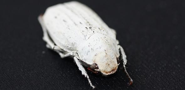
The blue-rayed limpet is a tiny mollusk that lives in kelp beds along the coasts of Norway, Iceland, the United Kingdom, Portugal, and the Canary Islands. These diminutive organisms — as small as a fingernail — might escape notice entirely, if not for a very conspicuous feature: bright blue dotted lines that run in parallel along the length of their translucent shells. Depending on the angle at which light hits, a limpet’s shell can flash brilliantly even in murky water.

Now scientists at MIT and Harvard University have identified two optical structures within the limpet’s shell that give its blue-striped appearance. The structures are configured to reflect blue light while absorbing all other wavelengths of incoming light. The researchers speculate that such patterning may have evolved to protect the limpet, as the blue lines resemble the color displays on the shells of more poisonous soft-bodied snails.

A Feb. 26, 2015 MIT news release (also on EurekAlert), which originated the news item, explains why this discovery is special,

The findings, reported this week in the journal Nature Communications, represent the first evidence of an organism using mineralized structural components to produce optical displays. While birds, butterflies, and beetles can display brilliant blues, among other colors, they do so with organic structures, such as feathers, scales, and plates. The limpet, by contrast, produces its blue stripes through an interplay of inorganic, mineral structures, arranged in such a way as to reflect only blue light.

The researchers say such natural optical structures may serve as a design guide for engineering color-selective, controllable, transparent displays that require no internal light source and could be incorporated into windows and glasses.

“Let’s imagine a window surface in a car where you obviously want to see the outside world as you’re driving, but where you also can overlay the real world with an augmented reality that could involve projecting a map and other useful information on the world that exists on the other side of the windshield,” says co-author Mathias Kolle, an assistant professor of mechanical engineering at MIT. “We believe that the limpet’s approach to displaying color patterns in a translucent shell could serve as a starting point for developing such displays.”

The news release then reveals how this research came about,

Kolle, whose research is focused on engineering bioinspired, optical materials — including color-changing, deformable fibers — started looking into the optical features of the limpet when his brother Stefan, a marine biologist now working at Harvard, brought Kolle a few of the organisms in a small container. Stefan Kolle was struck by the mollusk’s brilliant patterning, and recruited his brother, along with several others, to delve deeper into the limpet shell’s optical properties.

To do this, the team of researchers — which also included Ling Li and Christine Ortiz at MIT and James Weaver and Joanna Aizenberg at Harvard — performed a detailed structural and optical analysis of the limpet shells. They observed that the blue stripes first appear in juveniles, resembling dashed lines. The stripes grow more continuous as a limpet matures, and their shade varies from individual to individual, ranging from deep blue to turquoise.

The researchers scanned the surface of a limpet’s shell using scanning electron microscopy, and found no structural differences in areas with and without the stripes — an observation that led them to think that perhaps the stripes arose from features embedded deeper in the shell.

To get a picture of what lay beneath, the researchers used a combination of high-resolution 2-D and 3-D structural analysis to reveal the 3-D nanoarchitecture of the photonic structures embedded in the limpets’ translucent shells.

What they found was revealing: In the regions with blue stripes, the shells’ top and bottom layers were relatively uniform, with dense stacks of calcium carbonate platelets and thin organic layers, similar to the shell structure of other mollusks. However, about 30 microns beneath the shell surface the researchers noted a stark difference. In these regions, the researchers found that the regular plates of calcium carbonate morphed into two distinct structural features: a multilayered structure with regular spacing between calcium carbonate layers resembling a zigzag pattern, and beneath this, a layer of randomly dispersed, spherical particles.

The researchers measured the dimensions of the zigzagging plates, and found the spacing between them was much wider than the more uniform plates running through the shell’s unstriped sections. They then examined the potential optical roles of both the multilayer zigzagging structure and the spherical particles.

Kolle and his colleagues used optical microscopy, spectroscopy, and diffraction microscopy to quantify the blue stripe’s light-reflection properties. They then measured the zigzagging structures and their angle with respect to the shell surface, and determined that this structure is optimized to reflect blue and green light.

The researchers also determined that the disordered arrangement of spherical particles beneath the zigzag structures serves to absorb transmitted light that otherwise could de-saturate the reflected blue color.

From these results, Kolle and his team deduced that the zigzag pattern acts as a filter, reflecting only blue light. As the rest of the incoming light passes through the shell, the underlying particles absorb this light — an effect that makes a shell’s stripes appear even more brilliantly blue.

And, for those who can never get enough detail, the news release provides a bit more than the video,

The team then sought to tackle a follow-up question: What purpose do the blue stripes serve? The limpets live either concealed at the base of kelp plants, or further up in the fronds, where they are visually exposed. Those at the base grow a thicker shell with almost no stripes, while their blue-striped counterparts live higher on the plant.

Limpets generally don’t have well-developed eyes, so the researchers reasoned that the blue stripes must not serve as a communication tool, attracting one organism to another. Rather, they think that the limpet’s stripes may be a defensive mechanism: The mollusk sits largely exposed on a frond, so a plausible defense against predators may be to appear either invisible or unappetizing. The researchers determined that the latter is more likely the case, as the limpet’s blue stripes resemble the patterning of poisonous marine snails that also happen to inhabit similar kelp beds.

Kolle says the group’s work has revealed an interesting insight into the limpet’s optical properties, which may be exploited to engineer advanced transparent optical displays. The limpet, he points out, has evolved a microstructure in its shell to satisfy an optical purpose without overly compromising the shell’s mechanical integrity. Materials scientists and engineers could take inspiration from this natural balancing act.

“It’s all about multifunctional materials in nature: Every organism — no matter if it has a shell, or skin, or feathers — interacts in various ways with the environment, and the materials with which it interfaces to the outside world frequently have to fulfill multiple functions simultaneously,” Kolle says. “[Engineers] are more and more focusing on not only optimizing just one single property in a material or device, like a brighter screen or higher pixel density, but rather on satisfying several … design and performance criteria simultaneously. We can gain inspiration and insight from nature.”

Peter Vukusic, an associate professor of physics at the University of Exeter in the United Kingdom, says the researchers “have done an exquisite job” in uncovering the optical mechanism behind the limpet’s conspicuous appearance.

“By using multiple and complementary analysis techniques they have elucidated, in glorious detail, the many structural and physiological factors that have given rise to the optical signature of this highly evolved system,” says Vukusic, who was not involved in the study. “The animal’s complex morphology is highly interesting for photonics scientists and technologists interested in manipulating light and creating specialized appearances.”

Here’s a link to and a citation for the paper,

A highly conspicuous mineralized composite photonic architecture in the translucent shell of the blue-rayed limpet by Ling Li, Stefan Kolle, James C. Weaver, Christine Ortiz, Joanna Aizenberg & Mathias Kolle. Nature Communications 6, Article number: 6322 doi:10.1038/ncomms7322 Published 26 February 2015

This article is open access.

White beetles and complex photonic nanostructures

At least one species of white beetles which have excited scientists with their complex nanostructures are native to Southeast Asia according to an Aug. 15, 2014 news item on Nanowerk,

The physical properties of the ultra-white scales on certain species of beetle could be used to make whiter paper, plastics and paints, while using far less material than is used in current manufacturing methods.

The Cyphochilus beetle, which is native to South-East Asia, is whiter than paper, thanks to ultra-thin scales which cover its body. A new investigation of the optical properties of these scales has shown that they are able to scatter light more efficiently than any other biological tissue known, which is how they are able to achieve such a bright whiteness.

An Aug. 15, 2014 University of Cambridge press release (also on EurekAlert), which originated the news item, describes the properties needed to create the optical conditions necessary for the colour white to be seen,

Animals produce colours for several purposes, from camouflage to communication, to mating and thermoregulation. Bright colours are usually produced using pigments, which absorb certain wavelengths of light and reflect others, which our eyes then perceive as colour.

To appear as white, however, a tissue needs to reflect all wavelengths of light with the same efficiency. The ultra-white Cyphochilus and L. Stigma beetles produce this colouration by exploiting the geometry of a dense complex network of chitin – a molecule similar in structure to cellulose, which is found throughout nature, including in the shells of molluscs, the exoskeletons of insects and the cell walls of fungi. The chitin filaments are just a few billionths of a metre thick, and on their own are not particularly good at reflecting light.

The research, a collaboration between the University of Cambridge and the European Laboratory for non-Linear Spectroscopy in Italy has shown that the beetles have optimised their internal structure in order to produce maximum white with minimum material, like a painter who needs to whiten a wall with a very small quantity of paint. This efficiency is particularly important for insects that fly, as it makes them lighter.

Here’s what the Cyphochilus beetle looks like,

Cyphochilus beetle Credit: Lorenzo Cortese and Silvia Vignolini

Cyphochilus beetle Credit: Lorenzo Cortese and Silvia Vignolini Courtesy University of Cambridge

The press release goes on to describe the beetle’s optical properties in greater detail,

Over millions of years of evolution the beetles have developed a compressed network of chitin filaments. This network is directionally-dependent, or anisotropic, which allows high intensities of reflected light for all colours at the same time, resulting in a very intense white with very little material.

“Current technology is not able to produce a coating as white as these beetles can in such a thin layer,” said Dr Silvia Vignolini of the University’s Cavendish Laboratory, who led the research. “In order to survive, these beetles need to optimise their optical response but this comes with the strong constraint of using as little material as possible in order to save energy and to keep the scales light enough in order to fly. Curiously, these beetles succeed in this task using chitin, which has a relatively low refractive index.”

The secret lies in the beetles’ nanostructures,

Exactly how this could be possible remained unclear up to now. The researchers studied how light propagates in the white scales, quantitatively measuring their scattering strength for the first time and demonstrating that they scatter light more efficiently than any other low-refractive-index material yet known.

“These scales have a structure that is truly complex since it gives rise to something that is more than the sum of its parts,” said co-author Dr Matteo Burresi of the Italian National Institute of Optics in Florence. “Our simulations show that a randomly packed collection of its constituent elements by itself is not sufficient to achieve the degree of brightness that we observe.”

Here’s a link to and a citation for the paper,

Bright-White Beetle Scales Optimise Multiple Scattering of Light by Matteo Burresi, Lorenzo Cortese, Lorenzo Pattelli, Mathias Kolle, Peter Vukusic, Diederik S. Wiersma, Ullrich Steiner, & Silvia Vignolini.  Scientific Reports 4, Article number: 6075 doi:10.1038/srep06075 Published 15 August 2014

This paper is open access.

Structure of color

AGELESS BRILLIANCE: Although the pigment-derived leaf color of this decades-old specimen of the African perennial Pollia condensata has faded, the fruit still maintains its intense metallic-blue iridescence.COURTESY OF P.J. RUDALL [downloaded from http://www.the-scientist.com/?articles.view/articleNo/34200/title/Color-from-Structure/]

AGELESS BRILLIANCE: Although the pigment-derived leaf color of this decades-old specimen of the African perennial Pollia condensata has faded, the fruit still maintains its intense metallic-blue iridescence.COURTESY OF P.J. RUDALL [downloaded from http://www.the-scientist.com/?articles.view/articleNo/34200/title/Color-from-Structure/]

Hard to believe those berries were collected more than four decades ago, according to Cristina Luiggi in her Feb. 1, 2013 article, Color from Structure, for The Scientist magazine. Her focus is on biological nanostructures and it is a fascinating article which I urge you to read in its entirety if you have the time and this kind of thing interests you. As you can see, the pictures are great.

Here are a few excerpts from the piece,

Colors may be evolution’s most beautiful accident. Spontaneous mutations that perturbed the arrangement of structural components, such as cellulose, collagen, chitin, and keratin, inadvertently created nanoscale landscapes that catch light in the most vibrantly diverse ways—producing iridescent greens, fiery reds, brilliant blues, opalescent whites, glossy silvers, and ebony blacks.

Structural colors, in contrast to those produced by pigments or dyes, arise from the physical interaction of light with biological nanostructures. These color-creating structures likely developed as an important phenotype during the Cambrian explosion more than 500 million years ago, when organisms developed the first eyes and the ability to detect light, color, shade, and contrast. “As soon as you had visual predators, there were organisms that were either trying to distract, avoid, or communicate with those predators using structural coloration,” says Yale University evolutionary ornithologist Richard Prum.

Ever since, structural coloration has evolved multiple times across the tree of life, as a wide range of organisms developed ways to fine-tune the geometry of some of the most abundant (and often colorless) biomaterials on Earth, engineering grooves, pockets, and films that scatter light waves and cause them to interfere with each other in ways we humans happen to find aesthetically pleasing.

Here’s why color derived from structure doesn’t fade, from Luiggi’s article,

Pigments and dyes are molecules that produce colors by the selective absorption and reflection of specific wavelengths of electromagnetic radiation. Structural colors, on the other hand, rely exclusively on the shape of the material and not its chemical properties. While pigments and dyes degrade and their colors fade over time, some types of structural coloration, which rely on the same materials that make up tree bark, insect exoskeletons, and claws or nails, can persist hundreds, thousands, and even millions of years after the death of the organism.

Structural color can be found in a lot of plant life,

Although there are only a handful of known examples of structural colors in fruits, there are plenty to be found in the leaves and petals of plants. Within every family of flowering plants, there is at least one species that displays structural colors.

“The presence of structural colors, especially in flowers, is likely used by pollinators to spot the position of the flower and to recognize it better,” Vignolini [Silvia Vignolini, a physics postdoc at the University of Cambridge] explains. But in some plants, the evolutionary purpose of structural coloration is harder to pin down. The leaves of the low-lying tropical spikemoss Selaginella willdenowii, for example, produce blue-green iridescence when young and growing in the shade, and tend to lose the structural coloration with age and when exposed to high levels of light. The iridescence is achieved by cells in the leaves’ upper epidermis, which contain a few layers of cellulose microfibrils packed with different amounts of water. This ultrastructure is often absent in the leaves of the same species growing in direct sunlight. Researchers hypothesize that the spikemoss turns off its iridescence by changing the water content of the leaves’ cell walls, says Heather Whitney, a research fellow at the University of Bristol who studies iridescence in plants.

This capability is not limited to plants. Insects (jewel beetles and the morpho butterfly are often cited) and fish also have evolved to include structural color as protective or attractive devices, from Luiggi’s article,

The brightest living tissues on the planet are found in fish. Under ideal conditions, for example, the silvery scales of the European sardine and the Atlantic herring can act like near-perfect mirrors—reflecting up to 90 percent of incoming light. It is an irony of nature that these shiniest of structures are not meant to be flaunted, but are intended as camouflage.

“When you’re out in the open water, if you drop down below 10 to 30 meters, in any direction you look, the intensity of light is the same,” explains Nicholas Roberts, a physicist at the University of Bristol who specializes in bio-optics. At that depth, a perfect reflector, or mirror, would seem invisible, because light is equally reflected from all sides and angles.

It will be interesting to see if there’s any future discussion of the giant squid in the context of structural color since, according to very recent research (as per my Feb. 1, 2013 posting), it appears to be covered in gold leaf when observed in its habitat.

Luiggi’s article starts with an ornithologist and circles back in a discussion about the difficulty of creating nanostructures, soft matter condensed physics, and birds,

To create structural colors, organisms must master architecture at the nanoscale—the size of visible-light wavelengths. “But it turns out that biology doesn’t do a good job of creating nanostructures,” Prum says.

Instead, organisms create the initial conditions that allow those nanostructures to grow using self-organizing physical processes. Thus, organisms exploit what’s known as soft condensed matter physics, or “the physics of squishy stuff,” as Prum likes to call it. This relatively new field of physics deals with materials that are right at the boundaries of hard solids, liquids, and gases.

“There’ve been huge advances in this field in the last 30 years which have created rich theories of how structure can arise at the nanoscale,” Prum says. “It has been very applicable to the understanding of how structural colors grow.”

Soft condensed matter physics has been particularly useful in understanding the production of the amorphous nanostructures that imbue the feathers of certain bird species with intensely vibrant hues. The blue color of the male Eastern bluebird (Sialia sialis), for example, is produced by the selective scattering of blue light from a complex nanostructure of b-keratin channels and air pockets in the hairlike branches called feather barbs that give the quill its lift. The size of the air pockets determines the wavelengths that are selectively amplified.

While there’s better understanding of the mechanisms involved in structural color, scientists are a long way from replicating the processes, from the article,

“The three-dimensional nature of the structures themselves is just so complex,” says Vukusic. [physicist Peter Vukusic, a professor of natural photonics at the University of Exeter, UK] “Were it to be a simple periodic system with a regular geometry, you could easily put that into a computer model and run simulations all day. But the problem is that they are never perfectly periodic.”

This article is open access so, as I noted earlier, all you need is the time. As of my Feb. 6, 2013 posting, there was some new research announced about scientists making observations about the structural color in peacock feathers and applying some of those ideas to develop better resolution in e-readers.