Tag Archives: pluripotent stem cells

Artificially-grown mini-brains (organoids)—without animal components— offer opportunities for neuroscience

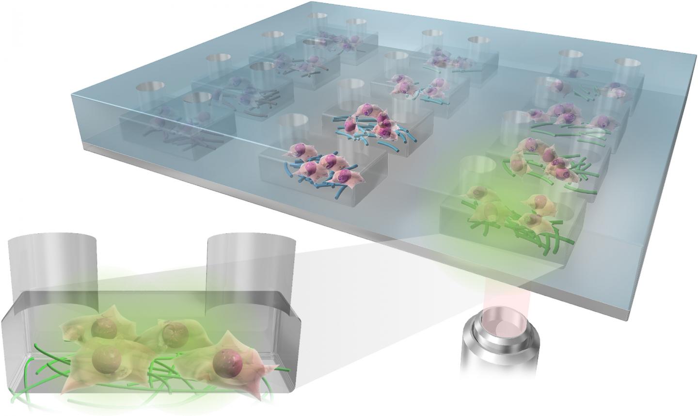
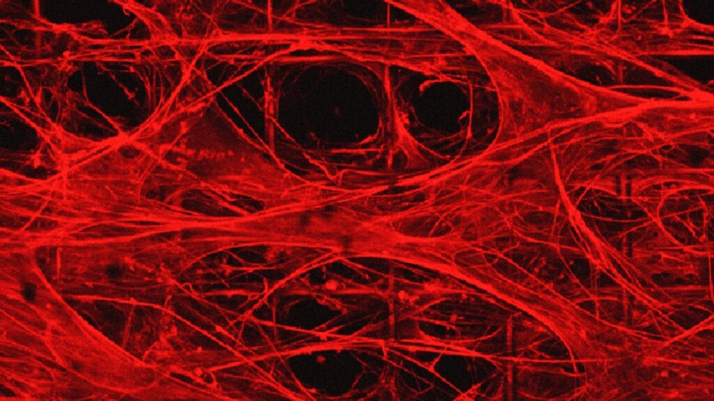
There’s a good (brief) description of how these fibres become organoids in the photo caption,

Engineered extracellular matrices composed of fibrillar fibronectin are suspended over a porous polymer framework and provide the niche for stem cells to attach, differentiate, and mature into organoids. Credit: Ayse Muñiz Courtesy: Michigan Medicine – University of Michigan

A July 13 ,2023 University of Michigan (Michigan Medicine) news release by Noah Fromson (also on EurekAlert) announces ‘kinder, gentler’ brain organoids. Coincidentally, these organoids more closely resemble human brains, Note: Links have been removed,

Researchers at University of Michigan developed a method to produce artificially grown miniature brains — called human brain organoids — free of animal cells that could greatly improve the way neurodegenerative conditions are studied and, eventually, treated.

Over the last decade of researching neurologic diseases, scientists have explored the use of human brain organoids as an alternative to mouse models. These self-assembled, 3D tissues derived from embryonic or pluripotent stem cells more closely model the complex brain structure compared to conventional two-dimensional cultures.

Until now, the engineered network of proteins and molecules that give structure to the cells in brain organoids, known as extracellular matrices, often used a substance derived from mouse sarcomas called Matrigel. That method suffers significant disadvantages, with a relatively undefined composition and batch-to-batch variability.

The latest U-M research, published in Annals of Clinical and Translational Neurology, offers a solution to overcome Matrigel’s weaknesses. Investigators created a novel culture method that uses an engineered extracellular matrix for human brain organoids — without the presence of animal components – and enhanced the neurogenesis of brain organoids compared to previous studies.

“This advancement in the development of human brain organoids free of animal components will allow for significant strides in the understanding of neurodevelopmental biology,” said senior author Joerg Lahann, Ph.D., director of the U-M Biointerfaces Institute and Wolfgang Pauli Collegiate Professor of Chemical Engineering at U-M.

“Scientists have long struggled to translate animal research into the clinical world, and this novel method will make it easier for translational research to make its way from the lab to the clinic.”

The foundational extracellular matrices of the research team’s brain organoids were comprised of human fibronectin, a protein that serves as a native structure for stem cells to adhere, differentiate and mature. They were supported by a highly porous polymer scaffold.

The organoids were cultured for months, while lab staff was unable to enter the building due to the COVID 19-pandemic.

Using proteomics, researchers found their brain organoids developed cerebral spinal fluid, a clear liquid that flows around healthy brain and spinal cords. This fluid more closely matched human adult CSF compared to a landmark study of human brain organoids developed in Matrigel.

“When our brains are naturally developing in utero, they are of course not growing on a bed of extracellular matrix produced by mouse cancer cells,” said first author Ayşe Muñiz, Ph.D., who was a graduate student in the U-M Macromolecular Science and Engineering Program at the time of the work.  

“By putting cells in an engineered niche that more closely resembles their natural environment, we predicted we would observe differences in organoid development that more faithfully mimics what we see in nature.”

The success of these xenogeneic-free human brain organoids opens the door for reprogramming with cells from patients with neurodegenerative diseases, says co-author Eva Feldman, M.D., Ph.D., director of the ALS Center of Excellence at U-M and James W. Albers Distinguished Professor of Neurology at U-M Medical School.

“There is a possibility to take the stem cells from a patient with a condition such as ALS or Alzheimer’s and, essentially, build an avatar mini brain of that patients to investigate possible treatments or model how their disease will progress,” Feldman said. “These models would create another avenue to predict disease and study treatment on a personalized level for conditions that often vary greatly from person to person.”

Here’s a link to and a citation for the paper,

Engineered extracellular matrices facilitate brain organoids from human pluripotent stem cells by Ayşe J. Muñiz, Tuğba Topal, Michael D. Brooks, Angela Sze, Do Hoon Kim, Jacob Jordahl, Joe Nguyen, Paul H. Krebsbach, Masha G. Savelieff, Eva L. Feldman, Joerg Lahann. Annals of Clinical and Translational Neurology DOI: https://doi.org/10.1002/acn3.51820 First published: 07 June 2023

This paper is open access.

Coelacanth (a living fish fossil) may provide clue to making artificial organs for transplantation

An ancient fish called a ‘living fossil’ has helped researchers understand the basics of stem cells. This will further stem cell research and be a step in the direction of creating artificial organs. The coelacanth fish is 400 million years old. Photo: Canva. Courtesy: university of Copenhagen

A December 12, 2022 University of Copenhagen press release (also on EurekAlert) describes work which may have an impact on organ transplants,

A beating heart. A complicated organ that pumps blood around the body of animals and humans. Not exactly something you associate with a Petri dish in a laboratory.

But that may change in the future, and save the lives of people whose own organs fail. And the research is now one step closer to that.

To design artificial organs you first have to understand stem cells and the genetic instructions that govern their remarkable properties.

Professor Joshua Mark Brickman at the Novo Nordisk Foundation Center for Stem Cell Medicine (reNEW) has unearthed the evolutionary origins of a master gene that acts on a network of genes instructing stem cells.

“The first step in stem cell research is to understand the gene regulatory network that supports so-called pluripotent stem cells. Understanding how their function was perfected in evolution can help provide knowledge about how to construct better stem cells,” says Joshua Mark Brickman.

Pluripotent stem cells are stem cells that can develop into all other cells. For example, heart cells. If we understand how the pluripotent stem cells develop into a heart, then we are one step closer to replicating this process in a laboratory.

What are stem cells?

Stem cells are non-specialized cells found in all multicellular organisms. Stem cells have two properties that distinguish them from other cell types. On the one hand, stem cells can undergo an unlimited number of cell divisions (mitoses), and on the other hand, stem cells have the ability to mature (differentiate) into several cell types.

A pluripotent stem cell is a cell that can develop into any other cell, such as a heart cell, hair cell or eye cell.

A ‘living fossil’ is the key to understanding stem cells

The pluripotent property of stem cells – meaning that the cells can develop into any other cell – is something that has traditionally been associated with mammals.

Now Joshua Mark Brickman and his colleagues have found that the master gene that controls stem cells and supports pluripotency also exists in a fish called coelacanth. In humans and mice this gene is called OCT4 and they found that the coelacanth version could replace the mammalian one in mouse stem cells.

In addition to the fact that the coelacanth is in a different class from mammals, it has also been called a ‘living fossil,’ since approximately 400 million years ago it developed into the form it has today. It has fins shaped like limbs and is therefore thought to resemble the first animals to move from the sea onto land.

“By studying its cells, you can go back in evolution, so to speak,” explains Assistant Professor Molly Lowndes.

Assistant Professor Woranop Sukparangsi continues: “The central factor controlling the gene network in stem cells is found in the coelacanth. This shows that the network already existed early in evolution, potentially as far back as 400 million years ago.”

And by studying the network in other species, such as this fish, the researchers can distill what the basic concepts that support a stem cell are.

“The beauty of moving back in evolution is that the organisms become simpler. For example, they have only one copy of some essential genes instead of many versions. That way, you can start to separate what is really important for stem cells and use that to improve how you grow stem cells in a dish,” says PhD student Elena Morganti.

Sharks, mice and kangaroos

In addition to the researchers finding out that the network around stem cells is much older than previously thought, and found in ancient species, they also learned how exactly evolution has modified the network of genes to support pluripotent stem cells.

The researchers looked at the stem cell genes from over 40 animals. For example sharks, mice and kangaroos. The animals were selected to provide a good sampling of the main branch points in evolution.

The researchers used artificial intelligence to build three-dimensional models of the different OCT4 proteins. The researchers could see that the general structure of the protein is maintained across evolution. While the regions of these proteins known to be important for stem cells do not change, species-specific differences in apparently unrelated regions of these proteins alter their orientation, potentially affecting how well it supports pluripotency.

“This a very exciting finding about evolution that would not have been possible prior to the advent of new technologies. You can see it as evolution cleverly thinking, we don not tinker with the ‘engine in the car’, but we can move the engine around and improve the drive train to see if it makes the car go faster,” says Joshua Mark Brickman.

The study is a collaborative project spanning Australia, Japan and Europe, with vital strategic partnerships with the groups of Sylvie Mazan at the Oceanological Observatory of Banyuls-sur-Mer in France and professor Guillermo Montoya at Novo Nordisk Foundation Center for Protein Research at University of Copenhagen.

Caption: Coelacanth-fish and other animals. Credit: By Woranop Sukparangsi Courtesy: University of Copenhagen

Here’s a link to and a citation for the paper,

Evolutionary origin of vertebrate OCT4/POU5 functions in supporting pluripotency by Woranop Sukparangsi, Elena Morganti, Molly Lowndes, Hélène Mayeur, Melanie Weisser, Fella Hammachi, Hanna Peradziryi, Fabian Roske, Jurriaan Hölzenspies, Alessandra Livigni, Benoit Gilbert Godard, Fumiaki Sugahara, Shigeru Kuratani, Guillermo Montoya, Stephen R. Frankenberg, Sylvie Mazan & Joshua M. Brickman. Nature Communications volume 13, Article number: 5537 (2022) DOI: https://doi.org/10.1038/s41467-022-32481-z Published: 21 September 2022

This paper is open access.

A new platform for culturing stem cells: a Multiplexed Artificial Cellular Microenvironment array

Japanese scientists have developed a more precise method for culturing stem cells according to a March 14, 2017 news item on Nanowerk,

A team of researchers in Japan has developed a new platform for culturing human pluripotent stem cells that provides far more control of culture conditions than previous tools by using micro and nanotechnologies.

The Multiplexed Artificial Cellular Microenvironment (MACME) array places nanofibres, mimicking cellular matrices, into fluid-filled micro-chambers of precise sizes, which mimic extracellular environments.

Caption: The Multiplexed Artificial Cellular Microenvironment (MACME) array, consisted with a microfluidic structure and nanofibre array for mimicking cellular microenvironments. Credit: Kyoto University iCeMS

A March 17, 2017 Kyoto University press release (also on EurekAlert), which originated the news item, explains the research in more detail,

Human pluripotent stems cells (hPSCs) hold great promise for tissue engineering, regenerative medicine and cell-based therapies because they can become any type of cell. The environment surrounding the cells plays a major role in determining what tissues they become, if they replicate into more cells, or die. However, understanding these interactions has been difficult because researchers have lacked tools that work on the appropriate scale.

Often, stem cells are cultured in a cell culture medium in small petri dishes. While factors such as medium pH levels and nutrients can be controlled, the artificial set up is on the macroscopic scale and does not allow for precise control of the physical environment surrounding the cells.

The MACME array miniaturizes this set up, culturing stem cells in rows of micro-chambers of cell culture medium. It also takes it a step further by placing nanofibers in these chambers to mimic the structures found around cells.

Led by Ken-ichiro Kamei of Kyoto University’s Institute for Integrated Cell-Material Sciences (iCeMS), the team tested a variety of nanofiber materials and densities, micro-chamber heights and initial stem cell densities to determine the best combination that encourages human pluripotent stem cells to replicate.

They stained the cells with several fluorescent markers and used a microscope to see if the cells died, replicated or differentiated into tissues.

Their analysis revealed that gelatin nanofibers and medium-sized chambers that create medium seed cell density provided the best environment for the stem cells to continue to multiply. The quantity and density of neighboring cells strongly influences cell survival.

The array is an “optimal and powerful approach for understanding how environmental cues regulate cellular functions,” the researchers conclude in a recently published paper in the journal Small.

This array appears to be the first time multiple kinds of extracellular environments can be mounted onto a single device, making it much easier to compare how different environments influence cells.

The MACME array could substantially reduce experiment costs compared to conventional tools, in part because it is low volume and requires less cell culture medium. The array does not require any special equipment and is compatible with both commonly used laboratory pipettes and automated pipette systems for performing high-throughput screening.

Here’s a link to and a citation for the paper,

Microfluidic-Nanofiber Hybrid Array for Screening of Cellular Microenvironments by Ken-ichiro Kamei, Yasumasa Mashimo, Momoko Yoshioka, Yumie Tokunaga, Christopher Fockenberg, Shiho Terada, Yoshie Koyama, Minako Nakajima, Teiko Shibata-Seki, Li Liu, Toshihiro Akaike, Eiry Kobatake, Siew-Eng How, Motonari Uesugi, and Yong Chen. Small DOI: 10.1002/smll.201603104 Version of Record online: 8 MAR 2017

© 2017 WILEY-VCH Verlag GmbH & Co. KGaA, Weinheim

This paper is behind a paywall.