Tag Archives: polymer nanoparticles

Monitoring the life of bacteria in microdroplets

Trying to establish better ways to test the effect of drugs on bacteria has led the Institute of Physical Chemistry of the Polish Academy of Sciences to develop a new monitoring technique. From a Jan.  11, 2017 news item on Nanowerk,

So far, however, there has been no quick or accurate method of assessing the oxygen conditions in individual microdroplets. This key obstacle has been overcome at the Institute of Physical Chemistry of the Polish Academy of Sciences.

Not in rows of large industrial tanks, nor on shelves laden with test tubes and beakers. The future of chemistry and biology is barely visible to the eye: it’s hundreds and thousands of microdroplets, whizzing through thin tubules of microfluidic devices. The race is on to find technologies that will make it possible to carry out controlled chemical and biological experiments in microdroplets. At the Institute of Physical Chemistry of the Polish Academy of Sciences (IPC PAS) in Warsaw a method of remote, yet rapid and accurate assessment of oxygen consumption by micro-organisms living in individual microdroplets has been demonstrated for the first time.

“Devices for the cultivation of bacteria in microdroplets have the chance to revolutionize work on the development of new antibiotics and the study of mechanisms responsible for the acquisition of drug resistance by bacteria. In one small microfluidic system it is possible to accommodate several hundred or even several thousand microdroplets – and to carry out a different experiment in each of them, for example with different types of microorganisms and at different concentrations of antibiotic in each drop,” describes Prof. Piotr Garstecki (IPC PAS), then explains: “For such studies to be possible, one has to provide the bacteria with conditions for development for even a few weeks. Thus, knowledge about the flow of oxygen to the droplets and the rate of its consumption by the microorganisms becomes extremely important. In our latest system we demonstrate how to read this key information.”

A Jan. 11, 2017 IPC PAS press release on EurekAlert, which originated the  news item, describes the work in more detail,

The bioreactors of the future are water droplets with culture medium suspended in a carrier liquid with which they are immiscible (usually this is oil). In the channel of the microfluidic device each droplet is longer than it is wide and it almost completely fills its lumen; sizes matched in this manner ensure that the drops do not swop places in the channel and throughout the duration of the experiment they can be identified without any problems. At the same time, there has to be a thin layer of oil maintained continuously between each microdroplet and the wall of the channel. Without this, the bacteria would be in direct contact with the walls of the channel so they would be able to settle on them and move from drop to drop. Unfortunately, when the microdroplet is stationary, with time it pushes out the oil separating it from the walls, laying it open to contamination. For this reason the drops must be kept in constant motion – even for weeks.

Growing bacteria need culture medium, and waste products need to be removed from their environment at an appropriate rate. Information about the bacterial oxygen consumption in individual droplets is therefore crucial to the operation of microbioreactors.

“It is immediately obvious where the problem lies. In each of the hundreds of moving droplets measurements need to be carried out at a frequency corresponding to the frequency of division of the bacteria or more, in practice at least once every 15 minutes. In addition, the measurement cannot cause any interference in the microdroplets,” says PhD student Michal Horka (IPC PAS), a co-author of the publication in the journal Analytical Chemistry.

Help was at hand for the Warsaw researchers from chemists from the Austrian Institute of Analytical Chemistry and Food Chemistry at the Graz University of Technology. They provided polymer nanoparticles with a phosphorescent dye, which after excitation emit light for longer the higher the concentration of oxygen in the surrounding solution (the nanoparticles underwent tests at the IPC PAS on bacteria in order to determine their possible toxicity – none was found).

Research on monitoring oxygen consumption in the droplets commenced with the preparation of an aqueous solution with the bacteria, the culture medium and a suitable quantity of nanoparticles. The mixture was injected into the microfluidic system constructed of tubing with Teflon connectors with correspondingly shaped channels. The first module formed droplets with a volume of approx. 4 microlitres, which were directed to the incubation tube wound on a spool. In the middle of its length there was another module, with detectors for measuring oxygen and absorbance.

“In the incubation part in one phase of the cycle the droplets flowed in one direction, in the second – in another, electronically controlled by means of suitable solenoid valves. All this looks seemingly simple enough, but in practice one of the biggest challenges was to ensure a smooth transition between the detection module and the tubing, so that bacterial contamination did not occur at the connections,” explains PhD student Horka.

During their passage through the detection module the droplets flowed under an optical sensor which measured the so-called optical density, which is the standard parameter used to evaluate the number of cells (the more bacteria in the droplets, the less light passes through them). In turn, the measurement of the duration of the phosphorescence of the nanoparticles, evaluating the concentration of oxygen in the microdroplets, was carried out using the Piccolo2 optical detector, provided by the Austrian group. This detector, which looks like a big pen drive, was connected directly to the USB port on the control computer. Comparing information from both sensors, IPC PAS researchers showed that the microfluidic device they had constructed made it possible to regularly and quickly monitor the metabolic activity of bacteria in the individual microdroplets.

“We carried out our tests both with bacteria floating in water singly – this is how the common Escherichia coli bacteria behave – as well as with those having a tendency to stick together in clumps – as is the case for tuberculosis bacilli or others belonging to the same family including Mycobacterium smegmatis which we studied. Evaluation of the rate of oxygen consumption by both species of microorganisms proved to be not only possible, but also reliable,” stresses PhD student Artur Ruszczak (IPC PAS).

The results of the research, funded by the European ERC Starting Grant (Polish side) and the Maria Sklodowska-Curie grant (Austrian side) are an important step in the process of building fully functional microfluidic devices for conducting biological experiments lasting many weeks. A system for culturing bacteria in microdroplets was developed at the IPC PAS a few years ago, however it was constructed on a polycarbonate plate. The maximum dimensions of the plate did not exceed 10 cm, which greatly limited the number of droplets; in addition, as a result of interaction with the polycarbonate, after four days the channels were contaminated with bacteria. Devices of Teflon modules and tubing would not have these disadvantages, and would be suitable for practical applications.

Here’s a link to and a citation for the paper,

Lifetime of Phosphorescence from Nanoparticles Yields Accurate Measurement of Concentration of Oxygen in Microdroplets, Allowing One To Monitor the Metabolism of Bacteria by Michał Horka, Shiwen Sun, Artur Ruszczak, Piotr Garstecki, and Torsten Mayr. Anal. Chem., 2016, 88 (24), pp 12006–12012 DOI: 10.1021/acs.analchem.6b03758 Publication Date (Web): November 23, 2016

Copyright © 2016 American Chemical Society

This paper is behind a paywall.
Nanomaterials and UV (ultraviolet) light for environmental cleanups

I think this is the first time I’ve seen anything about a technology that removes toxic materials from both water and soil; it’s usually one or the other. A July 22, 2015 news item on Nanowerk makes the announcement (Note: A link has been removed),

Many human-made pollutants in the environment resist degradation through natural processes, and disrupt hormonal and other systems in mammals and other animals. Removing these toxic materials — which include pesticides and endocrine disruptors such as bisphenol A (BPA) — with existing methods is often expensive and time-consuming.

In a new paper published this week in Nature Communications (“Nanoparticles with photoinduced precipitation for the extraction of pollutants from water and soil”), researchers from MIT [Massachusetts Institute of Technology] and the Federal University of Goiás in Brazil demonstrate a novel method for using nanoparticles and ultraviolet (UV) light to quickly isolate and extract a variety of contaminants from soil and water.

A July 21, 2015 MIT news release by Jonathan Mingle, which originated the news item, describes the inspiration and the research in more detail,

Ferdinand Brandl and Nicolas Bertrand, the two lead authors, are former postdocs in the laboratory of Robert Langer, the David H. Koch Institute Professor at MIT’s Koch Institute for Integrative Cancer Research. (Eliana Martins Lima, of the Federal University of Goiás, is the other co-author.) Both Brandl and Bertrand are trained as pharmacists, and describe their discovery as a happy accident: They initially sought to develop nanoparticles that could be used to deliver drugs to cancer cells.

Brandl had previously synthesized polymers that could be cleaved apart by exposure to UV light. But he and Bertrand came to question their suitability for drug delivery, since UV light can be damaging to tissue and cells, and doesn’t penetrate through the skin. When they learned that UV light was used to disinfect water in certain treatment plants, they began to ask a different question.

“We thought if they are already using UV light, maybe they could use our particles as well,” Brandl says. “Then we came up with the idea to use our particles to remove toxic chemicals, pollutants, or hormones from water, because we saw that the particles aggregate once you irradiate them with UV light.”

A trap for ‘water-fearing’ pollution

The researchers synthesized polymers from polyethylene glycol, a widely used compound found in laxatives, toothpaste, and eye drops and approved by the Food and Drug Administration as a food additive, and polylactic acid, a biodegradable plastic used in compostable cups and glassware.

Nanoparticles made from these polymers have a hydrophobic core and a hydrophilic shell. Due to molecular-scale forces, in a solution hydrophobic pollutant molecules move toward the hydrophobic nanoparticles, and adsorb onto their surface, where they effectively become “trapped.” This same phenomenon is at work when spaghetti sauce stains the surface of plastic containers, turning them red: In that case, both the plastic and the oil-based sauce are hydrophobic and interact together.

If left alone, these nanomaterials would remain suspended and dispersed evenly in water. But when exposed to UV light, the stabilizing outer shell of the particles is shed, and — now “enriched” by the pollutants — they form larger aggregates that can then be removed through filtration, sedimentation, or other methods.

The researchers used the method to extract phthalates, hormone-disrupting chemicals used to soften plastics, from wastewater; BPA, another endocrine-disrupting synthetic compound widely used in plastic bottles and other resinous consumer goods, from thermal printing paper samples; and polycyclic aromatic hydrocarbons, carcinogenic compounds formed from incomplete combustion of fuels, from contaminated soil.

The process is irreversible and the polymers are biodegradable, minimizing the risks of leaving toxic secondary products to persist in, say, a body of water. “Once they switch to this macro situation where they’re big clumps,” Bertrand says, “you won’t be able to bring them back to the nano state again.”

The fundamental breakthrough, according to the researchers, was confirming that small molecules do indeed adsorb passively onto the surface of nanoparticles.

“To the best of our knowledge, it is the first time that the interactions of small molecules with pre-formed nanoparticles can be directly measured,” they write in Nature Communications.

Nano cleansing

Even more exciting, they say, is the wide range of potential uses, from environmental remediation to medical analysis.

The polymers are synthesized at room temperature, and don’t need to be specially prepared to target specific compounds; they are broadly applicable to all kinds of hydrophobic chemicals and molecules.

“The interactions we exploit to remove the pollutants are non-specific,” Brandl says. “We can remove hormones, BPA, and pesticides that are all present in the same sample, and we can do this in one step.”

And the nanoparticles’ high surface-area-to-volume ratio means that only a small amount is needed to remove a relatively large quantity of pollutants. The technique could thus offer potential for the cost-effective cleanup of contaminated water and soil on a wider scale.

“From the applied perspective, we showed in a system that the adsorption of small molecules on the surface of the nanoparticles can be used for extraction of any kind,” Bertrand says. “It opens the door for many other applications down the line.”

This approach could possibly be further developed, he speculates, to replace the widespread use of organic solvents for everything from decaffeinating coffee to making paint thinners. Bertrand cites DDT, banned for use as a pesticide in the U.S. since 1972 but still widely used in other parts of the world, as another example of a persistent pollutant that could potentially be remediated using these nanomaterials. “And for analytical applications where you don’t need as much volume to purify or concentrate, this might be interesting,” Bertrand says, offering the example of a cheap testing kit for urine analysis of medical patients.

The study also suggests the broader potential for adapting nanoscale drug-delivery techniques developed for use in environmental remediation.

“That we can apply some of the highly sophisticated, high-precision tools developed for the pharmaceutical industry, and now look at the use of these technologies in broader terms, is phenomenal,” says Frank Gu, an assistant professor of chemical engineering at the University of Waterloo in Canada, and an expert in nanoengineering for health care and medical applications.

“When you think about field deployment, that’s far down the road, but this paper offers a really exciting opportunity to crack a problem that is persistently present,” says Gu, who was not involved in the research. “If you take the normal conventional civil engineering or chemical engineering approach to treating it, it just won’t touch it. That’s where the most exciting part is.”

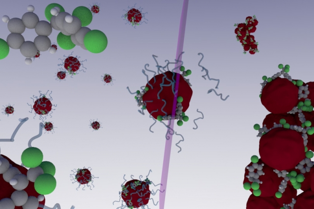
The researchers have made this illustration of their work available,

Nanoparticles that lose their stability upon irradiation with light have been designed to extract endocrine disruptors, pesticides, and other contaminants from water and soils. The system exploits the large surface-to-volume ratio of nanoparticles, while the photoinduced precipitation ensures nanomaterials are not released in the environment. Image: Nicolas Bertrand Courtesy: MIT

Nanoparticles that lose their stability upon irradiation with light have been designed to extract endocrine disruptors, pesticides, and other contaminants from water and soils. The system exploits the large surface-to-volume ratio of nanoparticles, while the photoinduced precipitation ensures nanomaterials are not released in the environment.
Image: Nicolas Bertrand Courtesy: MIT

Here’s a link to and a citation for the paper,

Nanoparticles with photoinduced precipitation for the extraction of pollutants from water and soil by Ferdinand Brandl, Nicolas Bertrand, Eliana Martins Lima & Robert Langer. Nature Communications 6, Article number: 7765 doi:10.1038/ncomms8765 Published 21 July 2015

This paper is open access.