Tag Archives: Qiaobing Xu

Precision targeting of the liver for gene editing

Apparently the magic is in the lipid nanoparticles. A March 1, 2021 news item on Nanowerk announced research into lipid nanoparticles as a means to deliver CRISPR (clustered regularly interspaced short palindromic repeats) to specific organs (Note: A link has been removed),

The genome editing technology CRISPR has emerged as a powerful new tool that can change the way we treat disease. The challenge when altering the genetics of our cells, however, is how to do it safely, effectively, and specifically targeted to the gene, tissue and organ that needs treatment.

Scientists at Tufts University and the Broad Institute of Harvard [University] and MIT [Massachusetts Institute of Technology] have developed unique nanoparticles comprised of lipids — fat molecules — that can package and deliver gene editing machinery specifically to the liver.

In a study published in the Proceedings of the National Academy of Sciences [PNAS] (“Lipid nanoparticle-mediated codelivery of Cas9 mRNA and single-guide RNA achieves liver-specific in vivo genome editing of Angptl3”), they have shown that they can use the lipid nanoparticles (LNPs) to efficiently deliver the CRISPR machinery into the liver of mice, resulting in specific genome editing and the reduction of blood cholesterol levels by as much as 57% — a reduction that can last for at least several months with just one shot.

A March 2, 2021 Tufts University news release (also on EurekAlert but published March 1, 2021), which originated the news item, provides greater insight into and technical detail about the research,

The problem of high cholesterol plagues more than 29 million Americans, according to the Centers for Disease Control and Prevention. The condition is complex and can originate from multiple genes as well as nutritional and lifestyle choices, so it is not easy to treat. The Tufts and Broad researchers, however, have modified one gene that could provide a protective effect against elevated cholesterol if it can be shut down by gene editing.

The gene that the researchers focused on codes for the angiopoietin-like 3 enzyme (Angptl3). That enzyme tamps down the activity of other enzymes – lipases – that help break down cholesterol. If researchers can knock out the Angptl3 gene, they can let the lipases do their work and reduce levels of cholesterol in the blood. It turns out that some lucky people have a natural mutation in their Angptl3 gene, leading to consistently low levels of triglycerides and low-density lipoprotein (LDL) cholesterol, commonly called “bad” cholesterol, in their bloodstream without any known clinical downsides.

“If we can replicate that condition by knocking out the angptl3 gene in others, we have a good chance of having a safe and long term solution to high cholesterol,” said Qiaobing Xu, associate professor of biomedical engineering at Tufts’ School of Engineering and corresponding author of the study. “We just have to make sure we deliver the gene editing package specifically to the liver so as not to create unwanted side effects.”

Xu’s team was able to do precisely that in mouse models. After a single injection of lipid nanoparticles packed with mRNA coding for CRISPR-Cas9 and a single-guide RNA targeting Angptl3, they observed a profound reduction in LDL cholesterol by as much as 57% and triglyceride levels by about 29 %, both of which remained at those lowered levels for at least 100 days. The researchers speculate that the effect may last much longer than that, perhaps limited only by the slow turnover of cells in the liver, which can occur over a period of about a year. The reduction of cholesterol and triglycerides is dose dependent, so their levels could be adjusted by injecting fewer or more LNPs in the single shot, the researchers said.

By comparison, an existing, FDA [US Food and Drug Administration]-approved version of CRISPR mRNA-loaded LNPs could only reduce LDL cholesterol by at most 15.7% and triglycerides by 16.3% when it was tested in mice, according to the researchers.

The trick to making a better LNP was in customizing the components – the molecules that come together to form bubbles around the mRNA. The LNPs are made up of long chain lipids that have a charged or polar head that is attracted to water, a carbon chain tail that points toward the middle of the bubble containing the payload, and a chemical linker between them. Also present are polyethylene glycol, and yes, even some cholesterol – which has a normal role in lipid membranes to make them less leaky – to hold their contents better.

The researchers found that the nature and relative ratio of these components appeared to have profound effects on the delivery of mRNA into the liver, so they tested LNPs with many combinations of heads, tails, linkers and ratios among all components for their ability to target liver cells. Because the in vitro potency of an LNP formulation rarely reflects its in vivo performance, they directly evaluated the delivery specificity and efficacy in mice that have a reporter gene in their cells that lights up red when genome editing occurs. Ultimately, they found a CRISPR mRNA-loaded LNP that lit up just the liver in mice, showing that it could specifically and efficiently deliver gene-editing tools into the liver to do their work.

The LNPs were built upon earlier work at Tufts, where Xu and his team developed LNPs with as much as 90% efficiency in delivering mRNA into cells. A unique feature of those nanoparticles was the presence of disulfide bonds between the long lipid chains. Outside the cells, the LNPs form a stable spherical structure that locks in their contents. When they are inside a cell, the environment within breaks the disulfide bonds to disassemble the nanoparticles. The contents are then quickly and efficiently released into the cell. By preventing loss outside the cell, the LNPs can have a much higher yield in delivering their contents.

“CRISPR is one of the most powerful therapeutic tools for the treatment of diseases with a genetic etiology. We have recently seen the first human clinical trail for CRISPR therapy enabled by LNP delivery to be administered systemically to edit genes inside the human body. Our LNP platform developed here holds great potential for clinical translation,” said Min Qiu, post-doctoral researcher in Xu’s lab at Tufts.  “We envision that with this LNP platform in hand, we could now make CRISPR a practical and safe approach to treat a broad spectrum of liver diseases or disorders,” said Zachary Glass, graduate student in the Xu lab. Qiu and Glass are co-first authors of the study.

Here’s a link to and a citation for the paper,

Lipid nanoparticle-mediated codelivery of Cas9 mRNA and single-guide RNA achieves liver-specific in vivo genome editing of Angptl3 by Min Qiu, Zachary Glass, Jinjin Chen, Mary Haas, Xin Jin, Xuewei Zhao, Xuehui Rui, Zhongfeng Ye, Yamin Li, Feng Zhang, and Qiaobing Xu. PNAS March 9, 2021 118 (10) e2020401118 DOI: https://doi.org/10.1073/pnas.2020401118

This paper appears to be behind a paywall.
CRISPR-CAS9 and gold

As so often happens in the sciences, now that the initial euphoria has expended itself problems (and solutions) with CRISPR ((clustered regularly interspaced short palindromic repeats))-CAAS9 are being disclosed to those of us who are not experts. From an Oct. 3, 2017 article by Bob Yirka for phys.org,

A team of researchers from the University of California and the University of Tokyo has found a way to use the CRISPR gene editing technique that does not rely on a virus for delivery. In their paper published in the journal Nature Biomedical Engineering, the group describes the new technique, how well it works and improvements that need to be made to make it a viable gene editing tool.

CRISPR-Cas9 has been in the news a lot lately because it allows researchers to directly edit genes—either disabling unwanted parts or replacing them altogether. But despite many success stories, the technique still suffers from a major deficit that prevents it from being used as a true medical tool—it sometimes makes mistakes. Those mistakes can cause small or big problems for a host depending on what goes wrong. Prior research has suggested that the majority of mistakes are due to delivery problems, which means that a replacement for the virus part of the technique is required. In this new effort, the researchers report that they have discovered just a such a replacement, and it worked so well that it was able to repair a gene mutation in a Duchenne muscular dystrophy mouse model. The team has named the new technique CRISPR-Gold, because a gold nanoparticle was used to deliver the gene editing molecules instead of a virus.

An Oct. 2, 2017 article by Abby Olena for The Scientist lays out the CRISPR-CAS9 problems the scientists are trying to solve (Note: Links have been removed),

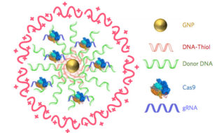
While promising, applications of CRISPR-Cas9 gene editing have so far been limited by the challenges of delivery—namely, how to get all the CRISPR parts to every cell that needs them. In a study published today (October 2) in Nature Biomedical Engineering, researchers have successfully repaired a mutation in the gene for dystrophin in a mouse model of Duchenne muscular dystrophy by injecting a vehicle they call CRISPR-Gold, which contains the Cas9 protein, guide RNA, and donor DNA, all wrapped around a tiny gold ball.

The authors have made “great progress in the gene editing area,” says Tufts University biomedical engineer Qiaobing Xu, who did not participate in the work but penned an accompanying commentary. Because their approach is nonviral, Xu explains, it will minimize the potential off-target effects that result from constant Cas9 activity, which occurs when users deliver the Cas9 template with a viral vector.

Duchenne muscular dystrophy is a degenerative disease of the muscles caused by a lack of the protein dystrophin. In about a third of patients, the gene for dystrophin has small deletions or single base mutations that render it nonfunctional, which makes this gene an excellent candidate for gene editing. Researchers have previously used viral delivery of CRISPR-Cas9 components to delete the mutated exon and achieve clinical improvements in mouse models of the disease.

“In this paper, we were actually able to correct [the gene for] dystrophin back to the wild-type sequence” via homology-directed repair (HDR), coauthor Niren Murthy, a drug delivery researcher at the University of California, Berkeley, tells The Scientist. “The other way of treating this is to do something called exon skipping, which is where you delete some of the exons and you can get dystrophin to be produced, but it’s not [as functional as] the wild-type protein.”

The research team created CRISPR-Gold by covering a central gold nanoparticle with DNA that they modified so it would stick to the particle. This gold-conjugated DNA bound the donor DNA needed for HDR, which the Cas9 protein and guide RNA bound to in turn. They coated the entire complex with a polymer that seems to trigger endocytosis and then facilitate escape of the Cas9 protein, guide RNA, and template DNA from endosomes within cells.

In order to do HDR, “you have to provide the cell [with] the Cas9 enzyme, guide RNA by which you target Cas9 to a particular part of the genome, and a big chunk of DNA, which will be used as a template to edit the mutant sequence to wild-type,” explains coauthor Irina Conboy, who studies tissue repair at the University of California, Berkeley. “They all have to be present at the same time and at the same place, so in our system you have a nanoparticle which simultaneously delivers all of those three key components in their active state.”

Olena’s article carries on to describe how the team created CRISPR-Gold and more.

Additional technical details are available in an Oct. 3, 2017 University of California at Berkeley news release by Brett Israel (also on EurekAlert), which originated the news item (Note: A link has been removed) ,

Scientists at the University of California, Berkeley, have engineered a new way to deliver CRISPR-Cas9 gene-editing technology inside cells and have demonstrated in mice that the technology can repair the mutation that causes Duchenne muscular dystrophy, a severe muscle-wasting disease. A new study shows that a single injection of CRISPR-Gold, as the new delivery system is called, into mice with Duchenne muscular dystrophy led to an 18-times-higher correction rate and a two-fold increase in a strength and agility test compared to control groups.

Diagram of CRISPR-Gold

CRISPR–Gold is composed of 15 nanometer gold nanoparticles that are conjugated to thiol-modified oligonucleotides (DNA-Thiol), which are hybridized with single-stranded donor DNA and subsequently complexed with Cas9 and encapsulated by a polymer that disrupts the endosome of the cell.

Since 2012, when study co-author Jennifer Doudna, a professor of molecular and cell biology and of chemistry at UC Berkeley, and colleague Emmanuelle Charpentier, of the Max Planck Institute for Infection Biology, repurposed the Cas9 protein to create a cheap, precise and easy-to-use gene editor, researchers have hoped that therapies based on CRISPR-Cas9 would one day revolutionize the treatment of genetic diseases. Yet developing treatments for genetic diseases remains a big challenge in medicine. This is because most genetic diseases can be cured only if the disease-causing gene mutation is corrected back to the normal sequence, and this is impossible to do with conventional therapeutics.

CRISPR/Cas9, however, can correct gene mutations by cutting the mutated DNA and triggering homology-directed DNA repair. However, strategies for safely delivering the necessary components (Cas9, guide RNA that directs Cas9 to a specific gene, and donor DNA) into cells need to be developed before the potential of CRISPR-Cas9-based therapeutics can be realized. A common technique to deliver CRISPR-Cas9 into cells employs viruses, but that technique has a number of complications. CRISPR-Gold does not need viruses.

In the new study, research lead by the laboratories of Berkeley bioengineering professors Niren Murthy and Irina Conboy demonstrated that their novel approach, called CRISPR-Gold because gold nanoparticles are a key component, can deliver Cas9 – the protein that binds and cuts DNA – along with guide RNA and donor DNA into the cells of a living organism to fix a gene mutation.

“CRISPR-Gold is the first example of a delivery vehicle that can deliver all of the CRISPR components needed to correct gene mutations, without the use of viruses,” Murthy said.

The study was published October 2 [2017] in the journal Nature Biomedical Engineering.

CRISPR-Gold repairs DNA mutations through a process called homology-directed repair. Scientists have struggled to develop homology-directed repair-based therapeutics because they require activity at the same place and time as Cas9 protein, an RNA guide that recognizes the mutation and donor DNA to correct the mutation.

To overcome these challenges, the Berkeley scientists invented a delivery vessel that binds all of these components together, and then releases them when the vessel is inside a wide variety of cell types, triggering homology directed repair. CRISPR-Gold’s gold nanoparticles coat the donor DNA and also bind Cas9. When injected into mice, their cells recognize a marker in CRISPR-Gold and then import the delivery vessel. Then, through a series of cellular mechanisms, CRISPR-Gold is released into the cells’ cytoplasm and breaks apart, rapidly releasing Cas9 and donor DNA.

Schematic of CRISPR-Gold's method of action

CRISPR-Gold’s method of action (Click to enlarge).

A single injection of CRISPR-Gold into muscle tissue of mice that model Duchenne muscular dystrophy restored 5.4 percent of the dystrophin gene, which causes the disease, to the wild- type, or normal, sequence. This correction rate was approximately 18 times higher than in mice treated with Cas9 and donor DNA by themselves, which experienced only a 0.3 percent correction rate.

Importantly, the study authors note, CRISPR-Gold faithfully restored the normal sequence of dystrophin, which is a significant improvement over previously published approaches that only removed the faulty part of the gene, making it shorter and converting one disease into another, milder disease.

CRISPR-Gold was also able to reduce tissue fibrosis – the hallmark of diseases where muscles do not function properly – and enhanced strength and agility in mice with Duchenne muscular dystrophy. CRISPR-Gold-treated mice showed a two-fold increase in hanging time in a common test for mouse strength and agility, compared to mice injected with a control.

“These experiments suggest that it will be possible to develop non-viral CRISPR therapeutics that can safely correct gene mutations, via the process of homology-directed repair, by simply developing nanoparticles that can simultaneously encapsulate all of the CRISPR components,” Murthy said.

CRISPR-Cas9

CRISPR in action: A model of the Cas9 protein cutting a double-stranded piece of DNA

The study found that CRISPR-Gold’s approach to Cas9 protein delivery is safer than viral delivery of CRISPR, which, in addition to toxicity, amplifies the side effects of Cas9 through continuous expression of this DNA-cutting enzyme. When the research team tested CRISPR-Gold’s gene-editing capability in mice, they found that CRISPR-Gold efficiently corrected the DNA mutation that causes Duchenne muscular dystrophy, with minimal collateral DNA damage.

The researchers quantified CRISPR-Gold’s off-target DNA damage and found damage levels similar to the that of a typical DNA sequencing error in a typical cell that was not exposed to CRISPR (0.005 – 0.2 percent). To test for possible immunogenicity, the blood stream cytokine profiles of mice were analyzed at 24 hours and two weeks after the CRISPR-Gold injection. CRISPR-Gold did not cause an acute up-regulation of inflammatory cytokines in plasma, after multiple injections, or weight loss, suggesting that CRISPR-Gold can be used multiple times safely, and that it has a high therapeutic window for gene editing in muscle tissue.

“CRISPR-Gold and, more broadly, CRISPR-nanoparticles open a new way for safer, accurately controlled delivery of gene-editing tools,” Conboy said. “Ultimately, these techniques could be developed into a new medicine for Duchenne muscular dystrophy and a number of other genetic diseases.”

A clinical trial will be needed to discern whether CRISPR-Gold is an effective treatment for genetic diseases in humans. Study co-authors Kunwoo Lee and Hyo Min Park have formed a start-up company, GenEdit (Murthy has an ownership stake in GenEdit), which is focused on translating the CRISPR-Gold technology into humans. The labs of Murthy and Conboy are also working on the next generation of particles that can deliver CRISPR into tissues from the blood stream and would preferentially target adult stem cells, which are considered the best targets for gene correction because stem and progenitor cells are capable of gene editing, self-renewal and differentiation.

“Genetic diseases cause devastating levels of mortality and morbidity, and new strategies for treating them are greatly needed,” Murthy said. “CRISPR-Gold was able to correct disease-causing gene mutations in vivo, via the non-viral delivery of Cas9 protein, guide RNA and donor DNA, and therefore has the potential to develop into a therapeutic for treating genetic diseases.”

The study was funded by the National Institutes of Health, the W.M. Keck Foundation, the Moore Foundation, the Li Ka Shing Foundation, Calico, Packer, Roger’s and SENS, and the Center of Innovation (COI) Program of the Japan Science and Technology Agency.

Here’s a link to and a citation for the paper,

Nanoparticle delivery of Cas9 ribonucleoprotein and donor DNA in vivo induces homology-directed DNA repair by Kunwoo Lee, Michael Conboy, Hyo Min Park, Fuguo Jiang, Hyun Jin Kim, Mark A. Dewitt, Vanessa A. Mackley, Kevin Chang, Anirudh Rao, Colin Skinner, Tamanna Shobha, Melod Mehdipour, Hui Liu, Wen-chin Huang, Freeman Lan, Nicolas L. Bray, Song Li, Jacob E. Corn, Kazunori Kataoka, Jennifer A. Doudna, Irina Conboy, & Niren Murthy. Nature Biomedical Engineering (2017) doi:10.1038/s41551-017-0137-2 Published online: 02 October 2017

This paper is behind a paywall.

Sutures that can gather data wirelessly

Are sutures which gather data hackable? It’s a little early to start thinking about that issue as this seems to be brand new research. A July 18, 2016 news item on ScienceDaily tells more,

For the first time, researchers led by Tufts University engineers have integrated nano-scale sensors, electronics and microfluidics into threads — ranging from simple cotton to sophisticated synthetics — that can be sutured through multiple layers of tissue to gather diagnostic data wirelessly in real time, according to a paper published online July 18 [2016] in Microsystems & Nanoengineering. The research suggests that the thread-based diagnostic platform could be an effective substrate for a new generation of implantable diagnostic devices and smart wearable systems.

A July 18, 2016 Tufts University news release (also on EurekAlert), which originated the news item, provides more detail,

The researchers used a variety of conductive threads that were dipped in physical and chemical sensing compounds and connected to wireless electronic circuitry to create a flexible platform that they sutured into tissue in rats as well as in vitro. The threads collected data on tissue health (e.g. pressure, stress, strain and temperature), pH and glucose levels that can be used to determine such things as how a wound is healing, whether infection is emerging, or whether the body’s chemistry is out of balance. The results were transmitted wirelessly to a cell phone and computer.

The three-dimensional platform is able to conform to complex structures such as organs, wounds or orthopedic implants.

While more study is needed in a number of areas, including investigation of long-term biocompatibility, researchers said initial results raise the possibility of optimizing patient-specific treatments.

“The ability to suture a thread-based diagnostic device intimately in a tissue or organ environment in three dimensions adds a unique feature that is not available with other flexible diagnostic platforms,” said Sameer Sonkusale, Ph.D., corresponding author on the paper and director of the interdisciplinary Nano Lab in the Department of Electrical and Computer Engineering at Tufts School of Engineering. “We think thread-based devices could potentially be used as smart sutures for surgical implants, smart bandages to monitor wound healing, or integrated with textile or fabric as personalized health monitors and point-of-care diagnostics.”

Until now, the structure of substrates for implantable devices has essentially been two-dimensional, limiting their usefulness to flat tissue such as skin, according to the paper. Additionally, the materials in those substrates are expensive and require specialized processing.

Here’s a link to and a citation for the paper,

A toolkit of thread-based microfluidics, sensors, and electronics for 3D tissue embedding for medical diagnostics by Pooria Mostafalu, Mohsen Akbari, Kyle A. Alberti, Qiaobing Xu, Ali Khademhosseini, & Sameer R. Sonkusale. Microsystems & Nanoengineering 2, Article number: 16039 (2016) doi:10.1038/micronano.2016.39 Published online 18 July 2016

This paper is open access.