Tag Archives: saliva

CRISPR-Cas12a as a new diagnostic tool

Similar to Cas9, Cas12a is has an added feature as noted in this February 15, 2018 news item on ScienceDaily,

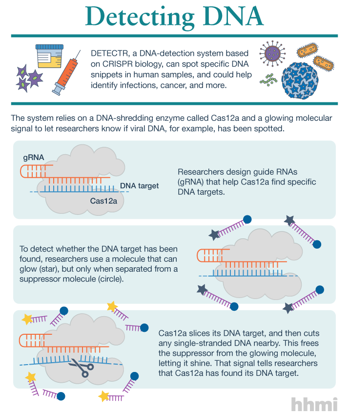
Utilizing an unsuspected activity of the CRISPR-Cas12a protein, researchers created a simple diagnostic system called DETECTR to analyze cells, blood, saliva, urine and stool to detect genetic mutations, cancer and antibiotic resistance and also diagnose bacterial and viral infections. The scientists discovered that when Cas12a binds its double-stranded DNA target, it indiscriminately chews up all single-stranded DNA. They then created reporter molecules attached to single-stranded DNA to signal when Cas12a finds its target.

A February 15, 2018 University of California at Berkeley (UC Berkeley) news release by Robert Sanders and which originated the news item, provides more detail and history,

CRISPR-Cas12a, one of the DNA-cutting proteins revolutionizing biology today, has an unexpected side effect that makes it an ideal enzyme for simple, rapid and accurate disease diagnostics.

blood in test tube

(iStock)

Cas12a, discovered in 2015 and originally called Cpf1, is like the well-known Cas9 protein that UC Berkeley’s Jennifer Doudna and colleague Emmanuelle Charpentier turned into a powerful gene-editing tool in 2012.

CRISPR-Cas9 has supercharged biological research in a mere six years, speeding up exploration of the causes of disease and sparking many potential new therapies. Cas12a was a major addition to the gene-cutting toolbox, able to cut double-stranded DNA at places that Cas9 can’t, and, because it leaves ragged edges, perhaps easier to use when inserting a new gene at the DNA cut.

But co-first authors Janice Chen, Enbo Ma and Lucas Harrington in Doudna’s lab discovered that when Cas12a binds and cuts a targeted double-stranded DNA sequence, it unexpectedly unleashes indiscriminate cutting of all single-stranded DNA in a test tube.

Most of the DNA in a cell is in the form of a double-stranded helix, so this is not necessarily a problem for gene-editing applications. But it does allow researchers to use a single-stranded “reporter” molecule with the CRISPR-Cas12a protein, which produces an unambiguous fluorescent signal when Cas12a has found its target.

“We continue to be fascinated by the functions of bacterial CRISPR systems and how mechanistic understanding leads to opportunities for new technologies,” said Doudna, a professor of molecular and cell biology and of chemistry and a Howard Hughes Medical Institute investigator.

DETECTR diagnostics

The new DETECTR system based on CRISPR-Cas12a can analyze cells, blood, saliva, urine and stool to detect genetic mutations, cancer and antibiotic resistance as well as diagnose bacterial and viral infections. Target DNA is amplified by RPA to make it easier for Cas12a to find it and bind, unleashing indiscriminate cutting of single-stranded DNA, including DNA attached to a fluorescent marker (gold star) that tells researchers that Cas12a has found its target.

The UC Berkeley researchers, along with their colleagues at UC San Francisco, will publish their findings Feb. 15 [2018] via the journal Science’s fast-track service, First Release.

The researchers developed a diagnostic system they dubbed the DNA Endonuclease Targeted CRISPR Trans Reporter, or DETECTR, for quick and easy point-of-care detection of even small amounts of DNA in clinical samples. It involves adding all reagents in a single reaction: CRISPR-Cas12a and its RNA targeting sequence (guide RNA), fluorescent reporter molecule and an isothermal amplification system called recombinase polymerase amplification (RPA), which is similar to polymerase chain reaction (PCR). When warmed to body temperature, RPA rapidly multiplies the number of copies of the target DNA, boosting the chances Cas12a will find one of them, bind and unleash single-strand DNA cutting, resulting in a fluorescent readout.

The UC Berkeley researchers tested this strategy using patient samples containing human papilloma virus (HPV), in collaboration with Joel Palefsky’s lab at UC San Francisco. Using DETECTR, they were able to demonstrate accurate detection of the “high-risk” HPV types 16 and 18 in samples infected with many different HPV types.

“This protein works as a robust tool to detect DNA from a variety of sources,” Chen said. “We want to push the limits of the technology, which is potentially applicable in any point-of-care diagnostic situation where there is a DNA component, including cancer and infectious disease.”

The indiscriminate cutting of all single-stranded DNA, which the researchers discovered holds true for all related Cas12 molecules, but not Cas9, may have unwanted effects in genome editing applications, but more research is needed on this topic, Chen said. During the transcription of genes, for example, the cell briefly creates single strands of DNA that could accidentally be cut by Cas12a.

The activity of the Cas12 proteins is similar to that of another family of CRISPR enzymes, Cas13a, which chew up RNA after binding to a target RNA sequence. Various teams, including Doudna’s, are developing diagnostic tests using Cas13a that could, for example, detect the RNA genome of HIV.

infographic about DETECTR system

(Infographic by the Howard Hughes Medical Institute)

These new tools have been repurposed from their original role in microbes where they serve as adaptive immune systems to fend off viral infections. In these bacteria, Cas proteins store records of past infections and use these “memories” to identify harmful DNA during infections. Cas12a, the protein used in this study, then cuts the invading DNA, saving the bacteria from being taken over by the virus.

The chance discovery of Cas12a’s unusual behavior highlights the importance of basic research, Chen said, since it came from a basic curiosity about the mechanism Cas12a uses to cleave double-stranded DNA.

“It’s cool that, by going after the question of the cleavage mechanism of this protein, we uncovered what we think is a very powerful technology useful in an array of applications,” Chen said.

Here’s a link to and a citation for the paper,

CRISPR-Cas12a target binding unleashes indiscriminate single-stranded DNase activity by Janice S. Chen, Enbo Ma, Lucas B. Harrington, Maria Da Costa, Xinran Tian, Joel M. Palefsky, Jennifer A. Doudna. Science 15 Feb 2018: eaar6245 DOI: 10.1126/science.aar6245

This paper is behind a paywall.

‘Potalyzer’ for roadside sobriety tests

Given the drive to legalize marijuana in Canada and in the US and the current crop of marijuana dispensaries in Vancouver (if nowhere else), this new ‘potalyzer’ test from Stanford University (California, US) seems quite timely and destined for popularity in police departments everywhere. From a Sept. 13, 2016 news item on Nanowerk,

This November [2016], several states will vote whether to legalize marijuana use, joining more than 20 states that already allow some form of cannabis use. This has prompted a need for effective tools for police to determine on the spot whether people are driving under the influence. Cars stopped while police interview drivers

Stanford researchers have devised a potential solution, applying magnetic nanotechnology, previously used as a cancer screen, to create what could be the first practical roadside test for marijuana intoxication.

While police are trying out potential tools, no device currently on the market has been shown to quickly provide a precise measurement of a driver’s marijuana intoxication as effectively as a breathalyzer gauges alcohol intoxication. THC, the drug’s most potent psychoactive agent, is commonly screened for in laboratory blood or urine tests – not very helpful for an officer in the field.

The Stanford device might function as a practical “potalyzer” because it can quickly detect not just the presence of THC in a person’s saliva, but also measure its concentration.

A Sept, 8, 2016 Stanford University news release by Carrie Kirby, which originated the news item, describes the technology in a little more detail,

Led by Shan Wang, a professor of materials science and engineering and of electrical engineering, the Stanford team created a mobile device that uses magnetic biosensors to detect tiny THC molecules in saliva. Officers could collect a spit sample with a cotton swab and read the results on a smartphone or laptop in as little as three minutes.

Researchers tackling the “potalyzer” problem have zeroed in on saliva because testing it is less invasive and because THC in saliva may correlate with impairment better than THC in urine or blood. The big challenge is that these spit tests may be called upon to detect superlatively tiny concentrations of THC. Some states have no set limit of THC in the body for drivers, while others set a limit of 0 or 5 nanograms (a billionth of a gram) per milliliter of blood.

Wang’s device can detect concentrations of THC in the range of 0 to 50 nanograms per milliliter of saliva. While there’s still no consensus on how much THC in a driver’s system is too much, previous studies have suggested a cutoff between 2 and 25 ng/mL, well within the capability of Wang’s device.

Repurposing biomedical tools

The researchers achieved such precision by harnessing the behavior of magnetism in nanoparticles, which measure just a few tens of billionths of a meter.

The Wang Group has been exploring magnetic nanotechnology for years, using it to attack such diverse problems as in vitro cancer diagnostics and magnetic information storage. In this case, they’re combining magnetic nanotechnology with the time-tested biochemical technique of the immunoassay. Immunoassays detect a certain molecule in a solution by introducing an antibody that will bind only to that molecule.

In the test, saliva is mixed with THC antibodies, which bind to any THC molecules in the sample. Then the sample is placed on a disposable chip cartridge, which contains magnetoresistive (GMR) sensors pre-coated with THC, and inserted into the handheld reader.

This sets in motion a “competition” between the THC pre-coated on the sensor and THC in the saliva to bind with the antibodies; the more THC in the saliva, the fewer antibodies will be available to bind to the THC on the sensor surface.

The number of antibodies bound to THC molecules on the sensor tells the device how many antibodies the THC in the sample used up, and therefore how many THC molecules were present in the sample.

Next, magnetic nanoparticles, specially made to bind only to the antibodies, are introduced to the sample. Each nanoparticle binds onto a THC-antibody pair like a sticky beacon, but only the molecules on the sensor surface will be close enough to trip the GMR biosensors in the reader. The device then uses Bluetooth to communicate results to the screen of a smartphone or laptop.

“To the best of our knowledge, this is the first demonstration that GMR biosensors are capable of detecting small molecules,” Wang wrote in a paper describing the device, published in Analytical Chemistry.

Beyond marijuana

The platform has potential usefulness beyond THC. Just as they do with THC, the GMR biosensors in the device could detect any small molecule, meaning that the platform could also test for morphine, heroin, cocaine or other drugs.

In fact, with 80 sensors built into it, the GMR biosensor chip could screen a single sample for multiple substances. The team has already tried screening for morphine with promising results.

Students are currently working on creating a user-friendly form factor for the device, which would need to go through field tests and be approved by regulators before it can be deployed by police.

Another thing that would have to happen before the device would be useful to law enforcement: State laws must set limits for the concentration of THC allowed in a driver’s saliva.

Here too, the Wang Group’s device could be helpful. For example, the next generation of the device could screen both the blood and saliva of a subject to establish an understanding of the correlation between blood THC level and saliva THC level at the same degree of intoxication.

Here’s a link to and a citation for the paper,

Small Molecule Detection in Saliva Facilitates Portable Tests of Marijuana Abuse by Jung-Rok Lee, Joohong Choi, Tyler O. Shultz, and Shan X. Wang. Anal. Chem., 2016, 88 (15), pp 7457–7461 DOI: 10.1021/acs.analchem.6b01688 Publication Date (Web): July 19, 2016

Copyright © 2016 American Chemical Society

This paper is behind a paywall.

Monitoring your saliva via mouth guard and smart phone

I first came across the notion that saliva instead of blood and urine could be used to assess and monitor health in a presentation abstract for the 2004 American Association for the Advancement of Science (AAAS) annual meeting held in Seattle, Washington (as per my Feb. 15, 2011 posting). There have been a few ‘saliva’ health monitoring projects mentioned here over the years but this proof-of-concept version seems like it has the potential to get to the marketplace. An August 31, 2015 news item on Nanowerk features a ‘saliva’ health monitoring project from the University of California at San Diego (UCSD),

Engineers at the University of California, San Diego, have developed a mouth guard that can monitor health markers, such as lactate, cortisol and uric acid, in saliva and transmit the information wirelessly to a smart phone, laptop or tablet.
The technology, which is at a proof-of-concept stage, could be used to monitor patients continuously without invasive procedures, as well as to monitor athletes’ performance or stress levels in soldiers and pilots. In this study, engineers focused on uric acid, which is a marker related to diabetes and to gout. Currently, the only way to monitor the levels of uric acid in a patient is to draw blood.

An August 31, 2015 UCSD news release (also on EurekAlert), which originated the news item, describes the research and the mouth guard in more detail,

In this study, researchers showed that the mouth guard sensor could offer an easy and reliable way to monitor uric acid levels. The mouth guard has been tested with human saliva but hasn’t been tested in a person’s mouth.

Researchers collected saliva samples from healthy volunteers and spread them on the sensor, which produced readings in a normal range. Next, they collected saliva from a patient who suffers from hyperuricemia, a condition characterized by an excess of uric acid in the blood. The sensor detected more than four times as much uric acid in the patient’s saliva than in the healthy volunteers.

The patient also took Allopurinol, which had been prescribed by a physician to treat their condition. Researchers were able to document a drop in the levels of uric acid over four or five days as the medication took effect. In the past, the patient would have needed blood draws to monitor levels and relied instead on symptoms to start and stop his medication.

Fabrication and design

Wang’s team created a screen-printed sensor using silver, Prussian blue ink and uricase, an enzyme that reacts with uric acid. Because saliva is extremely complex and contains many different biomarkers, researchers needed to make sure that the sensors only reacted with the uric acid. Nanoengineers set up the chemical equivalent of a two-step authentication system. The first step is a series of chemical keyholes, which ensures that only the smallest biochemicals get inside the sensor. The second step is a layer of uricase trapped in polymers, which reacts selectively with uric acid. The reaction between acid and enzyme generates hydrogen peroxide, which is detected by the Prussian blue ink.  That information is then transmitted to an electronic board as electrical signals via metallic strips that are part of the sensor.

The electronic board, developed by Mercier’s team, uses small chips that sense the output of the sensors, digitizes this output and then wirelessly transmits data to a smart phone, tablet or laptop. The entire electronic board occupies an area slightly larger than a U.S. penny.

Next steps

The next step is to embed all the electronics inside the mouth guard so that it can actually be worn. Researchers also will have to test the materials used for the sensors and electronics to make sure that they are indeed completely biocompatible. The next iteration of the mouth guard is about a year out, Mercier estimates.

“All the components are there,” he said. “It’s just a matter of refining the device and working on its stability.”

Wang and Mercier lead the Center for Wearable Sensors at UC San Diego, which has made a series of breakthroughs in the field, including temporary tattoos that monitor glucose, ultra-miniaturized energy-processing chips and pens filled with high-tech inks for Do It Yourself chemical sensors.

Here’s a link to and a citation for the paper,

Wearable salivary uric acid mouthguard biosensor with integrated wireless electronics by Jayoung Kim, Somayeh Imani, William R. de Araujo, Julian Warchall, Gabriela Valdés-Ramírez, Thiago R.L.C. Paixão, Patrick P. Mercier, & Joseph Wang. Biosensors and Bioelectronics Volume 74, 15 December 2015, Pages 1061–1068 doi:10.1016/j.bios.2015.07.039

This paper is behind a paywall.

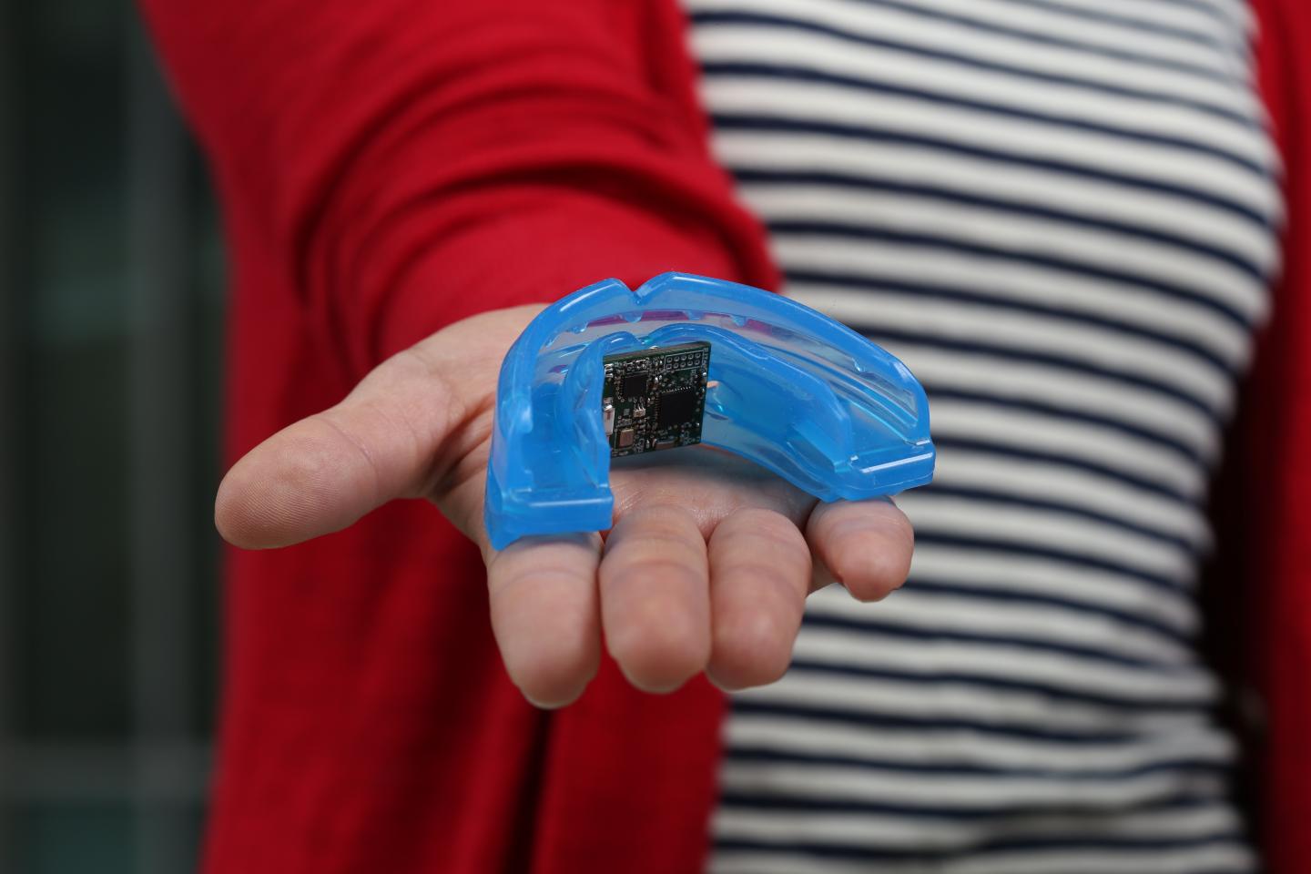
Here’s an image of UCSD’s proposed mouth guard,

The mouth guard sensor offers an easy and reliable way to monitor uric acid levels in human saliva. Credit: Jacobs School of Engineering, UC San Diego

The mouth guard sensor offers an easy and reliable way to monitor uric acid levels in human saliva. Credit: Jacobs School of Engineering, UC San Diego

Could nanoparticles in your mouthwash affect for your cells?

The first news item I’m going to highlight was posted on Nanowerk, March 8, 2012 and is focused on the use of silver nanoparticles in mouthwashes and dentures to prevent yeast infections,

Yeasts which cause hard-to-treat mouth infections are killed using silver nanoparticles in the laboratory, scientists have found. These yeast infections, caused by Candida albicans and Candida glabrata target the young, old and immuno-compromised. Professor Mariana Henriques, University of Minho [Portugal], and her colleagues hope to test silver nanoparticles in mouthwash and dentures as a potential preventative measure against these infections.

Professor Henriques and her team, who published their research in the Society for Applied Microbiology’s journal Letters in Applied Microbiology(“Silver nanoparticles: influence of stabilizing agent and diameter on antifungal activity against Candida albicans and Candida glabrata biofilms”), looked at the use of different sizes of silver nanoparticles to determine their anti-fungal properties …

The scientists used artificial biofilms in conditions which mimic those of saliva as closely as possible. They then added different sizes and concentrations of silver nanoparticles and found that different sizes of nanoparticles were equally effective at killing the yeasts. Due to the diversity of the sizes of nanoparticles demonstrating anti-fungal properties the researchers hope this will enable the nanoparticles to be used in many different applications.

Some researchers have expressed concerns around the safety of nanoparticle use but the authors stress this research is at an early stage and extensive safety trials will be carried out before any product reaches the market. [emphasis mine]

Following on the notion of safety and gargling silver nanoparticles, coincidentally, there was another news item also dated March 8, 2012 on Nanowerk, this one about the impact that nanoparticles may have on nutrient uptake,

Nanoparticles are everywhere. From cosmetics and clothes, to soda and snacks. But as versatile as they are, nanoparticles also have a downside, say researchers at Binghamton University and Cornell University in a recent paper published in the journal Nature Nanotechnology (“Oral exposure to polystyrene nanoparticles affects iron absorption”). These tiny particles, even in low doses, could have a big impact on our long-term health.

According to lead author of the article, Gretchen Mahler, assistant professor of bioengineering at Binghamton University, much of the existing research on the safety of nanoparticles has been on the direct health effects. But what Mahler, Michael L. Shuler of Cornell University and a team of researchers really wanted to know was what happens when someone gets constant exposure in small doses – the kind you’d get if you were taken a drug or supplement that included nanoparticles in some form. [e.g. silver nanoparticles in your mouthwash or on your dentures]

“We thought that the best way to measure the more subtle effects of this kind of intake was to monitor the reaction of intestinal cells,” said Mahler. “And we did this in two ways: in vitro, through human intestinal-lining cells that we had cultured in the lab; and in vivo, through the intestinal linings of live chickens. Both sets of results pointed to the same thing – that exposure to nanoparticles influences the absorption of nutrients into the bloodstream.”

As for why the researchers focused on iron and tested polystyrene nanoparticles (from the news item),

The uptake of iron, an essential nutrient, was of particular interest due to the way it is absorbed and processed through the intestines. The way Mahler and the team tested this was to use polystyrene nanoparticles because of its easily traceable fluorescent properties.

“What we found was that for brief exposures, iron absorption dropped by about 50 percent,” said Mahler. “But when we extended that period of time, absorption actually increased by about 200 percent. It was very clear – nanoparticles definitely affects iron uptake and transport.”

While acute oral exposure caused disruptions to intestinal iron transport, chronic exposure caused a remodeling of the intestinal villi – the tiny, finger-like projections that are vital to the intestine’s ability to absorb nutrients – making them larger and broader, thus allowing iron to enter the bloodstream much faster.

As to whether these changes are good or bad the researchers don’t speculate. They do have plans for more testing,

calcium,
copper,
zinc, and
fat-soluble vitamins A, D, E and K

They don’t mention any changes in the types of nanoparticles they might be testing in future.

In any event, our bodies have changed a lot over the centuries, you just have to visit a pyramid in Egypt or a museum that holds medieval armour to observe that humans were once much shorter than we are today.