Tag Archives: SEAS

What is a multiregional brain-on-a-chip?

In response to having created a multiregional brain-on-a-chip, there’s an explanation from the team at Harvard University (which answers my question) in a Jan. 13, 2017 Harvard John A. Paulson School of Engineering and Applied Sciences news release (also on EurekAlert) by Leah Burrows,

Harvard University researchers have developed a multiregional brain-on-a-chip that models the connectivity between three distinct regions of the brain. The in vitro model was used to extensively characterize the differences between neurons from different regions of the brain and to mimic the system’s connectivity.

“The brain is so much more than individual neurons,” said Ben Maoz, co-first author of the paper and postdoctoral fellow in the Disease Biophysics Group in the Harvard John A. Paulson School of Engineering and Applied Sciences (SEAS). “It’s about the different types of cells and the connectivity between different regions of the brain. When modeling the brain, you need to be able to recapitulate that connectivity because there are many different diseases that attack those connections.”

“Roughly twenty-six percent of the US healthcare budget is spent on neurological and psychiatric disorders,” said Kit Parker, the Tarr Family Professor of Bioengineering and Applied Physics Building at SEAS and Core Faculty Member of the Wyss Institute for Biologically Inspired Engineering at Harvard University. “Tools to support the development of therapeutics to alleviate the suffering of these patients is not only the human thing to do, it is the best means of reducing this cost.”

Researchers from the Disease Biophysics Group at SEAS and the Wyss Institute modeled three regions of the brain most affected by schizophrenia — the amygdala, hippocampus and prefrontal cortex.

They began by characterizing the cell composition, protein expression, metabolism, and electrical activity of neurons from each region in vitro.

“It’s no surprise that neurons in distinct regions of the brain are different but it is surprising just how different they are,” said Stephanie Dauth, co-first author of the paper and former postdoctoral fellow in the Disease Biophysics Group. “We found that the cell-type ratio, the metabolism, the protein expression and the electrical activity all differ between regions in vitro. This shows that it does make a difference which brain region’s neurons you’re working with.”

Next, the team looked at how these neurons change when they’re communicating with one another. To do that, they cultured cells from each region independently and then let the cells establish connections via guided pathways embedded in the chip.

The researchers then measured cell composition and electrical activity again and found that the cells dramatically changed when they were in contact with neurons from different regions.

“When the cells are communicating with other regions, the cellular composition of the culture changes, the electrophysiology changes, all these inherent properties of the neurons change,” said Maoz. “This shows how important it is to implement different brain regions into in vitro models, especially when studying how neurological diseases impact connected regions of the brain.”

To demonstrate the chip’s efficacy in modeling disease, the team doped different regions of the brain with the drug Phencyclidine hydrochloride — commonly known as PCP — which simulates schizophrenia. The brain-on-a-chip allowed the researchers for the first time to look at both the drug’s impact on the individual regions as well as its downstream effect on the interconnected regions in vitro.

The brain-on-a-chip could be useful for studying any number of neurological and psychiatric diseases, including drug addiction, post traumatic stress disorder, and traumatic brain injury.

“To date, the Connectome project has not recognized all of the networks in the brain,” said Parker. “In our studies, we are showing that the extracellular matrix network is an important part of distinguishing different brain regions and that, subsequently, physiological and pathophysiological processes in these brain regions are unique. This advance will not only enable the development of therapeutics, but fundamental insights as to how we think, feel, and survive.”

Here’s an image from the researchers,

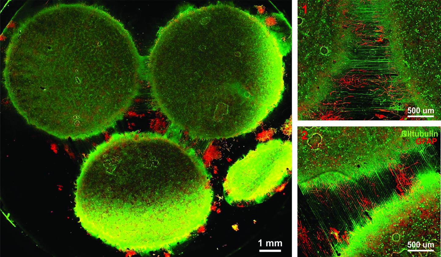
Caption: Image of the in vitro model showing three distinct regions of the brain connected by axons. Credit: Disease Biophysics Group/Harvard University

Here’s a link to and a citation for the paper,

Neurons derived from different brain regions are inherently different in vitro: A novel multiregional brain-on-a-chip by Stephanie Dauth, Ben M Maoz, Sean P Sheehy, Matthew A Hemphill, Tara Murty, Mary Kate Macedonia, Angie M Greer, Bogdan Budnik, Kevin Kit Parker. Journal of Neurophysiology Published 28 December 2016 Vol. no. [?] , DOI: 10.1152/jn.00575.2016

This paper is behind a paywall and they haven’t included the vol. no. in the citation I’ve found.

Tune your windows for privacy

Caption: With an applied voltage, the nanowires on either side of the glass become attracted to each other and move toward each other, squeezing and deforming the soft elastomer. Because the nanowires are scattered unevenly across the surface, the elastomer deforms unevenly. That uneven roughness causes light to scatter, turning the glass opaque. Credit: David Clarke/Harvard SEAS [School of Engineering and Applied Sciences]

Right now, this is my favourite science illustration. A March 14, 2016 news item on Nanowerk announces Harvard’s new technology that can turn a clear window into an opaque one at the touch of a switch,

Say goodbye to blinds.

Researchers at the Harvard John A. Paulson School of Engineering and Applied Sciences have developed a technique that can quickly change the opacity of a window, turning it cloudy, clear or somewhere in between with the flick of a switch.

Tunable windows aren’t new but most previous technologies have relied on electrochemical reactions achieved through expensive manufacturing. This technology, developed by David Clarke, the Extended Tarr Family Professor of Materials, and postdoctoral fellow Samuel Shian, uses geometry [to] adjust the transparency of a window.

A March 14, 2016 Harvard University news release (also on EurekAlert) by Leah Burrows, which originated the news item, describes the technology in more detail,

The tunable window is comprised of a sheet of glass or plastic, sandwiched between transparent, soft elastomers sprayed with a coating of silver nanowires, too small to scatter light on their own.

But apply an electric voltage and things change quickly.

With an applied voltage, the nanowires on either side of the glass are energized to move toward each other, squeezing and deforming the soft elastomer. Because the nanowires are distributed unevenly across the surface, the elastomer deforms unevenly. The resulting uneven roughness causes light to scatter, turning the glass opaque.

The change happens in less than a second.

It’s like a frozen pond, said Shian.

“If the frozen pond is smooth, you can see through the ice. But if the ice is heavily scratched, you can’t see through,” said Shian.

Clarke and Shian found that the roughness of the elastomer surface depended on the voltage, so if you wanted a window that is only light clouded, you would apply less voltage than if you wanted a totally opaque window.

“Because this is a physical phenomenon rather than based on a chemical reaction, it is a simpler and potentially cheaper way to achieve commercial tunable windows,” said Clarke.

Current chemical-based controllable windows use vacuum deposition to coat the glass, a process that deposits layers of a material molecule by molecule. It’s expensive and painstaking. In Clarke and Shian’s method, the nanowire layer can be sprayed or peeled onto the elastomer, making the technology scalable for larger architectural projects.

Next the team is working on incorporating thinner elastomers, which would require lower voltages, more suited for standard electronical supplies.

Here’s a link to and a citation for the paper,

Electrically tunable window device by Samuel Shian and David R. Clarke. Optics Letters Vol. 41, Issue 6, pp. 1289-1292 (2016) •doi: 10.1364/OL.41.001289

This is an open access paper.

SLIPS (Slippery Liquid-Infused Porous Surfaces) technology repels blood and bacteria from medical devices

Researchers at Harvard University’s Wyss Institute for Biologically Inspired Engineering have developed a coating for medical devices that helps to address some of these devices’ most  troublesome aspects. From an Oct. 12, 2014 news item on ScienceDaily,

From joint replacements to cardiac implants and dialysis machines, medical devices enhance or save lives on a daily basis. However, any device implanted in the body or in contact with flowing blood faces two critical challenges that can threaten the life of the patient the device is meant to help: blood clotting and bacterial infection.

A team of Harvard scientists and engineers may have a solution. They developed a new surface coating for medical devices using materials already approved by the Food and Drug Administration (FDA). The coating repelled blood from more than 20 medically relevant substrates the team tested — made of plastic to glass and metal — and also suppressed biofilm formation in a study reported in Nature Biotechnology. But that’s not all.

The team implanted medical-grade tubing and catheters coated with the material in large blood vessels in pigs, and it prevented blood from clotting for at least eight hours without the use of blood thinners such as heparin. Heparin is notorious for causing potentially lethal side-effects like excessive bleeding but is often a necessary evil in medical treatments where clotting is a risk.

“Devising a way to prevent blood clotting without using anticoagulants is one of the holy grails in medicine,” said Don Ingber, M.D., Ph.D., Founding Director of Harvard’s Wyss Institute for Biologically Inspired Engineering and senior author of the study. Ingber is also the Judah Folkman Professor of Vascular Biology at Harvard Medical School and Boston Children’s Hospital, as well as professor of bioengineering at Harvard School of Engineering and Applied Sciences (SEAS).

An Oct. 12, 2014 Wyss Institute news release (also on EurekAlert), which originated the news item, describes the inspiration for this work,

The idea for the coating evolved from SLIPS, a pioneering surface technology developed by coauthor Joanna Aizenberg, Ph.D., who is a Wyss Institute Core Faculty member and the Amy Smith Berylson Professor of Materials Science at Harvard SEAS. SLIPS stands for Slippery Liquid-Infused Porous Surfaces. Inspired by the slippery surface of the carnivorous pitcher plant, which enables the plant to capture insects, SLIPS repels nearly any material it contacts. The liquid layer on the surface provides a barrier to everything from ice to crude oil and blood.

“Traditional SLIPS uses porous, textured surface substrates to immobilize the liquid layer whereas medical surfaces are mostly flat and smooth – so we further adapted our approach by capitalizing on the natural roughness of chemically modified surfaces of medical devices,” said Aizenberg, who leads the Wyss Institute’s Adaptive Materials platform. “This is yet another incarnation of the highly customizable SLIPS platform that can be designed to create slippery, non-adhesive surfaces on any material.”

The Wyss team developed a super-repellent coating that can be adhered to existing, approved medical devices. In a two-step surface-coating process, they chemically attached a monolayer of perfluorocarbon, which is similar to Teflon. Then they added a layer of liquid perfluorocarbon, which is widely used in medicine for applications such as liquid ventilation for infants with breathing challenges, blood substitution, eye surgery, and more. The team calls the tethered perfluorocarbon plus the liquid layer a Tethered-Liquid Perfluorocarbon surface, or TLP for short.

In addition to working seamlessly when coated on more than 20 different medical surfaces and lasting for more than eight hours to prevent clots in a pig under relatively high blood flow rates without the use of heparin, the TLP coating achieved the following results:

  • TLP-treated medical tubing was stored for more than a year under normal temperature and humidity conditions and still prevented clot formation
  • The TLP surface remained stable under the full range of clinically relevant physiological shear stresses, or rates of blood flow seen in catheters and central lines, all the way up to dialysis machines
  • It repelled the components of blood that cause clotting (fibrin and platelets)
  • When bacteria called Pseudomonas aeruginosa were grown in TLP-coated medical tubing for more than six weeks, less than one in a billion bacteria were able to adhere. Central lines coated with TLP significantly reduce sepsis from Central-Line Mediated Bloodstream Infections (CLABSI). (Sepsis is a life-threatening blood infection caused by bacteria, and a significant risk for patients with implanted medical devices.)

Out of sheer curiosity, the researchers even tested a TLP-coated surface with a gecko – the superstar of sticking whose footpads contain many thousands of hairlike structures with tremendous adhesive strength. The gecko was unable to hold on.

“We were wonderfully surprised by how well the TLP coating worked, particularly in vivo without heparin,” said one of the co-lead authors, Anna Waterhouse, Ph.D., a Wyss Institute Postdoctoral Fellow. “Usually the blood will start to clot within an hour in the extracorporeal circuit, so our experiments really demonstrate the clinical relevance of this new coating.”

While most of the team’s demonstrations were performed on medical devices such as catheters and perfusion tubing using relatively simple setups, they say there is a lot more on the horizon.

“We feel this is just the beginning of how we might test this for use in the clinic,” said co-lead author Daniel Leslie, Ph.D., a Wyss Institute Staff Scientist, who aims to test it on more complex systems such as dialysis machines and ECMO, a machine used in the intensive care unit to help critically ill patients breathe.

I first featured SLIPS technology in a Jan. 15, 2014 post about its possible use for stain-free, self-cleaning clothing. This Wyss Institute video about the latest work featuring the use of  SLIPS technology in medical devices also describes its possible use in pipelines and airplanes,

You can find research paper with this link,

A bioinspired omniphobic surface coating on medical devices prevents thrombosis and biofouling by Daniel C Leslie, Anna Waterhouse, Julia B Berthet, Thomas M Valentin, Alexander L Watters, Abhishek Jain, Philseok Kim, Benjamin D Hatton, Arthur Nedder, Kathryn Donovan, Elana H Super, Caitlin Howell, Christopher P Johnson, Thy L Vu, Dana E Bolgen, Sami Rifai, Anne R Hansen, Michael Aizenberg, Michael Super, Joanna Aizenberg, & Donald E Ingber. Nature Biotechnology (2014) doi:10.1038/nbt.3020 Published online 12 October 2014

This paper is behind a paywall but there is a free preview available via ReadCube Access.

Asthma on a chip

Harvard University’s Wyss Institute for Biologically Inspired Engineering has found a way to mimic the lung’s muscle action when an asthma attack is being experienced according to a Sept. 23, 2014 news item on Nanowerk,

The majority of drugs used to treat asthma today are the same ones that were used 50 years ago. New drugs are urgently needed to treat this chronic respiratory disease, which causes nearly 25 million people in the United States alone to wheeze, cough, and find it difficult at best to take a deep breath.

But finding new treatments is tough: asthma is a patient-specific disease, so what works for one person doesn’t necessarily work for another, and the animal models traditionally used to test new drug candidates often fail to mimic human responses–costing tremendous money and time.

Hope for healthier airways may be on the horizon thanks to a Harvard University team that has developed a human airway muscle-on-a-chip that could be used to test new drugs because it accurately mimics the way smooth muscle contracts in the human airway, under normal circumstances and when exposed to asthma triggers. [emphasis mine]

A Sept. 23, 2014 Wyss Institute news release (also on EurekAlert*), which originated the news item, provides more details about the technology and its advantages,

The chip, a soft polymer well that is mounted on a glass substrate, contains a planar array of microscale, engineered human airway muscles, designed to mimic the laminar structure of the muscular layers of the human airway.

To mimic a typical allergic asthma response, the team first introduced interleukin-13 (IL-13) to the chip. IL-13 is a natural protein often found in the airway of asthmatic patients that mediates the response of smooth muscle to an allergen.

Then they introduced acetylcholine, a neurotransmitter that causes smooth muscle to contract. Sure enough, the airway muscle on the chip hypercontracted – and the soft chip curled up – in response to higher doses of the neurotransmitter.

They achieved the reverse effect as well and triggered the muscle to relax using drugs called β-agonists, which are used in inhalers.

Significantly, they were able to measure the contractile stress of the muscle tissue as it responded to varying doses of the drugs, said lead author Alexander Peyton Nesmith, a Ph.D./M.D. student at Harvard SEAS and the University of Alabama at Birmingham. “Our chip offers a simple, reliable and direct way to measure human responses to an asthma trigger,” he said.

The team then investigated what happened on a cellular level in response to the IL-13 and confirmed, for example, that the smooth muscle cells grew larger in the presence of IL-13 over time – a structural hallmark of the airways in asthma patients as well. They also documented an increased alignment of actin fibers within smooth muscle cells, which is consistent with the muscle in the airway of asthma patients. Actin fibers are super-thin cellular components involved in muscle contraction.

Next they observed how IL-13 changes the expression of contractile proteins called RhoA proteins, which have been implicated in the asthmatic response, although the details of their activation and signaling have remained elusive. To do this they introduced a drug called HA1077, which is not currently used to treat asthmatic patients – but targets the RhoA pathway. It turns out that the drug made the asthmatic tissue on the chip less sensitive to the asthma trigger – and preliminary tests indicated that using a combined therapy of HA1077 plus a currently approved asthma drug worked better than the single drug alone.

“Asthma is one of the top reasons for trips to the emergency room – particularly for children, and a large segment of the asthmatic population doesn’t respond to currently available treatments,” said Wyss Institute Founding Director Don Ingber, M.D., Ph.D. “The airway muscle-on-a-chip provides an important and exciting new tool for discovering new therapeutic agents.”

The scientists have provided an illustration of healthy and asthmatic airways,

Schematic comparing a healthy airway (few immune cells, normal airway diameter) to an asthmatic airway (many immune cells, constricted airway). Credit: Harvard's Wyss Institute and Harvard SEAS [School of Engineering and Applied Sciences]

Schematic comparing a healthy airway (few immune cells, normal airway diameter) to an asthmatic airway (many immune cells, constricted airway). Credit: Harvard’s Wyss Institute and Harvard SEAS [School of Engineering and Applied Sciences]

Here’s link to and a citation for the paper,

Human airway musculature on a chip: an in vitro model of allergic asthmatic bronchoconstriction and bronchodilation by Alexander Peyton Nesmith, Ashutosh Agarwal, Megan Laura McCain and Kevin Kit Parker.Lab Chip, 2014,14, 3925-3936 DOI: 10.1039/C4LC00688G First published online 05 Aug 2014

This paper is open access provided you have registered yourself for free at the site.

* EurekAlert link added Sept. 24, 2014.

Let’s make our turbine blades really big (greater than 75 metres) with new nanocomposite

The is a story about balsa wood, wind farms, turbine blades, and nanocomposites according to a June 25, 2014 news item on ScienceDaily,

In wind farms across North America and Europe, sleek turbines equipped with state-of-the-art technology convert wind energy into electric power. But tucked inside the blades of these feats of modern engineering is a decidedly low-tech core material: balsa wood.

Like other manufactured products that use sandwich panel construction to achieve a combination of light weight and strength, turbine blades contain carefully arrayed strips of balsa wood from Ecuador, which provides 95 percent of the world’s supply.

For centuries, the fast-growing balsa tree has been prized for its light weight and stiffness relative to density. But balsa wood is expensive and natural variations in the grain can be an impediment to achieving the increasingly precise performance requirements of turbine blades and other sophisticated applications.

As turbine makers produce ever-larger blades — the longest now measure 75 meters, almost matching the wingspan of an Airbus A380 jetliner — they must be engineered to operate virtually maintenance-free for decades. In order to meet more demanding specifications for precision, weight, and quality consistency, manufacturers are searching for new sandwich construction material options.

Now, using a cocktail of fiber-reinforced epoxy-based thermosetting resins and 3D extrusion printing techniques, materials scientists at the Harvard School of Engineering and Applied Sciences (SEAS) and the Wyss Institute for Biologically Inspired Engineering have developed cellular composite materials of unprecedented light weight and stiffness.

A June 25, 2014 Harvard University news release (also on EurekAlert), which originated the news item, goes on to describe the new technology in more detail while throwing 3D printing into the mix,

Until now, 3D printing has been developed for thermo plastics and UV-curable resins—materials that are not typically considered as engineering solutions for structural applications. “By moving into new classes of materials like epoxies, we open up new avenues for using 3D printing to construct lightweight architectures,” says principal investigator Jennifer A. Lewis, the Hansjörg Wyss Professor of Biologically Inspired Engineering at Harvard SEAS. “Essentially, we are broadening the materials palate for 3D printing.”

“Balsa wood has a cellular architecture that minimizes its weight since most of the space is empty and only the cell walls carry the load. It therefore has a high specific stiffness and strength,” explains Lewis, who in addition to her role at Harvard SEAS is also a Core Faculty Member at the Wyss Institute. “We’ve borrowed this design concept and mimicked it in an engineered composite.”

Lewis and Brett G. Compton, a former postdoctoral fellow in her group, developed inks of epoxy resins, spiked with viscosity-enhancing nanoclay platelets and a compound called dimethyl methylphosphonate, and then added two types of fillers: tiny silicon carbide “whiskers” and discrete carbon fibers. Key to the versatility of the resulting fiber-filled inks is the ability to control the orientation of the fillers.

The direction that the fillers are deposited controls the strength of the materials (think of the ease of splitting a piece of firewood lengthwise versus the relative difficulty of chopping on the perpendicular against the grain).

Lewis and Compton have shown that their technique yields cellular composites that are as stiff as wood, 10 to 20 times stiffer than commercial 3D-printed polymers, and twice as strong as the best printed polymer composites. The ability to control the alignment of the fillers means that fabricators can digitally integrate the composition, stiffness, and toughness of an object with its design.

“This paper demonstrates, for the first time, 3D printing of honeycombs with fiber-reinforced cell walls,” said Lorna Gibson, a professor of materials science and mechanical engineering at the Massachusetts Institute of Technology and one of world’s leading experts in cellular composites, who was not involved in this research. “Of particular significance is the way that the fibers can be aligned, through control of the fiber aspect ratio—the length relative to the diameter—and the nozzle diameter. This marks an important step forward in designing engineering materials that mimic wood, long known for its remarkable mechanical properties for its weight.”

“As we gain additional levels of control in filler alignment and learn how to better integrate that orientation into component design, we can further optimize component design and improve materials efficiency,” adds Compton, who is now a staff scientist in additive manufacturing at Oak Ridge National Laboratory. “Eventually, we will be able to use 3D printing technology to change the degree of fiber filler alignment and local composition on the fly.”

The work could have applications in many fields, including the automotive industry where lighter materials hold the key to achieving aggressive government-mandated fuel economy standards. According to one estimate, shedding 110 pounds from each of the 1 billion cars on the road worldwide could produce $40 billion in annual fuel savings.

3D printing has the potential to radically change manufacturing in other ways too. Lewis says the next step will be to test the use of thermosetting resins to create different kinds of architectures, especially by exploiting the technique of blending fillers and precisely aligning them. This could lead to advances not only in structural materials, but also in conductive composites.

Previously, Lewis has conducted groundbreaking research in the 3D printing of tissue constructs with vasculature and lithium-ion microbatteries.

Here’s a link to and citation for the paper,

3D-Printing of Lightweight Cellular Composites by Brett G. Compton and Jennifer A. Lewis. Advanced Materials DOI: 10.1002/adma.201401804 Article first published online: 18 JUN 2014

© 2014 WILEY-VCH Verlag GmbH & Co. KGaA, Weinheim

This paper is behind a paywall.

US Air Force wants to merge classical and quantum physics

The US Air Force wants to merge classical and quantum physics for practical purposes according to a May 5, 2014 news item on Azonano,

The Air Force Office of Scientific Research has selected the Harvard School of Engineering and Applied Sciences (SEAS) to lead a multidisciplinary effort that will merge research in classical and quantum physics and accelerate the development of advanced optical technologies.

Federico Capasso, Robert L. Wallace Professor of Applied Physics and Vinton Hayes Senior Research Fellow in Electrical Engineering, will lead this Multidisciplinary University Research Initiative [MURI] with a world-class team of collaborators from Harvard, Columbia University, Purdue University, Stanford University, the University of Pennsylvania, Lund University, and the University of Southampton.

The grant is expected to advance physics and materials science in directions that could lead to very sophisticated lenses, communication technologies, quantum information devices, and imaging technologies.

“This is one of the world’s strongest possible teams,” said Capasso. “I am proud to lead this group of people, who are internationally renowned experts in their fields, and I believe we can really break new ground.”

A May 1, 2014 Harvard University School of Engineering and Applied Sciences news release, which originated the news item, provides a description of project focus: nanophotonics and metamaterials along with some details of Capasso’s work in these areas (Note: Links have been removed),

The premise of nanophotonics is that light can interact with matter in unusual ways when the material incorporates tiny metallic or dielectric features that are separated by a distance shorter than the wavelength of the light. Metamaterials are engineered materials that exploit these phenomena, producing strange effects, enabling light to bend unnaturally, twist into a vortex, or disappear entirely. Yet the fabrication of thick, or bulk, metamaterials—that manipulate light as it passes through the material—has proven very challenging.

Recent research by Capasso and others in the field has demonstrated that with the right device structure, the critical manipulations can actually be confined to the very surface of the material—what they have dubbed a “metasurface.” These metasurfaces can impart an instantaneous shift in the phase, amplitude, and polarization of light, effectively controlling optical properties on demand. Importantly, they can be created in the lab using fairly common fabrication techniques.

At Harvard, the research has produced devices like an extremely thin, flat lens, and a material that absorbs 99.75% of infrared light. But, so far, such devices have been built to order—brilliantly suited to a single task, but not tunable.

This project, however,is focused on the future (Note: Links have been removed),

“Can we make a rapidly configurable metasurface so that we can change it in real time and quickly? That’s really a visionary frontier,” said Capasso. “We want to go all the way from the fundamental physics to the material building blocks and then the actual devices, to arrive at some sort of system demonstration.”

The proposed research also goes further. A key thrust of the project involves combining nanophotonics with research in quantum photonics. By exploiting the quantum effects of luminescent atomic impurities in diamond, for example, physicists and engineers have shown that light can be captured, stored, manipulated, and emitted as a controlled stream of single photons. These types of devices are essential building blocks for the realization of secure quantum communication systems and quantum computers. By coupling these quantum systems with metasurfaces—creating so-called quantum metasurfaces—the team believes it is possible to achieve an unprecedented level of control over the emission of photons.

“Just 20 years ago, the notion that photons could be manipulated at the subwavelength scale was thought to be some exotic thing, far fetched and of very limited use,” said Capasso. “But basic research opens up new avenues. In hindsight we know that new discoveries tend to lead to other technology developments in unexpected ways.”

The research team includes experts in theoretical physics, metamaterials, nanophotonic circuitry, quantum devices, plasmonics, nanofabrication, and computational modeling. Co-principal investigator Marko Lončar is the Tiantsai Lin Professor of Electrical Engineering at Harvard SEAS. Co-PI Nanfang Yu, Ph.D. ’09, developed expertise in metasurfaces as a student in Capasso’s Harvard laboratory; he is now an assistant professor of applied physics at Columbia. Additional co-PIs include Alexandra Boltasseva and Vladimir Shalaev at Purdue, Mark Brongersma at Stanford, and Nader Engheta at the University of Pennsylvania. Lars Samuelson (Lund University) and Nikolay Zheludev (University of Southampton) will also participate.

The bulk of the funding will support talented graduate students at the lead institutions.

The project, titled “Active Metasurfaces for Advanced Wavefront Engineering and Waveguiding,” is among 24 planned MURI awards selected from 361 white papers and 88 detailed proposals evaluated by a panel of experts; each award is subject to successful negotiation. The anticipated amount of the Harvard-led grant is up to $6.5 million for three to five years.

For anyone who’s not familiar (that includes me, anyway) with MURI awards, there’s this from Wikipedia (Note: links have been removed),

Multidisciplinary University Research Initiative (MURI) is a basic research program sponsored by the US Department of Defense (DoD). Currently each MURI award is about $1.5 million a year for five years.

I gather that in addition to the Air Force, the Army and the Navy also award MURI funds.

Mesenchymal condensation (a process embryos use to begin forming a variety of organs, including teeth, cartilage, bone, muscle, tendon, and kidney) for complex 3D tissue engineering

It seems that there are three strategies for creating complex 3D tissues and until now scientists have used only two of the three. From a March 5, 2014 news item on ScienceDaily,

A bit of pressure from a new shrinking, sponge-like gel is all it takes to turn transplanted unspecialized cells into cells that lay down minerals and begin to form teeth.

The bioinspired gel material could one day help repair or replace damaged organs, such as teeth and bone, and possibly other organs as well, scientists from the Wyss Institute for Biologically Inspired Engineering at Harvard University, Harvard School of Engineering and Applied Sciences (SEAS), and Boston Children’s Hospital report recently in Advanced Materials.

“Tissue engineers have long raised the idea of using synthetic materials to mimic the inductive power of the embryo,” said Don Ingber, M.D., Ph.D., Founding Director of the Wyss Institute, …, Professor of Bioengineering at SEAS, and senior author of the study. “We’re excited about this work because it shows that it really is possible.”

The March 5, 2014 Wyss Institute news release, which originated the news item, delves into the nature of the research,

Embryonic tissues have the power to drive cells and tissues to specialize and form organs. To do that, they employ biomolecules called growth factors to stimulate growth; gene-activating chemicals that cause the cells to specialize, and mechanical forces that modulate cell responses to these other factors.

But so far tissue engineers who want to build organs in the laboratory have employed only two of the three strategies — growth factors and gene-activating chemicals. Perhaps as a result, they have not yet succeeded in producing complex three-dimensional tissues.

A few years ago, Ingber and Tadanori Mammoto, M.D., Ph.D., Instructor in Surgery at Boston Children’s Hospital and Harvard Medical School, investigated a process called mesenchymal condensation that embryos use to begin forming a variety of organs, including teeth, cartilage, bone, muscle, tendon, and kidney.

In mesenchymal condensation, two adjacent tissue layers — loosely packed connective-tissue cells called mesenchyme and sheet-like tissue called an epithelium that covers it — exchange biochemical signals. This exchange causes the mesenchymal cells to squeeze themselves tightly into a small knot directly below where the new organ will form.

Here’s a video from the Wyss Institute illustrating the squeezing process,

When the temperature rises to just below body temperature, this biocompatible gel shrinks dramatically within minutes, bringing tooth-precursor cells (green) closer together. Credit: Basma Hashmi

Getting back to the research (from the news release),

By examining tissues isolated from the jaws of embryonic mice, Mammoto and Ingber showed that when the compressed mesenchymal cells turn on genes that stimulate them to generate whole teeth composed of mineralized tissues, including dentin and enamel.

Inspired by this embryonic induction mechanism, Ingber and Basma Hashmi, a Ph.D. candidate at SEAS who is the lead author of the current paper, set out to develop a way to engineer artificial teeth by creating a tissue-friendly material that accomplishes the same goal. Specifically, they wanted a porous sponge-like gel that could be impregnated with mesenchymal cells, then, when implanted into the body, induced to shrink in 3D to physically compact the cells inside it.

To develop such a material, Ingber and Hashmi teamed up with researchers led by Joanna Aizenberg, Ph.D., a Wyss Institute Core Faculty member who leads the Institute’s Adaptive Materials Technologies platform. Aizenberg is the Amy Smith Berylson Professor of Materials Science at SEAS and Professor of Chemistry and Chemical Biology at Harvard University.

They chemically modified a special gel-forming polymer called PNIPAAm that scientists have used to deliver drugs to the body’s tissues. PNIPAAm gels have an unusual property: they contract abruptly when they warm.

But they do this at a lukewarm temperature, whereas the researchers wanted them to shrink specifically at 37°C — body temperature — so that they’d squeeze their contents as soon as they were injected into the body. Hashmi worked with Lauren Zarzar, Ph.D., a former SEAS graduate student who’s now a postdoctoral associate at Massachusetts Institute of Technology, for more than a year, modifying PNIPAAm and testing the resulting materials. Ultimately, they developed a polymer that forms a tissue-friendly gel with two key properties: cells stick to it, and it compresses abruptly when warmed to body temperature.

As an initial test, Hashmi implanted mesenchymal cells in the gel and warmed it in the lab. Sure enough, when the temperature reached 37°C, the gel shrank within 15 minutes, causing the cells inside the gel to round up, shrink, and pack tightly together.

“The reason that’s cool is that the cells are alive,” Hashmi said. “Usually when this happens, cells are dead or dying.”

Not only were they alive — they activated three genes that drive tooth formation.

To see if the shrinking gel also worked its magic in the body, Hashmi worked with Mammoto to load mesenchymal cells into the gel, then implant the gel beneath the mouse kidney capsule — a tissue that is well supplied with blood and often used for transplantation experiments.

The implanted cells not only expressed tooth-development genes — they laid down calcium and minerals, just as mesenchymal cells do in the body as they begin to form teeth.

“They were in full-throttle tooth-development mode,” Hashmi said.

The researchers have future plans (from the news release),

In the embryo, mesenchymal cells can’t build teeth alone — they need to be combined with cells that form the epithelium. In the future, the scientists plan to test whether the shrinking gel can stimulate both tissues to generate an entire functional tooth.

Here’s a link to and a citation for the paper about the successful attempt to stimulate mesenchymal cells into the beginnings of tooth formation,

Developmentally-Inspired Shrink-Wrap Polymers for Mechanical Induction of Tissue Differentiation by Basma Hashmi, Lauren D. Zarzar, Tadanori Mammoto, Akiko Mammoto, Amanda Jiang, Joanna Aizenberg, and Donald E. Ingber. Advanced Materials Article first published online: 18 FEB 2014 DOI: 10.1002/adma.201304995

© 2014 WILEY-VCH Verlag GmbH & Co. KGaA, Weinheim

This paper is behind a paywall.

Chameleon materials

Harvard’s School of Engineering and Applied Sciences researchers discovered some unexpected properties when testing a new coating according to an Oct. 22, 2013 news item on Azonano,

Active camouflage has taken a step forward at the Harvard School of Engineering and Applied Sciences (SEAS), with a new coating that intrinsically conceals its own temperature to thermal cameras.

In a laboratory test, a team of applied physicists placed the device on a hot plate and watched it through an infrared camera as the temperature rose. Initially, it behaved as expected, giving off more infrared light as the sample was heated: at 60 degrees Celsius it appeared blue-green to the camera; by 70 degrees it was red and yellow. At 74 degrees it turned a deep red—and then something strange happened. The thermal radiation plummeted. At 80 degrees it looked blue, as if it could be 60 degrees, and at 85 it looked even colder. Moreover, the effect was reversible and repeatable, many times over.

The Oct. 21, 2013 Harvard University news release (also on EurekAlert), which originated the news item, discusses the potential for this discovery and describes the process of discovery in more detail (Note: A link has been removed),

Principal investigator Federico Capasso, Robert L. Wallace Professor of Applied Physics and Vinton Hayes Senior Research Fellow in Electrical Engineering at Harvard SEAS, predicts that with only small adjustments the coating could be used as a new type of thermal camouflage or as a kind of encrypted beacon to allow soldiers to covertly communicate their locations in the field.

The secret to the technology lies within a very thin film of vanadium oxide, an unusual material that undergoes dramatic electronic changes when it reaches a particular temperature. At room temperature, for example, pure vanadium oxide is electrically insulating, but at slightly higher temperatures it transitions to a metallic, electrically conductive state. During that transition, the optical properties change, too, which means special temperature-dependent effects—like infrared camouflage—can also be achieved.

The insulator-metal transition has been recognized in vanadium oxide since 1959. However, it is a difficult material to work with: in bulk crystals, the stress of the transition often causes cracks to develop and can shatter the sample. Recent advances in materials synthesis and characterization—especially those by coauthor Shriram Ramanathan, Associate Professor of Materials Science at Harvard SEAS—have allowed the creation of extremely pure samples of thin-film vanadium oxide, enabling a burst of new science and engineering to take off in just the last few years.

“Thanks to these very stable samples that we’re getting from Prof. Ramanathan’s lab, we now know that if we introduce small changes to the material, we can dramatically change the optical phenomena we observe,” explains lead author Mikhail Kats, a graduate student in Capasso’s group at Harvard SEAS. “By introducing impurities or defects in a controlled way via processes known as doping, modifying, or straining the material, it is possible to create a wide range of interesting, important, and predictable behaviors.”

By doping vanadium oxide with tungsten, for example, the transition temperature can be brought down to room temperature, and the range of temperatures over which the strange thermal radiation effect occurs can be widened. Tailoring the material properties like this, with specific outcomes in mind, may enable engineering to advance in new directions.

The researchers say a vehicle coated in vanadium oxide tiles could potentially mimic its environment like a chameleon, appearing invisible to an infrared camera with only very slight adjustments to the tiles’ actual temperature—a far more efficient system than the approaches in use today.

Tuned differently, the material could become a component of a secret beacon, displaying a particular thermal signature on cue to an infrared surveillance camera. Capasso’s team suggests that the material could be engineered to operate at specific wavelengths, enabling simultaneous use by many individually identifiable soldiers.

And, because thermal radiation carries heat, the researchers believe a similar effect could be employed to deliberately speed up or slow down the cooling of structures ranging from houses to satellites.

The Harvard team’s most significant contribution is the discovery that nanoscale structures that appear naturally in the transition region of vanadium oxide can be used to provide a special level of tunability, which can be used to suppress thermal radiation as the temperature rises. The researchers refer to such a spontaneously structured material as a “natural, disordered metamaterial.”

“To artificially create such a useful three-dimensional structure within a material is extremely difficult,” says Capasso. “Here, nature is giving us what we want for free. By taking these natural metamaterials and manipulating them to have all the properties we want, we are opening up a new area of research, a completely new direction of work. We can engineer new devices from the bottom up.”

Here’s an image, from the scientists, illustrating the material’s thermal camouflage (or chameleon) properties,

A new coating intrinsically conceals its own temperature to thermal cameras. (Image courtesy of Mikhail Kats.)

A new coating intrinsically conceals its own temperature to thermal cameras. (Image courtesy of Mikhail Kats.)

Here’s a link to and a citation for the research paper,

Vanadium Dioxide as a Natural Disordered Metamaterial: Perfect Thermal Emission and Large Broadband Negative Differential Thermal Emittance by Mikhail A. Kats, Romain Blanchard, Shuyan Zhang, Patrice Genevet, Changhyun Ko, Shriram Ramanathan, and Federico Capasso. Phys. Rev. X » Volume 3 » Issue 4  or Phys. Rev. X 3, 041004 (2013) DOI:10.1103/PhysRevX.3.041004

This paper is published in an open access journal according to the Harvard news release,

About Physical Review X

Launched in August 2011, PRX (http://prx.aps.org) is an open-access, peer-reviewed publication of the American Physical Society (www.aps.org), a non-profit membership organization working to advance and diffuse the knowledge of physics through its outstanding research journals, scientific meetings, and education, outreach, advocacy and international activities. APS represents 50,000 members, including physicists in academia, national laboratories and industry in the United States and throughout the world.

Chart junk: rethinking science data visualization

Which of these visualizations will you remember later? (Images courtesy of Michelle Borkin, Harvard SEAS.)

Which of these visualizations will you remember later? (Images courtesy of Michelle Borkin, Harvard SEAS.)

This chart of data visualization images accompanies an Oct. 16, 2013 news item on ScienceDaily concerning some research into what makes some charts more memorable than others,

It’s easy to spot a “bad” data visualization — one packed with too much text, excessive ornamentation, gaudy colors, and clip art. Design guru Edward Tufte derided such decorations as redundant at best, useless at worst, labeling them “chart junk.” Yet a debate still rages among visualization experts: Can these reviled extra elements serve a purpose?

Taking a scientific approach to design, researchers from Harvard University and Massachusetts Institute of Technology are offering a new take on that debate. The same design elements that attract so much criticism, they report, can also make a visualization more memorable.

Detailed results were presented this week at the IEEE Information Visualization (InfoVis) conference in Atlanta, hosted by the Institute of Electrical and Electronics Engineers.

The Oct. 16, 2013 School of Engineering and Applied Sciences (SEAS) Harvard University news release (also on EurekAlert), which originated the news item, details some of the ways in which the researchers attempted to study data visualizations and memorability (Note: Links from the news release to be found on the SEAS website have been removed),

For lead author Michelle Borkin, a doctoral student at the Harvard School of Engineering and Applied Sciences (SEAS), memorability has a particular importance:

“I spend a lot of my time reading these scientific papers, so I have to wonder, when I walk away from my desk, what am I going to remember? Which of the figures and visualizations in these publications are going to stick with me?”

But it’s more than grad-school anxiety. Working at the interface of computer science and psychology, Borkin specializes in the visual representation of data, looking for the best ways to communicate and interpret complex information. The applications of her work have ranged from astronomy to medical diagnostics and may already help save lives.

Her adviser, Hanspeter Pfister, An Wang Professor of Computer Science at Harvard SEAS, was intrigued by the chart junk debate, which has flared up on design blogs and at visualization conferences year after year.

Together, they turned to Aude Oliva, a principal research scientist at MIT’s Computer Science and Artificial Intelligence Lab, and a cognitive psychologist by training. Oliva’s lab has been studying visual memory for about six years now. Her team has found that in photographs, faces and human-centric scenes are typically easy to remember; landscapes are not.

“All of us are sensitive to the same kinds of images, and we forget the same kind as well,” Oliva says. “We like to believe our memories are unique, that they’re like the soul of a person, but in certain situations it’s as if we have the same algorithm in our heads that is going to be sensitive to a particular type of image. So when you find a result like this in photographs, you want to know: is it generalizable to many types of materials—words, sound, images, graphs?”

“Speaking with [Pfister] and his group, it became very exciting, the idea that we could study what makes a visualization memorable or not,” Oliva recalls. “If it turned out to be the same for everyone, we thought this would be a win-win result.”

For Oliva’s group, it would provide more evidence of cognitive similarities in the brain’s visual processing, from person to person. For Pfister’s group, it could suggest that certain design principles make visualizations inherently more memorable than others.

With Harvard students Azalea A. Vo ’13 and Shashank Sunkavalli SM ’13, as well as MIT graduate students Zoya Bylinskii and Phillip Isola, the team designed a large-scale study—in the form of an online game—to rigorously measure the memorability of a wide variety of visualizations. They collected more than 5,000 charts and graphics from scientific papers, design blogs, newspapers, and government reports and manually categorized them by a wide range of attributes. Serving them up in brief glimpses—just one second each—to participants via Amazon Mechanical Turk, the researchers tested the influence of features like color, density, and content themes on users’ ability to recognize which ones they had seen before.

The results meshed well with Oliva’s previous results, but added several new insights.

“A visualization will be instantly and overwhelmingly more memorable if it incorporates an image of a human-recognizable object—if it includes a photograph, people, cartoons, logos—any component that is not just an abstract data visualization,” says Pfister. “We learned that any time you have a graphic with one of those components, that’s the most dominant thing that affects the memorability.”

Visualizations that were visually dense proved memorable, as did those that used many colors. Other results were more surprising.

“You’d think the types of charts you’d remember best are the ones you learned in school—the bar charts, pie charts, scatter plots, and so on,” Borkin says. “But it was the opposite.”

Unusual types of charts, like tree diagrams, network diagrams, and grid matrices, were actually more memorable.

“If you think about those types of diagrams—for example, tree diagrams that show relationships between species, or diagrams that explain a molecular chemical process—every one of them is going to be a little different, but the branching structures feel very natural to us,” explains Borkin. “That combination of the familiar and the unique seems to influence the memorability.”

The best type of chart to use will always depend on the data, but for designers who are required to work within a certain style—for example, to achieve a recognizable consistency within a magazine—the results may be reassuring.

“A graph can be simple or complex, and they both can be memorable,” explains Oliva. “You can make something familiar either by keeping it simple or by having a little story around it. It’s not really that you should choose to use one color or many, or to include additional ornaments or not. If you need to keep it simple because it’s the style your boss likes or the style of your publication, you can still find a way to make it memorable.”

At this stage, however, the team hesitates to issue any sweeping design guidelines for an obvious reason: memorability isn’t the only thing that matters. Visualizations must also be accurate, easy to comprehend, aesthetically pleasing, and appropriate to the context.

“A memorable visualization is not necessarily a good visualization,” Borkin cautions. “As a community we need to keep asking these types of questions: What makes a visualization engaging? What makes it comprehensible?”

As for the chart junk, she says diplomatically, “I think it’s going to be an ongoing debate.”

I believe Michelle Borkin is the lead author of an unpublished (as yet) paper submitted to the 2013 IEEE Information Visualization (InfoVis) conference, which means I can’t offer a link to or a citation for the paper.

A strange state of light

Apparently combining a hologram with subwavelength structures at a scale of just tens of nanometers can lead to ‘strange’ light. From the Aug. 20, 2013 news item on Nanowerk,

Applied physicists at the Harvard School of Engineering and Applied Sciences (SEAS) have demonstrated that they can change the intensity, phase, and polarization of light rays using a hologram-like design decorated with nanoscale structures.

As a proof of principle, the researchers have used it to create an unusual state of light called a radially polarized beam, which—because it can be focused very tightly—is important for applications like high-resolution lithography and for trapping and manipulating tiny particles like viruses.

The Aug. 20, 2013 Harvard University news release by Manny Marone, which originated the news item, further describes the device and the effect (Note: A link has been removed),

This is the first time a single, simple device has been designed to control these three major properties of light at once. (Phase describes how two waves interfere to either strengthen or cancel each other, depending on how their crests and troughs overlap; polarization describes the direction of light vibrations; and the intensity is the brightness.)

“Our lab works on using nanotechnology to play with light,” says Patrice Genevet, a research associate at Harvard SEAS and co-lead author of a paper published this month in Nano Letters. “In this research, we’ve used holography in a novel way, incorporating cutting-edge nanotechnology in the form of subwavelength structures at a scale of just tens of nanometers.” One nanometer equals one billionth of a meter.

Using these novel nanostructured holograms, the Harvard researchers have converted conventional, circularly polarized laser light into radially polarized beams at wavelengths spanning the technologically important visible and near-infrared light spectrum.

“When light is radially polarized, its electromagnetic vibrations oscillate inward and outward from the center of the beam like the spokes of a wheel,” explains Capasso [Federico Capasso, professor of applied physics]. “This unusual beam manifests itself as a very intense ring of light with a dark spot in the center.”

“It is noteworthy,” Capasso points out, “that the same nanostructured holographic plate can be used to create radially polarized light at so many different wavelengths. Radially polarized light can be focused much more tightly than conventionally polarized light, thus enabling many potential applications in microscopy and nanoparticle manipulation.”

The new device resembles a normal hologram grating with an additional, nanostructured pattern carved into it. Visible light, which has a wavelength in the hundreds of nanometers, interacts differently with apertures textured on the ‘nano’ scale than with those on the scale of micrometers or larger. By exploiting these behaviors, the modular interface can bend incoming light to adjust its intensity, phase, and polarization.

Here’s a link to and a citation for the published paper,

Nanostructured Holograms for Broadband Manipulation of Vector Beams by Jiao Lin, Patrice Genevet, Mikhail A. Kats, Nicholas Antoniou, and Federico Capasso. Nano Lett., Article ASAP DOI: 10.1021/nl402039y Publication Date (Web): August 5, 2013
Copyright © 2013 American Chemical Society

This article is behind a paywall.

I last wrote about Federico Carpasso’s work in an Oct. 16, 2013 posting, Harvard researchers look deeply into oily puddles as they rethink thin films and optical loss.