Tag Archives: sensing

Combining optical technology with nanocomposite films at Oregon State University (OSU)

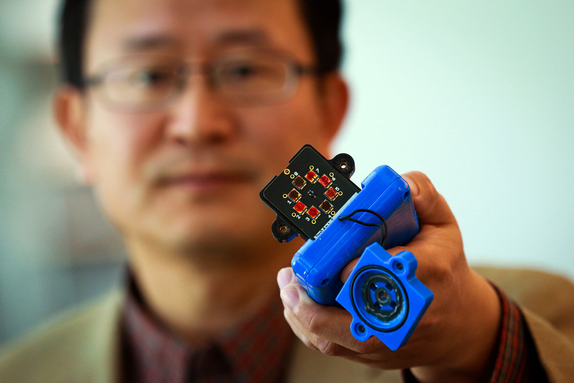
There is a lot of pressure in the US to commercialize nanotechnology-enabled products—a perfectly understandable stance after investing over $22B since 2000. Engineers at Oregon State University (OSU) are hoping to attract industry partners to improve and commercialize their gas sensors (from an April 2, 2015 OSU news release also on EurekAlert),

Engineers have combined innovative optical technology with nanocomposite thin-films to create a new type of sensor that is inexpensive, fast, highly sensitive and able to detect and analyze a wide range of gases.

The technology might find applications in everything from environmental monitoring to airport security or testing blood alcohol levels. The sensor is particularly suited to detecting carbon dioxide, and may be useful in industrial applications or systems designed to store carbon dioxide underground, as one approach to greenhouse gas reduction.

Oregon State University has filed for a patent on the invention, developed in collaboration with scientists at the National Energy Technology Lab or the U.S. Department of Energy, and with support from that agency. The findings were just reported in the Journal of Materials Chemistry C.

University researchers are now seeking industrial collaborators to further perfect and help commercialize the system.

“Optical sensing is very effective in sensing and identifying trace-level gases, but often uses large laboratory devices that are terribly expensive and can’t be transported into the field,” said Alan Wang, a photonics expert and an assistant professor in the OSU School of Electrical Engineering and Computer Science.

“By contrast, we use optical approaches that can be small, portable and inexpensive,” Wang said. “This system used plasmonic nanocrystals that act somewhat like a tiny lens, to concentrate a light wave and increase sensitivity.”

This approach is combined with a metal-organic framework of thin films, which can rapidly adsorb gases within material pores, and be recycled by simple vacuum processes. After the thin film captures the gas molecules near the surface, the plasmonic materials act at a near-infrared range, help magnify the signal and precisely analyze the presence and amounts of different gases.

“By working at the near-infrared range and using these plasmonic nanocrystals, there’s an order of magnitude increase in sensitivity,” said Chih-hung Chang, an OSU professor of chemical engineering. “This type of sensor should be able to quickly tell exactly what gases are present and in what amount.”

That speed, precision, portability and low cost, the researchers said, should allow instruments that can be used in the field for many purposes. The food industry, for industry, uses carbon dioxide in storage of fruits and vegetables, and the gas has to be kept at certain levels.

Gas detection can be valuable in finding explosives, and new technologies such as this might find application in airport or border security. Various gases need to be monitored in environmental research, and there may be other uses in health care, optimal function of automobile engines, and prevention of natural gas leakage.

The paper can be found here,

Plasmonics-enhanced metal–organic framework nanoporous films for highly sensitive near-infrared absorption by Ki-Joong Kim, Xinyuan Chong, Peter B. Kreider, Guoheng Ma,  Paul R. Ohodnicki, John P. Baltrus, Alan X. Wang, and Chih-Hung Chang. J. Mater. Chem. C, 2015,3, 2763-2767 DOI: 10.1039/C4TC02846E First published online 09 Feb 2015

It is behind a paywall.

Bomb-sniffing and other sniffing possibilities from Utah (US state)

A Nov. 4, 2014 news item on Phys.org features some research in Utah on the use of carbon nanotubes for sensing devices,

University of Utah engineers have developed a new type of carbon nanotube material for handheld sensors that will be quicker and better at sniffing out explosives, deadly gases and illegal drugs.

A carbon nanotube is a cylindrical material that is a hexagonal or six-sided array of carbon atoms rolled up into a tube. Carbon nanotubes are known for their strength and high electrical conductivity and are used in products from baseball bats and other sports equipment to lithium-ion batteries and touchscreen computer displays.

Vaporsens, a university spin-off company, plans to build a prototype handheld sensor by year’s end and produce the first commercial scanners early next year, says co-founder Ling Zang, a professor of materials science and engineering and senior author of a study of the technology published online Nov. 4 [2014] in the journal Advanced Materials.

The new kind of nanotubes also could lead to flexible solar panels that can be rolled up and stored or even “painted” on clothing such as a jacket, he adds.

Here’s Ling Zang holding a prototype of the device,

Ling Zang, a University of Utah professor of materials science and engineering, holds a prototype detector that uses a new type of carbon nanotube material for use in handheld scanners to detect explosives, toxic chemicals and illegal drugs. Zang and colleagues developed the new material, which will make such scanners quicker and more sensitive than today’s standard detection devices. Ling’s spinoff company, Vaporsens, plans to produce commercial versions of the new kind of scanner early next year. Courtesy: University of Utah

Ling Zang, a University of Utah professor of materials science and engineering, holds a prototype detector that uses a new type of carbon nanotube material for use in handheld scanners to detect explosives, toxic chemicals and illegal drugs. Zang and colleagues developed the new material, which will make such scanners quicker and more sensitive than today’s standard detection devices. Ling’s spinoff company, Vaporsens, plans to produce commercial versions of the new kind of scanner early next year. Courtesy: University of Utah

A Nov. 4, 2014 University of Utah news release (also on EurekAlert), which originated the news item, provides more detail about the research,

Zang and his team found a way to break up bundles of the carbon nanotubes with a polymer and then deposit a microscopic amount on electrodes in a prototype handheld scanner that can detect toxic gases such as sarin or chlorine, or explosives such as TNT.

When the sensor detects molecules from an explosive, deadly gas or drugs such as methamphetamine, they alter the electrical current through the nanotube materials, signaling the presence of any of those substances, Zang says.

“You can apply voltage between the electrodes and monitor the current through the nanotube,” says Zang, a professor with USTAR, the Utah Science Technology and Research economic development initiative. “If you have explosives or toxic chemicals caught by the nanotube, you will see an increase or decrease in the current.”

By modifying the surface of the nanotubes with a polymer, the material can be tuned to detect any of more than a dozen explosives, including homemade bombs, and about two-dozen different toxic gases, says Zang. The technology also can be applied to existing detectors or airport scanners used to sense explosives or chemical threats.

Zang says scanners with the new technology “could be used by the military, police, first responders and private industry focused on public safety.”

Unlike the today’s detectors, which analyze the spectra of ionized molecules of explosives and chemicals, the Utah carbon-nanotube technology has four advantages:

• It is more sensitive because all the carbon atoms in the nanotube are exposed to air, “so every part is susceptible to whatever it is detecting,” says study co-author Ben Bunes, a doctoral student in materials science and engineering.

• It is more accurate and generates fewer false positives, according to lab tests.

• It has a faster response time. While current detectors might find an explosive or gas in minutes, this type of device could do it in seconds, the tests showed.

• It is cost-effective because the total amount of the material used is microscopic.

This study was funded by the Department of Homeland Security, Department of Defense, National Science Foundation and NASA. …

Here’s a link to and a citation for the research paper,

Photodoping and Enhanced Visible Light Absorption in Single-Walled Carbon Nanotubes Functionalized with a Wide Band Gap Oligomer by Benjamin R. Bunes, Miao Xu, Yaqiong Zhang, Dustin E. Gross, Avishek Saha, Daniel L. Jacobs, Xiaomei Yang, Jeffrey S. Moore, and Ling Zang. Advanced Materials DOI: 10.1002/adma.201404112 Article first published online: 4 NOV 2014

© 2014 WILEY-VCH Verlag GmbH & Co. KGaA, Weinheim

This paper is behind a paywall.

For anyone curious about Vaporsens, you can find more here.

The Danish ‘Mini-mouth and wine

Denmark is not the first country that pops to mind when there’s mention of a nanosensor that mimics what happens in your mouth when you drink wine but that’s where the device was developed. From a Sept. 17, 2014 news item on ScienceDaily,

When wine growers turn their grapes into wine, they need to control a number of processes to bring out the desired flavour in the product that ends up in the wine bottle. An important part of the taste is known in wine terminology as astringency, and it is characteristic of the dry sensation you get in your mouth when you drink red wine in particular. It is the tannins in the wine that bring out the sensation that — otherwise beyond compare — can be likened to biting into an unripe banana. It is mixed with lots of tastes in the wine and feels both soft and dry.

Researchers at the Interdisciplinary Nanoscience Centre (iNANO ), Aarhus University, have now developed a nanosensor that is capable of measuring the effect of astringency in your mouth when you drink wine.

A Sept. 17, 2014 Aarhus University (Denmark) press release (also on EurekAlert), which originated the news item, provides a general description of the sensor,

… To put it simply, the sensor is a kind of mini-mouth that uses salivary proteins to measure the sensation that occurs in your mouth when you drink wine. The researchers are looking at how the proteins change in the interaction with the wine, and they can use this to describe the effect of the wine.

There is great potential in this – both for the wine producers and for research into the medicine of the future. Indeed, it is the first time that a sensor has been produced that not only measures the amount of proteins and molecules in your mouth when you drink wine, but also measures the effect of wine – or other substances – entering your mouth.

The wine producers’ perspective is introduced (from the news release),

The sensor makes it possible for wine producers to control the development of astringency during wine production because they can measure the level of astringency in the wine right from the beginning of the process. This can currently only be achieved when the wine is ready and only by using a professional tasting panel – with the associated risk of human inaccuracy. Using the sensor, producers can work towards the desired sensation of dryness before the wine is ready.

“We don’t want to replace the wine taster. We just want a tool that is useful in wine production. When you produce wine, you know that the finished product should have a distinct taste with a certain level of astringency. If it doesn’t work, people won’t drink the wine,” says PhD student Joana Guerreiro, first author of the scientific article in ACS NANO, which presents the sensor and its prospects.

Better Understanding of Astringency

There are many different elements in wine that create astringency, and this makes it difficult to measure because there are so many parameters. The sensor turns this upside down by measuring the molecules in your mouth instead.

“The sensor expands our understanding of the concept of astringency. The sensation arises because of the interaction between small organic molecules in the wine and proteins in your mouth. This interaction gets the proteins to change their structure and clump together. Until now, the focus has been on the clumping together that takes place fairly late in the process. With the sensor, we’ve developed a method that mimics the binding and change in the structure of the proteins, i.e. the early part of the process. It’s a more sensitive method, and it reproduces the effect of the astringency better,” says Joana Guerreiro.

There are also some technical details in the news release,

Quite specifically, the sensor is a small plate coated with nanoscale gold particles. On this plate, the researchers simulate what happens in your mouth by first adding some of the proteins contained in your saliva. After this they add the wine. The gold particles on the plate act as nano-optics and make it possible to focus a beam of light below the diffraction limit so as to precisely measure something that is very small – right down to 20 nanometres. This makes it possible to study and follow the proteins, and to see what effect the wine has. It is thereby possible to see the extent to which the small molecules have to bind together for the clumping effect on the protein to be set off.

The technique in itself is not new. What is new is using it to create a sensor that can measure an effect rather than just a number of molecules. In this case, the effect is the dry sensation you get in your mouth when you drink wine. However, it is also possible to use the sensor to measure other effects.

Here’s a look at the Mini-mouth,

PhD student Joana Guerreiro has taken part in developing a sensor, which - by using nanoscience - can measure how we experience the feeling of dryness in wine. Photo: Lars Kruse, Aarhus University.

PhD student Joana Guerreiro has taken part in developing a sensor, which – by using nanoscience – can measure how we experience the feeling of dryness in wine. Photo: Lars Kruse, Aarhus University.

Here’s a link to and a citation for the paper,

Multifunctional Biosensor Based on Localized Surface Plasmon Resonance for Monitoring Small Molecule–Protein Interaction by Joana Rafaela Lara Guerreiro, Maj Frederiksen, Vladimir E. Bochenkov, Victor De Freitas, Maria Goreti Ferreira Sales, and Duncan Steward Sutherland. ACS Nano, 2014, 8 (8), pp 7958–7967 DOI: 10.1021/nn501962y Publication Date (Web): July 8, 2014

Copyright © 2014 American Chemical Society

This paper is behind a paywall.

ETA Sept. 19, 2014: Dexter Johnson provides some insight into the field of ‘artificial mouths’ in his Sept. 18, 2014 posting (Nanoclast blog on the IEEE [Institute of Electrical and Electronics Engineers] about the work in Denmark.

Get yourself some e-whiskers for improved tactile sensing

E-whiskers are highly responsive tactile sensor networks made from carbon nanotubes and silver nanoparticles that resemble the whiskers of cats and other mammals. Courtesy: Berkeley Labs [downloaded from http://newscenter.lbl.gov/science-shorts/2014/01/20/e-whiskers/]

E-whiskers are highly responsive tactile sensor networks made from carbon nanotubes and silver nanoparticles that resemble the whiskers of cats and other mammals. Courtesy: Berkeley Labs [downloaded from http://newscenter.lbl.gov/science-shorts/2014/01/20/e-whiskers/]

A Jan. 21, 2014 news item on Azonano features work from researchers who have simulated the sensitivity of cat’s and rat’s whiskers by creating e-whiskers,

Researchers with Berkeley Lab and the University of California (UC) Berkeley have created tactile sensors from composite films of carbon nanotubes and silver nanoparticles similar to the highly sensitive whiskers of cats and rats. These new e-whiskers respond to pressure as slight as a single Pascal, about the pressure exerted on a table surface by a dollar bill. Among their many potential applications is giving robots new abilities to “see” and “feel” their surrounding environment.

The Jan. 20, 2014 Lawrence Berkeley National Laboratory (Berkeley Lab) ‘science short’ by Lynn Yarris, which originated the news item,  provides more information about the research,

“Whiskers are hair-like tactile sensors used by certain mammals and insects to monitor wind and navigate around obstacles in tight spaces,” says the leader of this research Ali Javey, a faculty scientist in Berkeley Lab’s Materials Sciences Division and a UC Berkeley professor of electrical engineering and computer science.  “Our electronic whiskers consist of high-aspect-ratio elastic fibers coated with conductive composite films of nanotubes and nanoparticles. In tests, these whiskers were 10 times more sensitive to pressure than all previously reported capacitive or resistive pressure sensors.”

Javey and his research group have been leaders in the development of e-skin and other flexible electronic devices that can interface with the environment. In this latest effort, they used a carbon nanotube paste to form an electrically conductive network matrix with excellent bendability. To this carbon nanotube matrix they loaded a thin film of silver nanoparticles that endowed the matrix with high sensitivity to mechanical strain.

“The strain sensitivity and electrical resistivity of our composite film is readily tuned by changing the composition ratio of the carbon nanotubes and the silver nanoparticles,” Javey says. “The composite can then be painted or printed onto high-aspect-ratio elastic fibers to form e-whiskers that can be integrated with different user-interactive systems.”

Javey notes that the use of elastic fibers with a small spring constant as the structural component of the whiskers provides large deflection and therefore high strain in response to the smallest applied pressures. As proof-of-concept, he and his research group successfully used their e-whiskers to demonstrate highly accurate 2D and 3D mapping of wind flow. In the future, e-whiskers could be used to mediate tactile sensing for the spatial mapping of nearby objects, and could also lead to wearable sensors for measuring heartbeat and pulse rate.

“Our e-whiskers represent a new type of highly responsive tactile sensor networks for real time monitoring of environmental effects,” Javey says. “The ease of fabrication, light weight and excellent performance of our e-whiskers should have a wide range of applications for advanced robotics, human-machine user interfaces, and biological applications.”

The researchers’ paper has been published in the Proceedings of the National Academy of Sciences and is titled: “Highly sensitive electronic whiskers based on patterned carbon nanotube and silver nanoparticle composite films.”

Here’s what the e-whiskers look like,

An array of seven vertically placed e-whiskers was used for 3D mapping of the wind by Ali Javey and his group [ Kuniharu Takei, Zhibin Yu, Maxwell Zheng, Hiroki Ota and Toshitake Takahashi].  Courtesy: Berkeley Lab

An array of seven vertically placed e-whiskers was used for 3D mapping of the wind by Ali Javey and his group [ Kuniharu Takei, Zhibin Yu, Maxwell Zheng, Hiroki Ota and Toshitake Takahashi]. Courtesy: Berkeley Lab

Doctor to patient: “Where would you like your carbon nanotubes implanted?”

A Nov. 3, 2013 news item on ScienceDaily offers some context, as well as, details for a sensing research project with medical applications being conducted at the Massachusetts Institute of Technology (MIT),

Nitric oxide (NO) is one of the most important signaling molecules in living cells, carrying messages within the brain and coordinating immune system functions. In many cancerous cells, levels are perturbed, but very little is known about how NO behaves in both healthy and cancerous cells.

“Nitric oxide has contradictory roles in cancer progression, and we need new tools in order to better understand it,” says Michael Strano, the Carbon P. Dubbs Professor of Chemical Engineering at MIT. “Our work provides a new tool for measuring this important molecule, and potentially others, in the body itself and in real time.”

Led by postdoc Nicole Iverson, Strano’s lab has built a sensor that can monitor NO in living animals for more than a year. The sensors, described in the Nov. 3 issue of Nature Nanotechnology, can be implanted under the skin and used to monitor inflammation — a process that produces NO. This is the first demonstration that nanosensors could be used within the body for this extended period of time.

The Nov. 3, 2013 MIT news release (also on EurekAlert) written by Anne Trafton, which originated the news item, describes carbon nanotubes and how they are being used as sensing devices by the research team,

Carbon nanotubes — hollow, one-nanometer-thick cylinders made of pure carbon — have drawn great interest as sensors. Strano’s lab has recently developed carbon nanotube sensors for a variety of molecules, including hydrogen peroxide and toxic agents such as the nerve gas sarin. Such sensors take advantage of carbon nanotubes’ natural fluorescence, by coupling them to a molecule that binds to a specific target. When the target is bound, the tubes’ fluorescence brightens or dims.

Strano’s lab has previously shown that carbon nanotubes can detect NO if the tubes are wrapped in DNA with a particular sequence. In the new paper, the researchers modified the nanotubes to create two different types of sensors: one that can be injected into the bloodstream for short-term monitoring, and another that is embedded in a gel so it can be implanted long-term under the skin.

To make the particles injectable, Iverson attached PEG, a biocompatible polymer that inhibits particle-clumping in the bloodstream. She found that when injected into mice, the particles can flow through the lungs and heart without causing any damage. Most of the particles accumulate in the liver, where they can be used to monitor NO associated with inflammation.

“So far we have only looked at the liver, but we do see that it stays in the bloodstream and goes to kidneys. Potentially we could study all different areas of the body with this injectable nanoparticle,” Iverson says.

The longer-term sensor consists of nanotubes embedded in a gel made from alginate, a polymer found in algae. Once this gel is implanted under the skin of the mice, it stays in place and remains functional for 400 days; the researchers believe it could last even longer. This kind of sensor could be used to monitor cancer or other inflammatory diseases, or to detect immune reactions in patients with artificial hips or other implanted devices, according to the researchers.

Once the sensors are in the body, the researchers shine a near-infrared laser on them, producing a near-infrared fluorescent signal that can be read using an instrument that can tell the difference between nanotubes and other background fluorescence.

There is research into how the sensor could be adapted for use in diabetics, from the news release,

Iverson is now working on adapting the technology to detect glucose, by wrapping different kinds of molecules around the nanotubes.

Most diabetic patients must prick their fingers several times a day to take blood glucose readings. While there are electrochemical glucose sensors available that can be attached to the skin, those sensors last only a week at most, and there is a risk of infection because the electrode pierces the skin.

Furthermore, Strano says, the electrochemical sensor technology is not accurate enough to be incorporated into the kind of closed-loop monitoring system that scientists are now working toward. This type of system would consist of a sensor that offers real-time glucose monitoring, connected to an insulin pump that would deliver insulin when needed, with no need for finger pricking or insulin injection by the patient.

“The current thinking is that every part of the closed-loop system is in place except for an accurate and stable sensor. There is considerable opportunity to improve upon devices that are now on the market so that a complete system can be realized,” Strano says.

Here’s a link to and a citation for the paper,

In vivo biosensing via tissue-localizable near-infrared-fluorescent single-walled carbon nanotubes by Nicole M. Iverson, Paul W. Barone, Mia Shandell, Laura J. Trudel, Selda Sen, Fatih Sen, Vsevolod Ivanov, Esha Atolia, Edgardo Farias, Thomas P. McNicholas, Nigel Reuel, Nicola M. A. Parry, Gerald N. Wogan & Michael S. Strano. Nature Nanotechnology (2013) doi:10.1038/nnano.2013.222 Published online 03 November 2013

There is a free preview of the article available via ReadCube Access otherwise this article is behind a paywall.

Paying attention to cricket hairs

Researchers at the University of Twente (Netherlands) have been working on cricket hairs or, rather, biomimicry projects based on cricket hairs for a few years now. There’s this Aug. 31, 2010 posting on ConsumerCourt.com which mentions the ‘cricket hair’ work in relationship to applications in cochlear implants (Note: A link has been removed),

Dutch scientists have recreated the tiny hairs found on a cricket that allow the insect to sense predators – a move that could lead to new cochlear implants for the hearing impaired.

A team of physicists at the University of Twente in the Netherlands, led by Gijs Krijnen and Remco Wiegerink, built a mechanical array with up to a few hundred artificial hairs, says a report in the university journal’s website.

The latest work by the University of Twente team is focused on cameras according to a Mar. 11, 2013 news item on ScienceDaily,

Crickets use sensitive hairs on their cerci (projections on the abdomen) to detect predators. For these insects, air currents carry information about the location of nearby predators and the direction in which they are moving. These University of Twente researchers wondered whether they could use the same principle to create a new kind of “camera,” capable of imaging entire flow patterns rather than measuring flows at a single point. They mimic the cricket hairs using microtechnology.

The hairs themselves are made of a type of epoxy, which is attached to a flexible suspended plate. That acts as a capacitor, whose capacitance varies with movement. Measuring that variation gives you information about the movement. Using an entire field or array of such fine hairs, it is possible to identify patterns in the flow, in much the same way as complete images are formed from the individual pixels detected by chips in cameras.

For those interested, here’s a citation and a link to the article,

A M K Dagamseh, R J Wiegerink, T S J Lammerink and G J M Krijnen (2012). Towards a high-resolution flow camera using artificial hair sensor arrays for flow pattern observations. Bioinspiration and Biomimetics, 7 (4) 046009 doi:10.1088/1748-3182/7/4/046009

This is an Institute of Physics science journal publication and the article, published in Sept. 2012, is open access.

Nanocanaries don’t die

It’s upsetting to think about the canaries in the mines singing to their heart’s content only to topple over and die when toxic gases make their presence felt during the mining process. The alternative, of course, is to sacrifice miners. Thankfully, choosing the lesser of two evils will no longer be necessary (actually, I don’t they’ve used canaries in quite a while) as scientists work on sensors that can detect any number of things not just toxic gases in the mines. The University of Massachusetts at Lowell is the latest to announce work on sensors (from the Nov. 15, 2012 news item on Nanowerk),

To detect the toxicity of engineered nanomaterials, such as carbon nanotubes, on living cells, electrical engineering Assoc. Prof. Joel Therrien — along with biology Prof. Susan Braunhut, chemistry Prof. Kenneth Marx and work environment Asst. Prof. Dhimiter Bello — has developed a “nanocanary,” the modern-day, high-tech equivalent of the canary in a coal mine that warned miners of dangerous buildups of toxic gases in the mine shaft.
The nanocanary is an ultrasensitive biosensor designed to continuously monitor tiny physiological changes in the live cells contained within it.

The Nov. 14, 2012 news release by Edwin L. Aguirre, which originated the news item, mentions a recent podcast by one of the researchers (Joel Therrien),

In a recent podcast produced by the Museum of Science in Boston, Therrien talked about the importance of studying how nano-sized particles affect human health and the environment as well as in the safe development of commercial nano products.

“Our biosensor has a wide range of applications, from testing for toxicity in nanomanufacturing to drug development and customized cancer therapeutics,” notes Therrien.

“In testing the toxicity of carbon nanotubes, for example, since the sensor can directly detect adverse effects on living cells, we are able to identify the threshold concentration at which carbon nanotubes lead to the cells’ death,” he explains. “The sensor can also be used to test the response of normal and cancerous cells to drug therapies. In the future, this technology may help guide oncologists in selecting the most appropriate drug for a cancer patient. We also see the potential for this to partially replace animals in testing drugs and other products.”

Therrien’s 16 min. podcast can be heard here.

Happy Canada Day!

This will be a short one. My recent paper, ‘Nanotechnology, storytelling, sensing, and materiality‘, gave me a chance to explore the impact that various sensing technologies used for the nanoscale might have on storytelling. In one of those happy coincidences that can occur, I came across a new sensing technique (although strictly speaking it’s not applied at the nanoscale) that incorporates light and sound on Nanowerk News here. The new technique has allowed researchers to create three-dimensional whole body visualizations of zebra fish. From Nanowerk News,

The real power of the technique, however, lies in specially developed mathematical formulas used to analyze the resulting acoustic patterns. An attached computer uses these formulas to evaluate and interpret the specific distortions caused by scales, muscles, bones and internal organs to generate a three-dimensional image. The result of this “multi-spectral opto-acoustic tomography”, or MSOT, is an image with a striking spatial resolution better than 40 micrometers (four hundredths of a millimeter). And best of all, the sedated fish wakes up and recovers without harm following the procedure.

This new technique, MSOT, has applications for medical research.

In tangentially related news, Rob Annan’s posting on the ‘Don’t leave Canada behind‘ blog (June 30, 2009) features a few comments about a recent article in the New York Times that suggests current funding structures inhibit innovative cancer research. The report was written about US funding but Annan offers some thoughts on the matter and points the way to more Canadian commentary as well as the New York Times article.

That’s it. Happy Canada Day.