Tag Archives: sharks

Virgin birth in a Sardinian aquarium and whistled languages could help us understand dolphins

A virgin birth story seems particularly apt at this time of the year (as I was taught the story, Jesus was born of a virgin birth on Christmas Day). As for the whistled language story, that’s pure self-indulgence.

Virgin shark birth

From an August 26, 2021 article by Harry Baker for Live Science (Note: Links have been removed),

A shark’s rare “virgin birth” in an Italian aquarium may be the first of its kind, scientists say.

The female baby smoothhound shark (Mustelus mustelus) — known as Ispera, or “hope” in *Sardinian* — was recently born at the Cala Gonone Aquarium in Sardinia to a mother that has spent the past decade sharing a tank with one other female and no males, Newsweek reported.

This rare phenomenon, known as parthenogenesis, is the result of females’ ability to self-fertilize their own eggs in extreme scenarios. Parthenogenesis has been observed in more than 80 vertebrate species — including sharks, fish and reptiles — but this may be the first documented occurrence in a smoothhound shark, according to Newsweek.

“It has been documented in quite a few species of sharks and rays now,” Demian Chapman, director of the sharks and rays conservation program at Mote Marine Laboratory & Aquarium in Florida, told Live Science. “But it is difficult to detect in the wild, so we really only know about it from captive animals,” said Chapman, who has led several studies on shark parthenogenesis.

A September 2, 2021 article by Louisa Wright for DW.com provides additional details (Note: Links have been removed),

To procreate, most species require an egg to be fertilized by a sperm. That’s the case with sharks, too. But some animals can produce offspring all by themselves. This is called parthenogenesis.

The term comes from the Greek words parthenos, meaning “virgin,” and genesis, meaning “origin.”

The case in Italy could be the first time this “immaculate conception” has occurred in smooth-hound sharks, at least in captivity.

… scientists still don’t know how often it happens, says Kevin Feldheim, a researcher at the Field Museum in Chicago, who researches the mating habits of sharks.”We don’t know how common it is and the handful of cases we have seen have mostly taken place in an aquarium setting,” Feldheim told DW.

One study from the Field Museum discovered parthenogenesis in a wild population of smalltooth sawfish, a type of ray. This was the first time a vertebrate (animals with backbones inside their body), which usually reproduces the conventional way with a mate, was found to reproduce asexually in the wild, Feldheim said.

Whistling could give insight into dolphin communication

A September 21, 2021 news item on phys.org announces research into how whistled languages might help us understand dolphins better,

Whistling while you work isn’t just a distraction for some people. More than 80 cultures employ a whistled form of their native language to communicate over long distances. A multidisciplinary team of scientists believe that some of these whistled languages can serve as a model for elucidating how information may be encoded in dolphin whistle communication. They made their case in a new paper published in the journal Frontiers in Psychology.

A September 21, 2021 Frontiers [open access publishers] news release on EurekAlert explains how whistled languages might provide a key to understanding dolphin communication,

Whistled human speech mostly evolved in places where people live in rugged terrain, such as mountains or dense forest, because the sounds carry much farther than ordinary speech or even shouting. While these whistled languages vary by region and culture, the basic principle is the same: People simplify words, syllable by syllable, into whistled melodies.

Trained whistlers can understand an amazing amount of information. In whistled Turkish, for example, common whistled sentences are understood up to 90 percent of the time. This ability to extract meaning from whistled speech has attracted linguists and other researchers interested in investigating the intricacies of how the human brain processes and even creates language.

The idea that human whistled speech could also be a model for how mammals like bottlenose dolphins communicate first emerged in the 1960s with work by René-Guy Busnel, a French researcher who pioneered the study of whistled languages. More recently, some of Busnel’s former colleagues have teamed up to explore the potential synergy between bottlenose dolphins and humans, which have largest brain relative to body size on the planet.

While humans and dolphins produce sounds and convey information differently, the structure and attributes found across human whistle languages may provide insights as to how bottlenose dolphins encode complex information, according to coauthor Dr Diana Reiss, a professor of psychology at Hunter College in the United States whose research focuses on understanding cognition and communication in dolphins and other cetaceans.

Lead author Dr Julien Meyer, a linguist in the Gipsa Lab at the French national research center (CNRS), offered this example: The ability of a listener to decode human language or whistled speech relies on the listener’s language competency, such as understanding phonemes, a unit of sound that can distinguish one word from another. However, images of sounds called sonograms are not always segmented by silences between these units in human whistled speech.

“By contrast, scientists trying to decode the whistled communication of dolphins and other whistling species often categorize whistles based on the silent intervals between whistles,” Reiss noted. In other words, researchers may need to rethink how they categorize whistled animal communication based on what the sonograms reveal about how information is conveyed structurally in human whistled speech.

Meyer, Reiss and coauthor Dr Marcelo Magnasco, a biophysicist and professor at Rockefeller University, plan to apply this and other insights discussed in their paper to develop new techniques to analyze dolphin whistles. They will leverage dolphin whistle data compiled by Reiss and Magnasco with a database on whistled speech that Meyer has been collecting since 2003 with the CNRS, the Collegium of Lyon, the Museu Paraense Emílio Goeldi in Brazil and several nonprofit research associations focused on whistled and instrumental speech (The World Whistles, Yo Silbo, Silbo herreño). 

“On these data, for example, we will develop new algorithms and test some hypotheses about combinatorial structure,” Meyer said, referring to the building blocks of language like phonemes that can be combined to impart meaning. 

Magnasco noted that scientists already use machine learning and AI to help track dolphins in videos and even to identify dolphin calls. However, Reiss said, to have an AI algorithm capable of “deciphering” dolphin whistle communication, “we would need to know what the minimum unit of meaningful sound is, how they are organized, and how they function.”

Here’s a link to and a citation for the paper,

The Relevance of Human Whistled Languages for the Analysis and Decoding of Dolphin Communication by Julien Meyer, Marcelo O. Magnasco, and Diana Reiss. Front. Psychol., 21 September 2021 DOI: https://doi.org/10.3389/fpsyg.2021.689501

This paper is open access.

*December 30, 2021: “The female baby smoothhound shark (Mustelus mustelus) — known as Ispera, or “hope” in Maltese …” was corrected to “hope” in Sardinian … .” When you think about it, it makes a lot more sense than naming a special baby shark in a language not native to where it was born. Thank you to Carla and her partner who is from Sardinia!*
4th USA Science & Engineering Festival on April 16 and 17, 2016

Thanks to David Bruggeman and a March 23, 2016 posting on his Pasco Phronesis blog for reminding me of this event (Note: Links have been removed),

The Fourth USA Science and Engineering Festival will return [if memory serves this festival takes place every two years] to Washington D.C. next month.  The big Expo is April 16th and 17th [2016] at the Walter Washington Convention Center and open to all.  There is a Sneak Peek taking place on the 15th, which is open to school groups (including homeschooled children) and military families.  Registration is required and now open.

Here’s more from the USA Science and Engineering Festival’s homepage,

Attendees will have access to over 3000 hands-on, interactive activities offered by more than 1000 leading science organizations from around the nation. Imagine chatting with Albert Einstein, flying a simulated jet fighter, being a crime scene investigator, building an underwater robot, taking a vacation in space or watching a science magician. …

The 2016 sneak peek mentioned in the excerpt from David’s post is shown here in a video of 2014 festival sneak peek event,

There aren’t many details about the 2016 programme but I did find this in a March 16, 2016 posting on the USA Science & Engineering festival blog,

Tracking Sharks with Chris Fischer on Stage at the Festival

OCEARCH is a recognized world leader in generating critical scientific data related to tracking (telemetry) and biological studies of keystone marine species such as great white sharks, in conjunction with conservation outreach and education at a measurable global scale. In a collaborative environment established by Founding Chairman and Expedition Leader Chris Fischer, OCEARCH shares real-time data through OCEARCH’s Global Shark Tracker, inspires current and future generations of explorers, scientists, and stewards of the ocean, and enables leading researchers and institutions to generate previously unattainable data. OCEARCH has completed 22 expeditions as of September 2015; by 2016, a total of 26 will be completed.

Meet Chris Fischer as he speaks about his expeditions and efforts to track white sharks and other ocean giants at the USA Science & Engineering Festival. …

The festival itself is free.

You may want to read David’s March 23, 2016 posting in its entirety as he highlights different aspects of the festival.

Synthetic microfish (nanoengineered and 3D printed) to inspire ‘smart’ microbots

An August 26, 2015 news item on Nanowerk features some microfish (they look like sharks to me) fabricated in University of California at San Diego (UCSD) laboratories,

Nanoengineers at the University of California, San Diego used an innovative 3D printing technology they developed to manufacture multipurpose fish-shaped microrobots — called microfish — that swim around efficiently in liquids, are chemically powered by hydrogen peroxide and magnetically controlled. These proof-of-concept synthetic microfish will inspire a new generation of “smart” microrobots that have diverse capabilities such as detoxification, sensing and directed drug delivery, researchers said.

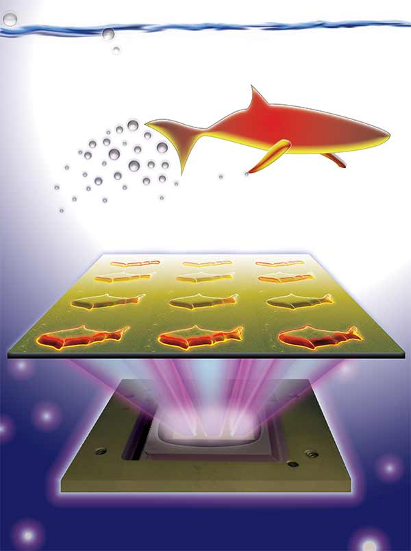
3D-printed microfish contain functional nanoparticles that enable them to be self-propelled, chemically powered and magnetically steered. The microfish are also capable of removing and sensing toxins. Image credit: J. Warner, UC San Diego Jacobs School of Engineering.

3D-printed microfish contain functional nanoparticles that enable them to be self-propelled, chemically powered and magnetically steered. The microfish are also capable of removing and sensing toxins. Image credit: J. Warner, UC San Diego Jacobs School of Engineering.

An August 25, 2015 UCSD news release (also on EurekAlert) by Liezel Labios, which originated the news item, provides more detail,

The technique used to fabricate the microfish provides numerous improvements over other methods traditionally employed to create microrobots with various locomotion mechanisms, such as microjet engines, microdrillers and microrockets. Most of these microrobots are incapable of performing more sophisticated tasks because they feature simple designs — such as spherical or cylindrical structures — and are made of homogeneous inorganic materials. In this new study, researchers demonstrated a simple way to create more complex microrobots.

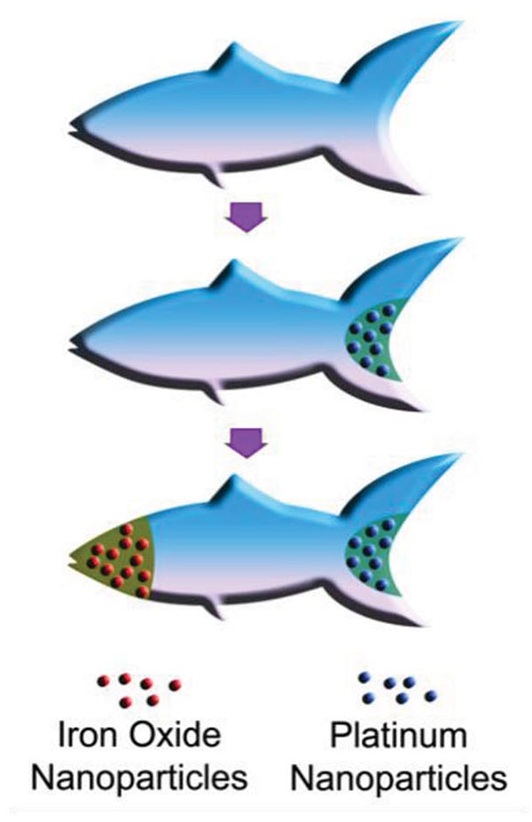
By combining Chen’s 3D printing technology with Wang’s expertise in microrobots, the team was able to custom-build microfish that can do more than simply swim around when placed in a solution containing hydrogen peroxide. Nanoengineers were able to easily add functional nanoparticles into certain parts of the microfish bodies. They installed platinum nanoparticles in the tails, which react with hydrogen peroxide to propel the microfish forward, and magnetic iron oxide nanoparticles in the heads, which allowed them to be steered with magnets.

Here’s an illustration of the platinum and iron oxide microfish,

Schematic illustration of the process of functionalizing the microfish. Platinum nanoparticles are first loaded into the tail of the fish for propulsion via reaction with hydrogen peroxide. Next, iron oxide nanoparticles are loaded into the head of the fish for magnetic control. Image credit: W. Zhu and J. Li, UC San Diego Jacobs School of Engineering.

Schematic illustration of the process of functionalizing the microfish. Platinum nanoparticles are first loaded into the tail of the fish for propulsion via reaction with hydrogen peroxide. Next, iron oxide nanoparticles are loaded into the head of the fish for magnetic control. Image credit: W. Zhu and J. Li, UC San Diego Jacobs School of Engineering.

Back to the news release,

“We have developed an entirely new method to engineer nature-inspired microscopic swimmers that have complex geometric structures and are smaller than the width of a human hair. With this method, we can easily integrate different functions inside these tiny robotic swimmers for a broad spectrum of applications,” said the co-first author Wei Zhu, a nanoengineering Ph.D. student in Chen’s research group at the Jacobs School of Engineering at UC San Diego.

As a proof-of-concept demonstration, the researchers incorporated toxin-neutralizing nanoparticles throughout the bodies of the microfish. Specifically, the researchers mixed in polydiacetylene (PDA) nanoparticles, which capture harmful pore-forming toxins such as the ones found in bee venom. The researchers noted that the powerful swimming of the microfish in solution greatly enhanced their ability to clean up toxins. When the PDA nanoparticles bind with toxin molecules, they become fluorescent and emit red-colored light. The team was able to monitor the detoxification ability of the microfish by the intensity of their red glow.

“The neat thing about this experiment is that it shows how the microfish can doubly serve as detoxification systems and as toxin sensors,” said Zhu.

“Another exciting possibility we could explore is to encapsulate medicines inside the microfish and use them for directed drug delivery,” said Jinxing Li, the other co-first author of the study and a nanoengineering Ph.D. student in Wang’s research group.

For anyone curious about the new 3D printing technique, the news release provides more information about that too,

The new microfish fabrication method is based on a rapid, high-resolution 3D printing technology called microscale continuous optical printing (μCOP), which was developed in Chen’s lab. Some of the benefits of the μCOP technology are speed, scalability, precision and flexibility. Within seconds, the researchers can print an array containing hundreds of microfish, each measuring 120 microns long and 30 microns thick. This process also does not require the use of harsh chemicals. Because the μCOP technology is digitized, the researchers could easily experiment with different designs for their microfish, including shark and manta ray shapes. [emphasis mine] “With our 3D printing technology, we are not limited to just fish shapes. We can rapidly build microrobots inspired by other biological organisms such as birds,” said Zhu.

The key component of the μCOP technology is a digital micromirror array device (DMD) chip, which contains approximately two million micromirrors. Each micromirror is individually controlled to project UV light in the desired pattern (in this case, a fish shape) onto a photosensitive material, which solidifies upon exposure to UV light. The microfish are built using a photosensitive material and are constructed one layer at a time, allowing each set of functional nanoparticles to be “printed” into specific parts of the fish bodies.

“This method has made it easier for us to test different designs for these microrobots and to test different nanoparticles to insert new functional elements into these tiny structures. It’s my personal hope to further this research to eventually develop surgical microrobots that operate safer and with more precision,” said Li.

Nice to see I can recognize a shark shape when I see one. Getting back to the research, yet again, here’s a link to and a citation for the paper.

3D-Printed Artificial Microfish by Wei Zhu, Jinxing Li, Yew J. Leong, Isaac Rozen, Xin Qu, Renfeng Dong, Zhiguang Wu, Wei Gao, Peter H. Chung, Joseph Wang, and Shaochen Chen. Advanced Materials Volume 27, Issue 30, pages 4411–4417, August 12, 2015 DOI: 10.1002/adma.201501372 Article first published online: 29 JUN 2015

© 2015 WILEY-VCH Verlag GmbH & Co. KGaA, Weinheim

This paper is behind a paywall.

Sharklet’s sharkskin-like material

It’s one of my favourite technologies but there hasn’t been much talk about Sharklet for the last few years. My Feb. 10, 2011 posting about it had this,

They used sharkskin as an example for making a ‘smarter’ material. Scientists have observed that nanoscale structures on a shark’s skin have antibacterial properties. This is especially important when we have a growing problem with bacteria that are antibiotic resistant. David Pogue’s (the program host) interviewed scientists at Sharklet and highlighted their work producing a plastic with nanostructures similar to those found on sharkskin for use in hospitals, restaurants, etc.  I found this on the Sharklet website (from a rotating graphic on the home page),

The World Health Organization calls antibiotic resistance a leading threat to human health.

Sharkjet provides a non-toxic approach to bacterial control and doesn’t create resistance.

The reason that the material does not create resistance is that it doesn’t kill the bacteria (antibiotics kill most bacteria but cannot kill all of them with the consequence that only the resistant survive and reproduce). Excerpted from Sharklet’s technology page,

While the Sharklet pattern holds great promise to improve the way humans co-exist with microorganisms, the pattern was developed far outside of a laboratory. In fact, Sharklet was discovered via a seemingly unrelated problem: how to keep algae from coating the hulls of submarines and ships. In 2002, Dr. Anthony Brennan, a materials science and engineering professor at the University of Florida, was visiting the U.S. naval base at Pearl Harbor in Oahu as part of Navy-sponsored research. The U.S. Office of Naval Research solicited Dr. Brennan to find new antifouling strategies to reduce use of toxic antifouling paints and trim costs associated with dry dock and drag.

The most recent news from Sharklet comes in a Sept. 16, 2014 news release on EurekAlert which refines the definition for Sharklet and provides research about the latest research on this material,

Transmission of bacterial infections, including MRSA and MSSA could be curbed by coating hospital surfaces with microscopic bumps that mimic the scaly surface of shark skin, according to research published in the open access journal Antimicrobial Resistance and Infection Control.

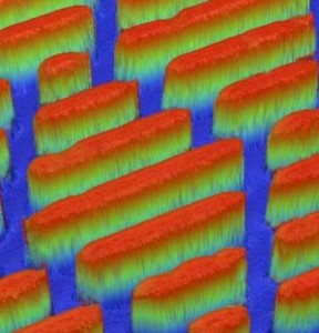
The study modelled how well different materials prevented the spread of human disease bacteria through touching, sneezes or spillages. The micropattern, named Sharklet™, is an arrangement of ridges formulated to resemble shark skin. The study showed that Sharklet harboured 94% less MRSA bacteria than a smooth surface, and fared better than copper, a leading antimicrobial material. The bacteria were less able to attach to Sharklet’s imperceptibly textured surface, suggesting it could reduce the spread of superbugs in hospital settings.

The surfaces in hospitals and healthcare settings are often rife with bacteria and patients are vulnerable to bacterial infection. Scientists are investigating the ability of different materials to prevent the spread of bacteria. Copper alloys are a popular option, as they are toxic to bacterial cells, interfering with their cellular processes and killing them. The Sharklet micropattern works differently – the size and composition of its microscopic features prevent bacteria from attaching to it. It mimics the unique qualities of shark skin, which, unlike other underwater surfaces, inhibits bacteria, because it is covered with a natural micropattern of tooth-like structures, called denticles.

Dr Ethan Mann, a research scientist at Sharklet Technologies, the manufacturer of the micropattern, says: “The Sharklet texture is designed to be manufactured directly into the surfaces of plastic products that surround patients in hospital, including environmental surfaces as well as medical devices. Sharklet does not introduce new materials or coatings – it simply alters the shape and texture of existing materials to create surface properties that are unfavorable for bacterial contamination.”

The researchers from Sharklet Technologies compared how well two types of infection-causing bacteria, methicillin-resistant or susceptible Staphylococcus aureus (MRSA and MSSA), fared at contaminating three surfaces – the Sharklet micropattern, a copper alloy, and a smooth control surface. They created experimental procedures to mimic common ways bacteria infect surfaces. Sneezing was mimicked by using a paint sprayer to spread the bacterial solution on 10 samples of each surface. To mimic infected patients touching the surfaces, velveteen cloth was put in contact with bacteria for 10s, and then placed on another set of each test surface for 10s. A third set of each surface was immersed in bacterial solution for an hour, then rinsed and dried, to mimic spills.

Surfaces were sampled for remaining contaminations either immediately following exposure to MSSA and MRSA or 90 minutes after being exposed. The Sharklet micropattern reduced transmission of MSSA by 97% compared to the smooth control, while copper was no better than the control. The micropattern also harboured 94% less MRSA bacteria than the control surface, while the copper had 80% less.

Dr Mann says: “Shark skin itself is not an antimicrobial surface, rather it seems highly adapted to resist attachment of living organisms such as algae and barnacles. Shark skin has a specific roughness and certain properties that deter marine organisms from attaching to the skin surface. We have learned much from nature in building this material texture for the future.”

Here’s an illustration the researchers have provided,

Caption: This is an image of the Sharklet micropattern, which mimics the denticles of shark skin. Credit: Mann et al.

Caption: This is an image of the Sharklet micropattern, which mimics the denticles of shark skin.
Credit: Mann et al.

Here’s a link to and a citation for the paper,

Surface micropattern limits bacterial contamination by Ethan E Mann, Dipankar Manna, Michael R Mettetal, Rhea M May, Elisa M Dannemiller, Kenneth K Chung, Anthony B Brennan, and Shravanthi T Reddy. Antimicrobial Resistance and Infection Control 2014, 3:28  doi:10.1186/2047-2994-3-28

This is an open access paper.

Bioluminscent sharks and their photon hunting abilities

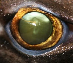
This is the eye of a velvet belly lanternshark. Credit: Dr. J. Mallefet (FNRS/UCL); CC-BY

This is the eye of a velvet belly lanternshark.
Credit: Dr. J. Mallefet (FNRS/UCL); CC-BY

The velvet belly is a bioluminscent shark, i.e., it projects some light. Here’s a description from its Wikipedia entry (Note: Links have been removed),

The velvet belly lanternshark (or simply velvet belly, Etmopterus spinax) is a species of dogfish shark in the family Etmopteridae. One of the most common deepwater sharks in the northeastern Atlantic Ocean, the velvet belly is found from Iceland and Norway to Gabon and South Africa at a depth of 70–2,490 m (230–8,170 ft). A small shark generally no more than 45 cm (18 in) long, the velvet belly is so named because its black underside is abruptly distinct from the brown coloration on the rest of its body. … Like other lanternsharks, the velvet belly is bioluminescent, with light-emitting photophores forming a species-specific pattern over its flanks and abdomen. These photophores are thought to function in counter-illumination, which camouflages the shark against predators. They may also play a role in social interactions.

An Aug. 6, 2014 news item on ScienceDaily highlights some recent featuring the velvet belly,

The eyes of deep-sea bioluminescent sharks have a higher rod density when compared to non-bioluminescent sharks, according to a study published August 6, 2014 in the open-access journal PLOS ONE by Julien M. Claes, postdoctoral researcher from the FNRS at Université catholique de Louvain (Belgium), and colleagues. This adaptation is one of many these sharks use to produce and perceive bioluminescent light in order to communicate, find prey, and camouflage themselves against predators.

An Aug. 6, 2014 news item on phys.org elucidates further,

The mesopelagic twilight zone, or about 200-1000 meters deep in the sea, is a vast, dim habitat, where, with increasing depth, sunlight is progressively replaced by point-like bioluminescent emissions. To better understand strategies used by bioluminescent predators inhabiting this region that help optimize photon capture, the authors of this study analyzed the eye shape, structure, and retinal cell mapping in the visual systems of five deep-sea bioluminescent sharks, including four Lanternsharks (Etmopteridae) and one kitefin shark (Dalatiidae).

The researchers found that the sharks’ eyes contained a translucent area present in the upper eye orbit of the lantern sharks, which might aid in adjusting counter-illumination, or in using bioluminescence to camouflage the fish. They also found several ocular specializations, such as a gap between the lens and iris that allows extra light to the retina, which was previously unknown in sharks. Comparisons with previous data on non-bioluminescent sharks reveals that bioluminescent sharks possess higher rod densities in their eyes, which might provide them with improved temporal resolution, particularly useful for bioluminescent communication during social interactions.

“Every bioluminescent signal needs to reach a target photoreceptor to be ecologically efficient. Here, we clearly found evidence that the visual system of bioluminescent sharks has co-evolved with their light-producing capability, even though more work is needed to understand the full story,” said Dr. Claes.

Here’s a link to and a citation for the paper,

Photon Hunting in the Twilight Zone: Visual Features of Mesopelagic Bioluminescent Sharks by Julien M. Claes, Julian C. Partridge, Nathan S. Hart, Eduardo Garza-Gisholt, Hsuan-Ching Ho, Jérôme Mallefet, and Shaun P. Collin. PLOS ONE DOI: 10.1371/journal.pone.0104213 Published: August 06, 2014

This study is open access as is the journal where it appears, PLOS (Public Library of Science) ONE.