Tag Archives: shigella

A city of science in Japan: Kawasaki (Kanagawa)

Happily, I’m getting more nanotechnology (for the most part) information from Japan. Given Japan’s prominence in this field of endeavour I’ve long felt FrogHeart has not adequately represented Japanese contributions. Now that I’m receiving English language translations, I hope to better address the situation.

This morning (March 26, 2015), there were two news releases from Kawasaki INnovation Gateway at SKYFRONT (KING SKYFRONT), Coastal Area International Strategy Office, Kawasaki City, Japan in my mailbox. Before getting on to the news releases, here’s a little about  the city of Kawasaki and about its innovation gateway. From the Kawasaki, Kanagawa entry in Wikipedia (Note: Links have been removed),

Kawasaki (川崎市 Kawasaki-shi?) is a city in Kanagawa Prefecture, Japan, located between Tokyo and Yokohama. It is the 9th most populated city in Japan and one of the main cities forming the Greater Tokyo Area and Keihin Industrial Area.

Kawasaki occupies a belt of land stretching about 30 kilometres (19 mi) along the south bank of the Tama River, which divides it from Tokyo. The eastern end of the belt, centered on JR Kawasaki Station, is flat and largely consists of industrial zones and densely built working-class housing, the Western end mountainous and more suburban. The coastline of Tokyo Bay is occupied by vast heavy industrial complexes built on reclaimed land.

There is a 2014 video about Kawasaki’s innovation gateway, which despite its 14 mins. 39 secs. running time I am embedding here. (Caution: They highlight their animal testing facility at some length.)

Now on to the two news releases. The first concerns research on gold nanoparticles that was published in 2014. From a March 26, 2015 Kawasaki INnovation Gateway news release,

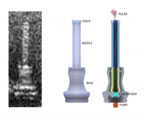
Gold nanoparticles size up to cancer treatment

Incorporating gold nanoparticles helps optimise treatment carrier size and stability to improve delivery of cancer treatment to cells.

Treatments that attack cancer cells through the targeted silencing of cancer genes could be developed using small interfering RNA molecules (siRNA). However delivering the siRNA into the cells intact is a challenge as it is readily degraded by enzymes in the blood and small enough to be eliminated from the blood stream by kidney filtration.  Now Kazunori Kataoka at the University of Tokyo and colleagues at Tokyo Institute of Technology have designed a protective treatment delivery vehicle with optimum stability and size for delivering siRNA to cells.

The researchers formed a polymer complex with a single siRNA molecule. The siRNA-loaded complex was then bonded to a 20 nm gold nanoparticle, which thanks to advances in synthesis techniques can be produced with a reliably low size distribution. The resulting nanoarchitecture had the optimum overall size – small enough to infiltrate cells while large enough to accumulate.

In an assay containing heparin – a biological anti-coagulant with a high negative charge density – the complex was found to release the siRNA due to electrostatic interactions. However when the gold nanoparticle was incorporated the complex remained stable. Instead, release of the siRNA from the complex with the gold nanoparticle could be triggered once inside the cell by the presence of glutathione, which is present in high concentrations in intracellular fluid. The glutathione bonded with the gold nanoparticles and the complex, detaching them from each other and leaving the siRNA prone to release.

The researchers further tested their carrier in a subcutaneous tumour model. The authors concluded that the complex bonded to the gold nanoparticle “enabled the efficient tumor accumulation of siRNA and significant in vivo gene silencing effect in the tumor, demonstrating the potential for siRNA-based cancer therapies.”

The news release provides links to the March 2015 newsletter which highlights this research and to the specific article and video,

March 2015 Issue of Kawasaki SkyFront iNewsletter: http://inewsletter-king-skyfront.jp/en/

Contents

Feature video on Professor Kataoka’s research : http://inewsletter-king-skyfront.jp/en/video_feature/vol_3/feature01/

Research highlights: http://inewsletter-king-skyfront.jp/en/research_highlights/vol_3/research01/

Here’s a link to and a citation for the paper,

Precise Engineering of siRNA Delivery Vehicles to Tumors Using Polyion Complexes and Gold Nanoparticles by Hyun Jin Kim, Hiroyasu Takemoto, Yu Yi, Meng Zheng, Yoshinori Maeda, Hiroyuki Chaya, Kotaro Hayashi, Peng Mi, Frederico Pittella, R. James Christie, Kazuko Toh, Yu Matsumoto, Nobuhiro Nishiyama, Kanjiro Miyata, and Kazunori Kataoka. ACS Nano, 2014, 8 (9), pp 8979–8991 DOI: 10.1021/nn502125h Publication Date (Web): August 18, 2014
Copyright © 2014 American Chemical Society

This article is behind a paywall.

The second March 26, 2015 Kawasaki INnovation Gateway news release concerns a DNA chip and food-borne pathogens,

Rapid and efficient DNA chip technology for testing 14 major types of food borne pathogens

Conventional methods for testing food-borne pathogens is based on the cultivation of pathogens, a process that is complicated and time consuming. So there is demand for alternative methods to test for food-borne pathogens that are simpler, quick and applicable to a wide range of potential applications.

Now Toshiba Ltd and Kawasaki City Institute for Public Health have collaborated in the development of a rapid and efficient automatic abbreviated DNA detection technology that can test for 14 major types of food borne pathogens. The so called ‘DNA chip card’ employs electrochemical DNA chips and overcomes the complicated procedures associated with genetic testing of conventional methods. The ‘DNA chip card’ is expected to find applications in hygiene management in food manufacture, pharmaceuticals, and cosmetics.

Details

The so-called automatic abbreviated DNA detection technology ‘DNA chip card’ was developed by Toshiba Ltd and in a collaboration with Kawasaki City Institute for Public Health, used to simultaneously detect 14 different types of food-borne pathogens in less than 90 minutes. The detection sensitivity depends on the target pathogen and has a range of 1E+01~05 cfu/mL.

Notably, such tests would usually take 4-5 days using conventional methods based on pathogen cultivation. Furthermore, in contrast to conventional DNA protocols that require high levels of skill and expertise, the ‘DNA chip card’ only requires the operator to inject nucleic acid, thereby making the procedure easier to use and without specialized operating skills.

Examples of pathogens associated with food poisoning that were tested with the “DNA chip card”

Enterohemorrhagic Escherichia coli

Salmonella

Campylobacter

Vibrio parahaemolyticus

Shigella

Staphylococcus aureus

Enterotoxigenic Escherichia coli

Enteroaggregative Escherichia coli

Enteropathogenic Escherichia coli

Clostridium perfringens

Bacillus cereus

Yersinia

Listeria

Vibrio cholerae

I think 14 is the highest number of tests I’ve seen for one of these chips. This chip is quite an achievement.

One final bit from the news release about the DNA chip provides a brief description of the gateway and something they call King SkyFront,

About KING SKYFRONT

The Kawasaki INnovation Gateway (KING) SKYFRONT is the flagship science and technology innovation hub of Kawasaki City. KING SKYFRONT is a 40 hectare area located in the Tonomachi area of the Keihin Industrial Region that spans Tokyo and Kanagawa Prefecture and Tokyo International Airport (also often referred to as Haneda Airport).

KING SKYFRONT was launched in 2013 as a base for scholars, industrialists and government administrators to work together to devise real life solutions to global issues in the life sciences and environment.

I find this emphasis on the city interesting. It seems that cities are becoming increasingly important and active where science research and development are concerned. Europe seems to have adopted a biannual event wherein a city is declared a European City of Science in conjunction with the EuroScience Open Forum (ESOF) conferences. The first such city was Dublin in 2012 (I believe the Irish came up with the concept themselves) and was later adopted by Copenhagen for 2014. The latest city to embrace the banner will be Manchester in 2016.

New molecular ruler could help with developing antibiotics

Researchers at the University of Utah have developed a molecular ruler which could help to determine the length at which a nanoscale needle is effective. From a March 17, 2015 news item on Azonano,

When a salmonella bacterium attacks a cell, it uses a nanoscopic needle to inject it with proteins to aid the infection. If the needle is too short, the cell won’t be infected. Too long, and the needle breaks. Now, University of Utah biologists report how a disposable molecular ruler or tape measure determines the length of the bacterial needle so it is just right.

The findings have potential long-term applications for developing new antibiotics against salmonella and certain other disease-causing bacteria, for designing bacteria that could inject cancer cells with chemotherapy drugs, and for helping people how to design machines at the nanoscopic or molecular scale.

A March 16, 2015 University of Utah news release, which originated the news item, provides some insight from the researchers,

“If you look at important pathogens – the bubonic plague bacterium, salmonella, shigella and plant pathogens like fire blight – they all use hypodermic-like needles to inject proteins that facilitate disease processes,” Hughes [University of Utah biology professor Kelly Hughes] says.

“Our work says that there is one mechanism – the molecular ruler – to explain how the lengths are controlled for needles in gram-negative bacteria and for hooks on flagella [the U-joints in propellers bacteria use to move] in all bacteria,” he adds.

In their study, Wee [University of Utah doctoral student Daniel Wee] and Hughes found that as a bacterial needle or “injectisome” grows, a molecular ruler – really, more like a gooey tape measure – is secreted from within the needle’s base. It oozes up through the tube-like needle, and when the bottom end of the ruler reaches the bottom end of the needle, the needle stops growing and begins to inject proteins into the target cell to help the infection process.

The biologists say the [US] National Institutes of Health-funded study refutes other theories for how salmonella and some other disease bacteria determine needle lengths.

The news release also explains how this finding could be made useful,

“What we understand from bacteria can help us build nanomachines and nanobots,” Hughes says, noting that bacterial flagella – the nanoscopic motor-and-propeller system they use to swim to dinner or to targets – are “the most sophisticated nanomachines in the universe.”

In one example, Swiss scientists are using the design of bacterial flagella as the basis for a nanobot that will be put inside the eye to do nanoscale surgery, he adds.

In addition to flagella, a number of disease-causing bacteria also have injectisomes, which also are built of proteins, as are most structures in living organisms.

“In the case of the needle, you have a structure that extends from the surface of the bacterium like a hypodermic,” Hughes says. “These needles are fragile. If one is too long, it will break off and be useless. If you make it too short, then it can’t get past the surface proteins on cells it needs to invade.”

By understanding how bacteria determine the length for their needles, it someday may be possible to engineer bacteria to inject chemotherapy drugs right into cancer cells.

“People would like to design bacteria that can get to cancer cells and inject poisons into just those cells and kill them, and not harm the rest of us,” Hughes says.

And by understanding how certain disease-causing bacteria build their injectisomes, new antibiotics might be developed in a decade or so to target and destroy the needles and thus deter bacterial infections. The rulers that help build flagella also might be attacked by drugs to prevent bacteria from reaching target cells, “so you can kill two birds with one stone by hitting the two machines at the same time,” Hughes says.

He says that approach might work against injectisome-equipped bacteria such as salmonella species that cause typhoid fever and food poisoning; shigella species that cause dysentery; the bubonic plague bacterium Yersinia pestis; disease-causing E. coli; sexually transmitted Chlamydia trachomatis; many plant pathogens; and Pseudomonas aeruginosa, which often infects burn patients and the lungs of cystic fibrosis patients.

Not usually my kind of thing, I find this quite fascinating (from the news release),

Bacteria secrete a molecular ruler to measure needle length

Bacterial injectisomes are incredibly small, measuring only 20 to 100 nanometers long. A nanometer is one billionth of a meter, and a meter is about 39 inches long. The width of a typical human hair often is given as 100 microns, so the maximum length of a bacterial needle, 100 nanometers, is one-thousandth of the width of a human hair.

Gram-negative, disease-causing bacteria “are very closely related species, so how do they subtly control the various needle lengths to be perfect?” Hughes asks. “In one case it might be 40 nanometers versus 55 nanometers. These are small sizes. So to do this, the bacteria developed molecular rulers to differentiate needles of different lengths.”

(Gram-negative bacteria are those with membranes lining both the inside and outside of their cell wall, while gram-positive bacteria have only an inner membrane.)

Like any cell, a bacterium is encased in a cell wall. So bacteria developed all kinds of secretions to make contact with and infect other cells: flagellar propellers to swim to food or target cells, docking structures to help bacteria stick to targets, and injectisomes to inject infection-promoting proteins into targets.

When a bacterium builds a needle, it first builds a base. “A series of proteins form a doughnut, and inside the doughnut hole, the actual secretion machine gets constructed,” Hughes says. “It’s the same for the flagella as it is for these needles.”

Next, proteins start assembling to form the needle or injectisome.

The new study demonstrated that in salmonella, the ruler or tape measure is secreted slowly through the channel of the growing needle. Once amino acids at the bottom end of the ruler pass through the base of the needle, they tell the bacterium that the needle is long enough and to stop growing. They also tell the needle to injecting virulence proteins into the target cell, and the molecular ruler is ejected, Wee says.

Here’s an image of what the injectisome looks like,

On the left is an electron microscope image of an injectisome, the nanoscopic needle that salmonella and certain other bacteria use to inject proteins into target cells as part of the infection process. The illustration at center depicts the exterior of the needle and its base. The cross-section at right shows the string-like molecular ruler that determines the length of salmonella’s bacteria needle, according to a new University of Utah study by doctoral student Daniel Wee and biology professor Kelly Hughes. Credit: Daniel Wee, University of Utah

On the left is an electron microscope image of an injectisome, the nanoscopic needle that salmonella and certain other bacteria use to inject proteins into target cells as part of the infection process. The illustration at center depicts the exterior of the needle and its base. The cross-section at right shows the string-like molecular ruler that determines the length of salmonella’s bacteria needle, according to a new University of Utah study by doctoral student Daniel Wee and biology professor Kelly Hughes. Credit: Daniel Wee, University of Utah

The news release also offers some specific details about the research,

How the study was performed

The new study used the Typhimurium strain of Salmonella enterica, which causes food poisoning. The researchers proved the molecular ruler determines needle length in salmonella by inserting amino acids from the plague bacterium’s molecular ruler genes into genes for salmonella’s molecular ruler, making rulers with seven different lengths.

Genetically engineered salmonella with seven ruler lengths were grown in a flask, their needles isolated, and the needle lengths measured under an electron microscope.

Wee found the ruler lengths correlated precisely with the lengths of the resulting needles or injectisomes, with each amino acid added to the ruler gene making the resulting needle 0.2 nanometers longer.

Previous studies found the molecular ruler determines the length of the hook or U-joint that helps turn flagella or propellers in many bacteria. Research also found the molecular ruler determines the length of both the flagellar hook and the needle in plague bacteria. But some researchers argued salmonella needle’s length was determined by some other mechanism:

– One theory holds that a molecular measuring cup in the needle’s base sends a cupful of needle components to assemble the needle, and the length of the needle is determined by the size of the cup. The new study disproved that by genetically removing the cup and showing that the injectisomes or needles still grew to correct lengths.

– Another theory says that as needle components assemble outside the needle’s base, a rod-shaped structure assembles inside the base to link the base and needle, and that when the rod is complete, needle assembly stops, thus determining needle length. But the Utah study found the rod and needle components are not made simultaneously, but compete with each other, so as more rod parts are made, fewer needle parts are made, giving an illusion that rod completion controls needle length.

Here’s a link to and a citation for the paper,

Molecular ruler determines needle length for the Salmonella Spi-1 injectisome by Daniel H. Wee and Kelly T. Hughes. Published online before print March 16, 2015, doi: 10.1073/pnas.1423492112 PNAS March 16, 2015

This paper is behind a paywall.