Tag Archives: skin

Sound waves for wearable patches that deliver drugs painlessly

While watching this video I started wondering if they were testing their research on students but that’s not the case; these wearable patches were tested on porcine (pig) skin, which is quite similar to human skin, Note: They tested a B vitamin called niacinamide so, it’s highly unlikely the pigs suffered from it,

An April 20, 2023 news item on ScienceDaily announces the research into using ultrasonic waves for drug delivery,

The skin is an appealing route for drug delivery because it allows drugs to go directly to the site where they’re needed, which could be useful for wound healing, pain relief, or other medical and cosmetic applications. However, delivering drugs through the skin is difficult because the tough outer layer of the skin prevents most small molecules from passing through it.

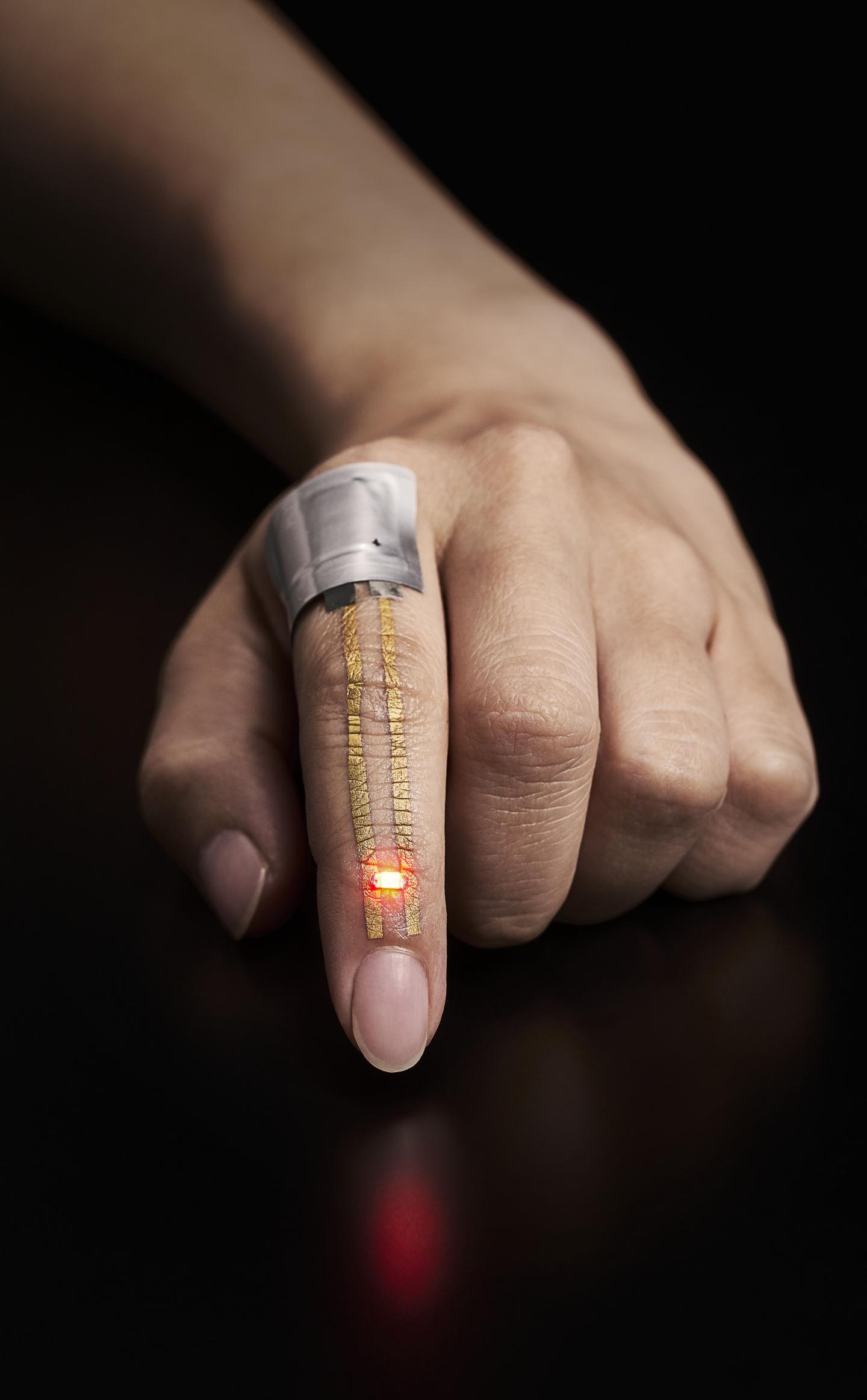
In hopes of making it easier to deliver drugs through the skin, MIT [Massachusetts Institute of Technology] researchers have developed a wearable patch that applies painless ultrasonic waves to the skin, creating tiny channels that drugs can pass through. This approach could lend itself to delivery of treatments for a variety of skin conditions, and could also be adapted to deliver hormones, muscle relaxants, and other drugs, the researchers say.

An April 20, 2023 Massachusetts Institute of Technology (MIT) news release (also on EurekAlert), which originated the news item, provides technical details about the research, Note: A link has been removed,

“The ease-of-use and high-repeatability offered by this system provides a game-changing alternative to patients and consumers suffering from skin conditions and premature skin aging,” says Canan Dagdeviren, an associate professor in MIT’s Media Lab and the senior author of the study. “Delivering drugs this way could offer less systemic toxicity and is more local, comfortable, and controllable.”

MIT research assistants Chia-Chen Yu and Aastha Shah are the lead authors of the paper, which appears in Advanced Materials, as part of the journal’s “Rising Stars” series, which showcases the outstanding work of researchers in the early stages of their independent careers. Other MIT authors include Research Assistant Colin Marcus and postdoc Md Osman Goni Nayeem. Nikta Amiri, Amit Kumar Bhayadia, and Amin Karami of the University of Buffalo are also authors of the paper.

A boost from sound waves

The researchers began this project as an exploration of alternative ways to deliver drugs. Most drugs are delivered orally or intravenously, but the skin is a route that could offer much more targeted drug delivery for certain applications.

“The main benefit with skin is that you bypass the whole gastrointestinal tract. With oral delivery, you have to deliver a much larger dose in order to account for the loss that you would have in the gastric system,” Shah says. “This is a much more targeted, focused modality of drug delivery.”

Ultrasound exposure has been shown to enhance the skin’s permeability to small-molecule drugs, but most of the existing techniques for performing this kind of drug delivery require bulky equipment. The MIT team wanted to come up with a way to perform this kind of transdermal drug delivery with a lightweight, wearable patch, which could make it easier to use for a variety of applications.

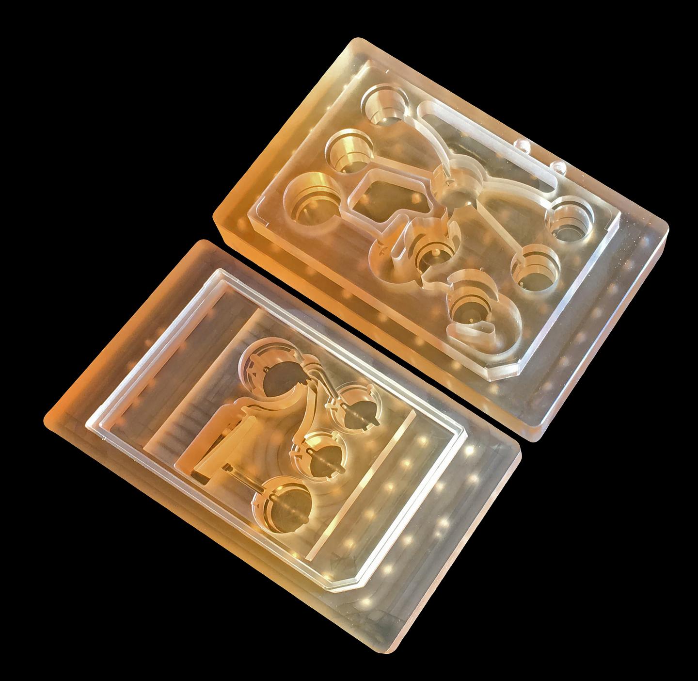
The device that they designed consists of a patch embedded with several disc-shaped piezoelectric transducers, which can convert electric currents into mechanical energy. Each disc is embedded in a polymeric cavity that contains the drug molecules dissolved in a liquid solution. When an electric current is applied to the piezoelectric elements, they generate pressure waves in the fluid, creating bubbles that burst against the skin. These bursting bubbles produce microjets of fluid that can penetrate through the skin’s tough outer layer, the stratum corneum.

“This works open the door to using vibrations to enhance drug delivery. There are several parameters that result in generation of different kinds of waveform patterns. Both mechanical and biological aspects of drug delivery can be improved by this new toolset,” Karami says.

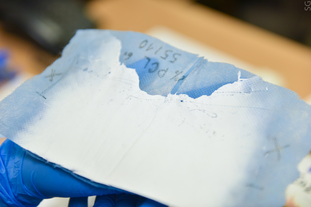
The patch is made of PDMS, a silicone-based polymer that can adhere to the skin without tape. In this study, the researchers tested the device by delivering a B vitamin called niacinamide, an ingredient in many sunscreens and moisturizers.

In tests using pig skin, the researchers showed that when they delivered niacinamide using the ultrasound patch, the amount of drug that penetrated the skin was 26 times greater than the amount that could pass through the skin without ultrasonic assistance.

The researchers also compared the results from their new device to microneedling, a technique sometimes used for transdermal drug delivery, which involves puncturing the skin with miniature needles. The researchers found that their patch was able to deliver the same amount of niacinamide in 30 minutes that could be delivered with microneedles over a six-hour period.

Local delivery

With the current version of the device, drugs can penetrate a few millimeters into the skin, making this approach potentially useful for drugs that act locally within the skin. These could include niacinamide or vitamin C, which is used to treat age spots or other dark spots on the skin, or topical drugs used to heal burns.

With further modifications to increase the penetration depth, this technique could also be used for drugs that need to reach the bloodstream, such as caffeine, fentanyl, or lidocaine. Dagdeviren also envisions that this kind of patch could be useful for delivering hormones such as progesterone. In addition, the researchers are now exploring the possibility of implanting similar devices inside the body to deliver drugs to treat cancer or other diseases.

The researchers are also working on further optimizing the wearable patch, in hopes of testing it soon on human volunteers. They also plan to repeat the lab experiments they did in this study, with larger drug molecules.

“After we characterize the drug penetration profiles for much larger drugs, we would then see which candidates, like hormones or insulin, can be delivered using this technology, to provide a painless alternative for those who are currently bound to self-administer injections on a daily basis,” Shah says.

Here’s a link to and a citation for the paper,

A Conformable Ultrasound Patch for Cavitation-Enhanced Transdermal Cosmeceutical Delivery by Chia-Chen Yu, Aastha Shah, Nikta Amiri, Colin Marcus, Md Osman Goni Nayeem, Amit Kumar Bhayadia, Amin Karami, Canan Dagdeviren. Advanced Materials Volume35, Issue 23 June 8, 2023 2300066 DOI: https://doi.org/10.1002/adma.202300066 First published online: 19 March 2023

This paper is open access.

Robots with living human skin tissue?

So far, it looks like they’ve managed a single robotic finger. I expect it will take a great deal more work before an entire robotic hand is covered in living skin. BTW, I have a few comments at the end of this post.

Caption: Illustration showing the cutting and healing process of the robotic finger (A), its anchoring structure (B) and fabrication process (C). Credit: ©2022 Takeuchi et al.

I have two news releases highlighting the work. This a June 9, 2022 Cell Press news release,

From action heroes to villainous assassins, biohybrid robots made of both living and artificial materials have been at the center of many sci-fi fantasies, inspiring today’s robotic innovations. It’s still a long way until human-like robots walk among us in our daily lives, but scientists from Japan are bringing us one step closer by crafting living human skin on robots. The method developed, presented June 9 in the journal Matter, not only gave a robotic finger skin-like texture, but also water-repellent and self-healing functions.

“The finger looks slightly ‘sweaty’ straight out of the culture medium,” says first author Shoji Takeuchi, a professor at the University of Tokyo, Japan. “Since the finger is driven by an electric motor, it is also interesting to hear the clicking sounds of the motor in harmony with a finger that looks just like a real one.”

Looking “real” like a human is one of the top priorities for humanoid robots that are often tasked to interact with humans in healthcare and service industries. A human-like appearance can improve communication efficiency and evoke likability. While current silicone skin made for robots can mimic human appearance, it falls short when it comes to delicate textures like wrinkles and lacks skin-specific functions. Attempts at fabricating living skin sheets to cover robots have also had limited success, since it’s challenging to conform them to dynamic objects with uneven surfaces.

“With that method, you have to have the hands of a skilled artisan who can cut and tailor the skin sheets,” says Takeuchi. “To efficiently cover surfaces with skin cells, we established a tissue molding method to directly mold skin tissue around the robot, which resulted in a seamless skin coverage on a robotic finger.”

To craft the skin, the team first submerged the robotic finger in a cylinder filled with a solution of collagen and human dermal fibroblasts, the two main components that make up the skin’s connective tissues. Takeuchi says the study’s success lies within the natural shrinking tendency of this collagen and fibroblast mixture, which shrank and tightly conformed to the finger. Like paint primers, this layer provided a uniform foundation for the next coat of cells—human epidermal keratinocytes—to stick to. These cells make up 90% of the outermost layer of skin, giving the robot a skin-like texture and moisture-retaining barrier properties.

The crafted skin had enough strength and elasticity to bear the dynamic movements as the robotic finger curled and stretched. The outermost layer was thick enough to be lifted with tweezers and repelled water, which provides various advantages in performing specific tasks like handling electrostatically charged tiny polystyrene foam, a material often used in packaging. When wounded, the crafted skin could even self-heal like humans’ with the help of a collagen bandage, which gradually morphed into the skin and withstood repeated joint movements.

“We are surprised by how well the skin tissue conforms to the robot’s surface,” says Takeuchi. “But this work is just the first step toward creating robots covered with living skin.” The developed skin is much weaker than natural skin and can’t survive long without constant nutrient supply and waste removal. Next, Takeuchi and his team plan to address those issues and incorporate more sophisticated functional structures within the skin, such as sensory neurons, hair follicles, nails, and sweat glands.

“I think living skin is the ultimate solution to give robots the look and touch of living creatures since it is exactly the same material that covers animal bodies,” says Takeuchi.

A June 10, 2022 University of Tokyo news release (also on EurekAlert but published June 9, 2022) covers some of the same ground while providing more technical details,

Researchers from the University of Tokyo pool knowledge of robotics and tissue culturing to create a controllable robotic finger covered with living skin tissue. The robotic digit had living cells and supporting organic material grown on top of it for ideal shaping and strength. As the skin is soft and can even heal itself, so could be useful in applications that require a gentle touch but also robustness. The team aims to add other kinds of cells into future iterations, giving devices the ability to sense as we do.

Professor Shoji Takeuchi is a pioneer in the field of biohybrid robots, the intersection of robotics and bioengineering. Together with researchers from around the University of Tokyo, he explores things such as artificial muscles, synthetic odor receptors, lab-grown meat, and more. His most recent creation is both inspired by and aims to aid medical research on skin damage such as deep wounds and burns, as well as help advance manufacturing.

“We have created a working robotic finger that articulates just as ours does, and is covered by a kind of artificial skin that can heal itself,” said Takeuchi. “Our skin model is a complex three-dimensional matrix that is grown in situ on the finger itself. It is not grown separately then cut to size and adhered to the device; our method provides a more complete covering and is more strongly anchored too.”

Three-dimensional skin models have been used for some time for cosmetic and drug research and testing, but this is the first time such materials have been used on a working robot. In this case, the synthetic skin is made from a lightweight collagen matrix known as a hydrogel, within which several kinds of living skin cells called fibroblasts and keratinocytes are grown. The skin is grown directly on the robotic component which proved to be one of the more challenging aspects of this research, requiring specially engineered structures that can anchor the collagen matrix to them, but it was worth it for the aforementioned benefits.

“Our creation is not only soft like real skin but can repair itself if cut or damaged in some way. So we imagine it could be useful in industries where in situ repairability is important as are humanlike qualities, such as dexterity and a light touch,” said Takeuchi. “In the future, we will develop more advanced versions by reproducing some of the organs found in skin, such as sensory cells, hair follicles and sweat glands. Also, we would like to try to coat larger structures.”

The main long-term aim for this research is to open up new possibilities in advanced manufacturing industries. Having humanlike manipulators could allow for the automation of things currently only achievable by highly skilled professionals. Other areas such as cosmetics, pharmaceuticals and regenerative medicine could also benefit. This could potentially reduce cost, time and complexity of research in these areas and could even reduce the need for animal testing.

Here’s a link to and a citation for the paper,

Living skin on a robot by Michio Kawai, Minghao Nie, Haruka Oda, Yuya Morimoto, Shoji Takeuchi. Matter DOI: https://doi.org/10.1016/j.matt.2022.05.019 Published:June 09, 2022

This paper appears to be open access.

There more images and there’s at least one video all of which can be found by clicking on the links to one or both of the news releases and to the paper. Personally, I found the images fascinating and …

Frankenstein, cyborgs, and more

The word is creepy. I find the robot finger images fascinating and creepy. The work brings to mind Frankenstein (by Mary Shelley) and The Island of Dr. Moreau (by H. G. Wells) both of which feature cautionary tales. Dr. Frankenstein tries to bring a dead ‘person’ assembled with parts from various corpses to life and Dr. Moreau attempts to create hybrids composed humans and animals. It’s fascinating how 19th century nightmares prefigure some of the research being performed now.

The work also brings to mind the ‘uncanny valley’, a term coined by Masahiro Mori, where people experience discomfort when something that’s not human seems too human. I have an excerpt from an essay that Mori wrote about the uncanny valley in my March 10, 2011 posting; scroll down about 50% of the way.) The diagram which accompanies it illustrates the gap between the least uncanny or the familiar (a healthy person, a puppet, etc.) and the most uncanny or the unfamiliar (a corpse, a zombie, a prosthetic hand).

Mori notes that the uncanny valley is not immovable; things change and the unfamiliar becomes familiar. Presumably, one day, I will no longer find robots with living skin to be creepy.

All of this changes the meaning (for me) of a term i coined for this site, ‘machine/flesh’. At the time, I was thinking of prosthetics and implants and how deeply they are being integrated into the body. But this research reverses the process. Now, the body (skin in this case) is being added to the machine (robot).

Combine a nonwoven nanotextile and unique compounds to treat skin infections

A September 30, 2021 news item on Nanowerk a new material for treating skin infections (Note: A link has been removed),

Researchers at the Institute of Organic Chemistry and Biochemistry of the CAS (IOCB Prague) and the Technical University of Liberec in collaboration with researchers from the Institute of Microbiology of the CAS, the Department of Burns Medicine of the Third Faculty of Medicine at Charles University (Czech Republic), and P. J. Šafárik University in Košice (Slovakia) have developed a novel antibacterial material combining nonwoven nanotextile and unique compounds with antibacterial properties (Scientific Reports, “Novel lipophosphonoxin-loaded polycaprolactone electrospun nanofiber dressing reduces Staphylococcus aureus induced wound infection in mice”).

A September 30, 2021 Institute of Organic Chemistry and Biochemistry of the Czech Academy of Sciences (IOCB Prague) press release (also on EurekAlert), which originated the news item, describes the work in more detail,

Because the number of bacterial strains resistant to common antibiotics is steadily increasing, there is a growing need for new substances with antibacterial properties. A very promising class of substances are the so-called lipophosphonoxins (LPPO) developed by the team of Dominik Rejman of IOCB Prague in collaboration with Libor Krásný of the Institute of Microbiology of the CAS.

“Lipophosphonoxins hold considerable promise as a new generation of antibiotics. They don’t have to penetrate the bacteria but instead act on the surface, where they disrupt the bacterial cell membrane. That makes them very efficient at destroying bacteria,” says Rejman.

“A big advantage of LPPO is the limited ability of bacteria to develop resistance to them. In an experiment lasting several weeks, we failed to find a bacteria resistant to these substances, while resistance to well-known antibiotics developed relatively easily,” explains Krásný.

The potential of LPPO is especially evident in situations requiring immediate targeted intervention, such as skin infections. Here, however, the substances must be combined with a suitable material that ensures their topical efficacy without the need to enter the circulatory system. This reduces the burden to the body and facilitates use.

One such suitable material is a polymer nanofiber developed by the team of David Lukáš of the Faculty of Science, Humanities and Education at the Technical University of Liberec. The researchers combined it with LPPO to prepare a new type of dressing material for bacteria-infected skin wounds. The material’s main benefit is that the antibacterial LPPO are released from it gradually and in relation to the presence and extent of infection.

“The research and development of the material NANO-LPPO is a continuation of the work carried out in a clinical trial on the NANOTARDIS medical device, which we recently successfully completed in collaboration with Regional Hospital Liberec, University Hospital Královské Vinohrady, and Bulovka University Hospital. With its morphological and physical-chemical properties, the device promotes the healing of clean acute wounds,” says Lukáš. “This collaboration with colleagues from IOCB Prague is really advancing the possibilities for use of functionalized nanofiber materials in the areas of chronic and infected wounds.”

“Enzymes decompose the nanomaterial into harmless molecules. The LPPO are an integral component of the material and are primarily released from it during this decomposition. Moreover, the process is greatly accelerated by the presence of bacteria, which produce lytic enzymes. This means that the more bacteria there are in the wound, the faster the material decomposes, which in turn releases more of the active substances into the affected site to promote healing and regeneration of soft tissues,” says Rejman in describing the action of the material.  

“Our experiments on mice confirmed the ability of NANO-LPPO to prevent infection in the wound and thus accelerate healing and regeneration. There was practically no spread of infection where we used the material. If clinical trials go well, this could be a breakthrough in the treatment of burns and other serious injuries where infection poses an acute threat and complication to treatment,” explains wound care specialist Peter Gál of the Department of Burns Medicine at Charles University’s Third Faculty of Medicine, the Faculty of Medicine at P. J. Šafárik University in Košice, and the East Slovak Institute for Cardiovascular Diseases.  

In terms of applications, NANO-LPPO is an interesting material for manufacturers of medicinal products and medical devices. Its commercialization is being coordinated through a collaborative effort between IOCB TECH, a subsidiary of IOCB Prague, and Charles University Innovations Prague, a subsidiary of Charles University, both of which were created for the purpose of transferring results of academic research to practice. The companies are currently seeking a suitable commercial partner.

Here’s a link to and a citation for the paper,

Novel lipophosphonoxin-loaded polycaprolactone electrospun nanofiber dressing reduces Staphylococcus aureus induced wound infection in mice by Duy Dinh Do Pham, Věra Jenčová, Miriam Kaňuchová, Jan Bayram, Ivana Grossová, Hubert Šuca, Lukáš Urban, Kristýna Havlíčková, Vít Novotný, Petr Mikeš, Viktor Mojr, Nikifor Asatiani, Eva Kuželová Košťáková, Martina Maixnerová, Alena Vlková, Dragana Vítovská, Hana Šanderová, Alexandr Nemec, Libor Krásný, Robert Zajíček, David Lukáš, Dominik Rejman & Peter Gál. Scientific Reports volume 11, Article number: 17688 (2021) DOI: https://doi.org/10.1038/s41598-021-96980-7 Published: 03 September 2021

This paper is open access.

Shape-conforming hydrogel and the body’s own healing mechanisms

A June 11, 2018 news item on ScienceDaily announces a development of interest to people with diabetes or those who treat them,

A simple scrape or sore might not cause alarm for most people. But for diabetic patients, an untreated scratch can turn into an open wound that could potentially lead to a limb amputation or even death.

A Northwestern University team has developed a new device, called a regenerative bandage, that quickly heals these painful, hard-to-treat sores without using drugs. During head-to-head tests, Northwestern’s bandage healed diabetic wounds 33 percent faster than one of the most popular bandages currently on the market.

A June 11, 2018 Northwestern University news release by Amanda Morris, which originated the news item, provides more detail,

“The novelty is that we identified a segment of a protein in skin that is important to wound healing, made the segment and incorporated it into an antioxidant molecule that self-aggregates at body temperature to create a scaffold that facilitates the body’s ability to regenerate tissue at the wound site,” said Northwestern’s Guillermo Ameer, who led the study. “With this newer approach, we’re not releasing drugs or outside factors to accelerate healing. And it works very well.”

Because the bandage leverages the body’s own healing power without releasing drugs or biologics, it faces fewer regulatory hurdles. This means patients could see it on the market much sooner.

The research was published today, June 11 [2018], in the Proceedings of the National Academy of Sciences. Although Ameer’s laboratory is specifically interested in diabetes applications, the bandage can be used to heal all types of open wounds.

An expert in biomaterials and regenerative engineering, Ameer is the Daniel Hale Williams Professor of Biomedical Engineering in the McCormick School of Engineering, professor of surgery in the Feinberg School of Medicine and director of Northwestern’s new Center for Advanced Regenerative Engineering (CARE).

The difference between a sore in a physically healthy person versus a diabetic patient? Diabetes can cause nerve damage that leads to numbness in the extremities. People with diabetes, therefore, might experience something as simple as a blister or small scratch that goes unnoticed and untreated because they cannot feel it to know it’s there. As high glucose levels also thicken capillary walls, blood circulation slows, making it more difficult for these wounds to heal. It’s a perfect storm for a small nick to become a limb-threatening — or life-threatening — wound.

The secret behind Ameer’s regenerative bandage is laminin, a protein found in most of the body’s tissues including the skin. Laminin sends signals to cells, encouraging them to differentiate, migrate and adhere to one another. Ameer’s team identified a segment of laminin — 12 amino acids in length — called A5G81 that is critical for the wound-healing process.

“This particular sequence caught our eye because it activates cellular receptors to get cells to adhere, migrate and proliferate,” Ameer said. “Then we cut up the sequence to find the minimum size that we needed for it to work.”

By using such a small fragment of laminin rather than the entire protein, it can be easily synthesized in the laboratory — making it more reproducible while keeping manufacturing costs low. Ameer’s team incorporated A5G81 into an antioxidant hydrogel bandage that it previously developed in the laboratory.

The bandage’s antioxidant nature counters inflammation. And the hydrogel is thermally responsive: It is a liquid when applied to the wound bed, then rapidly solidifies into a gel when exposed to body temperature. This phase change allows it to conform to the exact shape of the wound — a property that helped it out-perform other bandages on the market.

“Wounds have irregular shapes and depths. Our liquid can fill any shape and then stay in place,” Ameer said. “Other bandages are mostly based on collagen films or sponges that can move around and shift away from the wound site.”

Patients also must change bandages often, which can rip off the healing tissue and re-injure the site. Ameer’s bandage, however, can be rinsed off with cool saline, so the regenerating tissue remains undisturbed.

Not only will the lack of drugs or biologics make the bandage move to market faster, it also increases the bandage’s safety. So far, Ameer’s team has not noticed any adverse side effects in animal models. This is a stark difference from another product on the market, which contains a growth factor linked to cancer.

“It is not acceptable for patients who are trying to heal an open sore to have to deal with an increased risk of cancer,” Ameer said.

Next, Ameer’s team will continue to investigate the bandage in a larger pre-clinical model.

Here’s a link to and a citation for the paper,

Potent laminin-inspired antioxidant regenerative dressing accelerates wound healing in diabetes by Yunxiao Zhu, Zdravka Cankova, Marta Iwanaszko, Sheridan Lichtor, Milan Mrksich, and Guillermo A. Ameer. PNAS [Proceedings of the National Academy of Science] June 11, 2018. 201804262; published ahead of print June 11, 2018. https://doi.org/10.1073/pnas.1804262115

This paper is behind a paywall.

Body-on-a-chip (10 organs)

Also known as human-on-a-chip, the 10-organ body-on-a-chip was being discussed at the 9th World Congress on Alternatives to Animal Testing in the Life Sciences in 2014 in Prague, Czech Republic (see this July 1, 2015 posting for more). At the time, scientists were predicting success at achieving their goal of 10 organs on-a-chip in 2017 (the best at the time was four organs). Only a few months past that deadline, scientists from the Massachusetts Institute of Technology (MIT) seem to have announced a ’10 organ chip’ in a March 14, 2018 news item on ScienceDaily,

MIT engineers have developed new technology that could be used to evaluate new drugs and detect possible side effects before the drugs are tested in humans. Using a microfluidic platform that connects engineered tissues from up to 10 organs, the researchers can accurately replicate human organ interactions for weeks at a time, allowing them to measure the effects of drugs on different parts of the body.

Such a system could reveal, for example, whether a drug that is intended to treat one organ will have adverse effects on another.

A March 14, 2018 MIT news release (also on EurekAlert), which originated the news item, expands on the theme,

“Some of these effects are really hard to predict from animal models because the situations that lead to them are idiosyncratic,” says Linda Griffith, the School of Engineering Professor of Teaching Innovation, a professor of biological engineering and mechanical engineering, and one of the senior authors of the study. “With our chip, you can distribute a drug and then look for the effects on other tissues, and measure the exposure and how it is metabolized.”

These chips could also be used to evaluate antibody drugs and other immunotherapies, which are difficult to test thoroughly in animals because they are designed to interact with the human immune system.

David Trumper, an MIT professor of mechanical engineering, and Murat Cirit, a research scientist in the Department of Biological Engineering, are also senior authors of the paper, which appears in the journal Scientific Reports. The paper’s lead authors are former MIT postdocs Collin Edington and Wen Li Kelly Chen.

Modeling organs

When developing a new drug, researchers identify drug targets based on what they know about the biology of the disease, and then create compounds that affect those targets. Preclinical testing in animals can offer information about a drug’s safety and effectiveness before human testing begins, but those tests may not reveal potential side effects, Griffith says. Furthermore, drugs that work in animals often fail in human trials.

“Animals do not represent people in all the facets that you need to develop drugs and understand disease,” Griffith says. “That is becoming more and more apparent as we look across all kinds of drugs.”

Complications can also arise due to variability among individual patients, including their genetic background, environmental influences, lifestyles, and other drugs they may be taking. “A lot of the time you don’t see problems with a drug, particularly something that might be widely prescribed, until it goes on the market,” Griffith says.

As part of a project spearheaded by the Defense Advanced Research Projects Agency (DARPA), Griffith and her colleagues decided to pursue a technology that they call a “physiome on a chip,” which they believe could offer a way to model potential drug effects more accurately and rapidly. To achieve this, the researchers needed new equipment — a platform that would allow tissues to grow and interact with each other — as well as engineered tissue that would accurately mimic the functions of human organs.

Before this project was launched, no one had succeeded in connecting more than a few different tissue types on a platform. Furthermore, most researchers working on this kind of chip were working with closed microfluidic systems, which allow fluid to flow in and out but do not offer an easy way to manipulate what is happening inside the chip. These systems also require external pumps.

The MIT team decided to create an open system, which essentially removes the lid and makes it easier to manipulate the system and remove samples for analysis. Their system, adapted from technology they previously developed and commercialized through U.K.-based CN BioInnovations, also incorporates several on-board pumps that can control the flow of liquid between the “organs,” replicating the circulation of blood, immune cells, and proteins through the human body. The pumps also allow larger engineered tissues, for example tumors within an organ, to be evaluated.

Complex interactions

The researchers created several versions of their chip, linking up to 10 organ types: liver, lung, gut, endometrium, brain, heart, pancreas, kidney, skin, and skeletal muscle. Each “organ” consists of clusters of 1 million to 2 million cells. These tissues don’t replicate the entire organ, but they do perform many of its important functions. Significantly, most of the tissues come directly from patient samples rather than from cell lines that have been developed for lab use. These so-called “primary cells” are more difficult to work with but offer a more representative model of organ function, Griffith says.

Using this system, the researchers showed that they could deliver a drug to the gastrointestinal tissue, mimicking oral ingestion of a drug, and then observe as the drug was transported to other tissues and metabolized. They could measure where the drugs went, the effects of the drugs on different tissues, and how the drugs were broken down. In a related publication, the researchers modeled how drugs can cause unexpected stress on the liver by making the gastrointestinal tract “leaky,” allowing bacteria to enter the bloodstream and produce inflammation in the liver.

Kevin Healy, a professor of bioengineering and materials science and engineering at the University of California at Berkeley, says that this kind of system holds great potential for accurate prediction of complex adverse drug reactions.

“While microphysiological systems (MPS) featuring single organs can be of great use for both pharmaceutical testing and basic organ-level studies, the huge potential of MPS technology is revealed by connecting multiple organ chips in an integrated system for in vitro pharmacology. This study beautifully illustrates that multi-MPS “physiome-on-a-chip” approaches, which combine the genetic background of human cells with physiologically relevant tissue-to-media volumes, allow accurate prediction of drug pharmacokinetics and drug absorption, distribution, metabolism, and excretion,” says Healy, who was not involved in the research.

Griffith believes that the most immediate applications for this technology involve modeling two to four organs. Her lab is now developing a model system for Parkinson’s disease that includes brain, liver, and gastrointestinal tissue, which she plans to use to investigate the hypothesis that bacteria found in the gut can influence the development of Parkinson’s disease.

Other applications include modeling tumors that metastasize to other parts of the body, she says.

“An advantage of our platform is that we can scale it up or down and accommodate a lot of different configurations,” Griffith says. “I think the field is going to go through a transition where we start to get more information out of a three-organ or four-organ system, and it will start to become cost-competitive because the information you’re getting is so much more valuable.”

The research was funded by the U.S. Army Research Office and DARPA.

Caption: MIT engineers have developed new technology that could be used to evaluate new drugs and detect possible side effects before the drugs are tested in humans. Using a microfluidic platform that connects engineered tissues from up to 10 organs, the researchers can accurately replicate human organ interactions for weeks at a time, allowing them to measure the effects of drugs on different parts of the body. Credit: Felice Frankel

Here’s a link to and a citation for the paper,

Interconnected Microphysiological Systems for Quantitative Biology and Pharmacology Studies by Collin D. Edington, Wen Li Kelly Chen, Emily Geishecker, Timothy Kassis, Luis R. Soenksen, Brij M. Bhushan, Duncan Freake, Jared Kirschner, Christian Maass, Nikolaos Tsamandouras, Jorge Valdez, Christi D. Cook, Tom Parent, Stephen Snyder, Jiajie Yu, Emily Suter, Michael Shockley, Jason Velazquez, Jeremy J. Velazquez, Linda Stockdale, Julia P. Papps, Iris Lee, Nicholas Vann, Mario Gamboa, Matthew E. LaBarge, Zhe Zhong, Xin Wang, Laurie A. Boyer, Douglas A. Lauffenburger, Rebecca L. Carrier, Catherine Communal, Steven R. Tannenbaum, Cynthia L. Stokes, David J. Hughes, Gaurav Rohatgi, David L. Trumper, Murat Cirit, Linda G. Griffith. Scientific Reports, 2018; 8 (1) DOI: 10.1038/s41598-018-22749-0 Published online:

This paper which describes testing for four-, seven-, and ten-organs-on-a-chip, is open access. From the paper’s Discussion,

In summary, we have demonstrated a generalizable approach to linking MPSs [microphysiological systems] within a fluidic platform to create a physiome-on-a-chip approach capable of generating complex molecular distribution profiles for advanced drug discovery applications. This adaptable, reusable system has unique and complementary advantages to existing microfluidic and PDMS-based approaches, especially for applications involving high logD substances (drugs and hormones), those requiring precise and flexible control over inter-MPS flow partitioning and drug distribution, and those requiring long-term (weeks) culture with reliable fluidic and sampling operation. We anticipate this platform can be applied to a wide range of problems in disease modeling and pre-clinical drug development, especially for tractable lower-order (2–4) interactions.

Congratulations to the researchers!

“Living” bandages made from biocompatible anti-burn nanofibers

A February 16, 2018 news item on Nanowerk announces research from a Russian team about their work on “living” bandages,

In regenerative medicine, and particularly in burn therapy, the effective regeneration of damaged skin tissue and the prevention of scarring are usually the main goals. Scars form when skin is badly damaged, whether through a cut, burn, or a skin problem such as acne or fungal infection.

Scar tissue mainly consists of irreversible collagen and significantly differs from the tissue it replaces, having reduced functional properties. For example, scars on skin are more sensitive to ultraviolet radiation, are not elastic, and the sweat glands and hair follicles are not restored in the area.

The solution of this medical problem was proposed by the researchers from the NUST MISIS [National University of Science and Technology {formerly Moscow Institute of Steel and Alloys State Technological University})] Inorganic Nanomaterials Laboratory, led by PhD Anton Manakhov, a senior researcher. The team of nanotechnology scientists has managed to create multi-layer ‘bandages’ made of biodegradable fibers and multifunctional bioactive nanofilms, which [the bandages] prevent scarring and accelerate tissue regeneration.

A February 14, 2018 NUST MISIS press release, which originated the news item, provides more detail,

The addition of the antibacterial effect by the introduction of silver nanoparticles or joining antibiotics, as well as the increase of biological activity to the surface of hydrophilic groups (-COOH) and the blood plasma proteins have provided unique healing properties to the material.

A significant acceleration of the healing process, the successful regeneration of normal skin covering tissue, and the prevention of scarring on the site of burnt or damaged skin have been observed when applying these bandages made of the developed material to an injured area. The antibacterial components of multifunctional nanofibers decrease inflammation, and the blood plasma with an increased platelet level — vital and multi-purposed for every element in the healing process — stimulates the regeneration of tissues. The bandages should not be removed or changed during treatment as it may cause additional pain to the patient. After a certain period of time, the biodegradable fiber simply “dissolves” without any side effects.

“With the help of chemical bonds, we were able to create a stable layer containing blood plasma components (growth factors, fibrinogens, and other important proteins that promote cell growth) on a polycaprolactone base. The base fibers were synthesized by electroforming. Then, with the help of plasma treatment, to increase the material`s hydrophilic properties, a polymer layer containing carboxyl groups was applied to the surface. The resulting layer was enriched with antibacterial and protein components”, noted Elizabeth Permyakova, one of the project members and laboratory scientists.

The researchers have made images of their work available including this one,

Courtesy NUST MISS [downloaded from http://en.misis.ru/university/news/science/2018-02/5219/]

There is doesn’t appear to be an accompanying published paper.

Nanomesh for hypoallergenic wearable electronics

It stands to reason that sensors and monitoring devices held against the skin (wearable electronics) for long periods of time could provoke an allergic reaction. Scientists at the University of Tokyo have devised a possible solution according to a July 17, 2017 news item on ScienceDaily,

A hypoallergenic electronic sensor can be worn on the skin continuously for a week without discomfort, and is so light and thin that users forget they even have it on, says a Japanese group of scientists. The elastic electrode constructed of breathable nanoscale meshes holds promise for the development of noninvasive e-skin devices that can monitor a person’s health continuously over a long period.

Here’s an image illustrating the hypoallergenic electronics,

Caption: The electric current from a flexible battery placed near the knuckle flows through the conductor and powers the LED just below the fingernail. Credit: 2017 Someya Laboratory.

A University of Tokyo press release on EurekAlert, which originated the news item, expands on the theme,

Wearable electronics that monitor heart rate and other vital health signals have made headway in recent years, with next-generation gadgets employing lightweight, highly elastic materials attached directly onto the skin for more sensitive, precise measurements. However, although the ultrathin films and rubber sheets used in these devices adhere and conform well to the skin, their lack of breathability is deemed unsafe for long-term use: dermatological tests show the fine, stretchable materials prevent sweating and block airflow around the skin, causing irritation and inflammation, which ultimately could lead to lasting physiological and psychological effects.

“We learned that devices that can be worn for a week or longer for continuous monitoring were needed for practical use in medical and sports applications,” says Professor Takao Someya at the University of Tokyo’s Graduate School of Engineering whose research group had previously developed an on-skin patch that measured oxygen in blood.

In the current research, the group developed an electrode constructed from nanoscale meshes containing a water-soluble polymer, polyvinyl alcohol (PVA), and a gold layer–materials considered safe and biologically compatible with the body. The device can be applied by spraying a tiny amount of water, which dissolves the PVA nanofibers and allows it to stick easily to the skin–it conformed seamlessly to curvilinear surfaces of human skin, such as sweat pores and the ridges of an index finger’s fingerprint pattern.

The researchers next conducted a skin patch test on 20 subjects and detected no inflammation on the participants’ skin after they had worn the device for a week. The group also evaluated the permeability, with water vapor, of the nanomesh conductor–along with those of other substrates like ultrathin plastic foil and a thin rubber sheet–and found that its porous mesh structure exhibited superior gas permeability compared to that of the other materials.

Furthermore, the scientists proved the device’s mechanical durability through repeated bending and stretching, exceeding 10,000 times, of a conductor attached on the forefinger; they also established its reliability as an electrode for electromyogram recordings when its readings of the electrical activity of muscles were comparable to those obtained through conventional gel electrodes.

“It will become possible to monitor patients’ vital signs without causing any stress or discomfort,” says Someya about the future implications of the team’s research. In addition to nursing care and medical applications, the new device promises to enable continuous, precise monitoring of athletes’ physiological signals and bodily motion without impeding their training or performance.

Here’s a link to and a citation for the paper,

Inflammation-free, gas-permeable, lightweight, stretchable on-skin electronics with nanomeshes by Akihito Miyamoto, Sungwon Lee, Nawalage Florence Cooray, Sunghoon Lee, Mami Mori, Naoji Matsuhisa, Hanbit Jin, Leona Yoda, Tomoyuki Yokota, Akira Itoh, Masaki Sekino, Hiroshi Kawasaki, Tamotsu Ebihara, Masayuki Amagai, & Takao Someya. Nature Nanotechnology (2017) doi:10.1038/nnano.2017.125 Published online 17 July 2017

This paper is behind a paywall.

DNA sunscreen: the longer you wear it, the better it gets due to its sacrificial skin

Using this new sunscreen does mean slathering on salmon sperm, more or lees, (read the Methods section of the academic paper cited later in this post). Considering that you’ve likely eaten (insect parts in chocolate) and slathered on more discomfiting stuff already and this development gives you access to an all natural, highly effective sunscreen, if it ever makes its way out of the laboratory, it might not be so bad. From a July 26, 2017 article by Sarah Knapton for The Telegraph,

Sunscreen made from DNA [deoxyribonucleic acid] which acts like a second skin to prevent sun damage is on the horizon.

Scientists in the US have developed a film from the DNA of salmon which gets better at protecting the skin from ultraviolet light the more it is exposed to the Sun.

It also helps lock in moisture beneath the surface which is usually lost during tanning.

Exciting, yes? A July 27, 2017 Binghamton University news release (also on EurekAlert but dated July 26, 2017) provides more detail,

“Ultraviolet (UV) light can actually damage DNA, and that’s not good for the skin,” said Guy German, assistant professor of biomedical engineering at Binghamton University. “We thought, let’s flip it. What happens instead if we actually used DNA as a sacrificial layer? So instead of damaging DNA within the skin, we damage a layer on top of the skin.”

German and a team of researchers developed thin and optically transparent crystalline DNA films and irradiated them with UV light. They found that the more they exposed the film to UV light, the better the film got at absorbing it.

“If you translate that, it means to me that if you use this as a topical cream or sunscreen, the longer that you stay out on the beach, the better it gets at being a sunscreen,” said German.

As an added bonus, the DNA coatings are also hygroscopic, meaning that skin coated with the DNA films can store and hold water much more than uncoated skin. When applied to human skin, they are capable of slowing water evaporation and keeping the tissue hydrated for extended periods of time.

German intends to see next if these materials might be good as a wound covering for hostile environments where 1) you want to be able to see the wound healing without removing the dressing, 2) you want to protect the wound from the sun and 3) you want to keep the wound in a moist environment, known to promote faster wound healing rates.

“Not only do we think this might have applications for sunscreen and moisturizers directly, but if it’s optically transparent and prevents tissue damage from the sun and it’s good at keeping the skin hydrated, we think this might be potentially exploitable as a wound covering for extreme environments,” he said.

Here’s a link to and a citation for the paper,

Non-ionising UV light increases the optical density of hygroscopic self assembled DNA crystal films by Alexandria E. Gasperini, Susy Sanchez, Amber L. Doiron, Mark Lyles & Guy K. German. Scientific Reports 7, Article number: 6631 (2017) doi:10.1038/s41598-017-06884-8 Published online: 26 July 2017

This paper is open access.

The ultimate natural sunscreen

For those of us in the northern hemisphere, sunscreen season is on the horizon. While the “ultimate natural sunscreen” researchers from the University of California at San Diego (UCSD) have developed is a long way from the marketplace, this is encouraging news (from a May 17, 2017 news item on Nanowerk),

Chemists, materials scientists and nanoengineers at UC San Diego have created what may be the ultimate natural sunscreen.

In a paper published in the American Chemical Society journal ACS Central Science, they report the development of nanoparticles that mimic the behavior of natural melanosomes, melanin-producing cell structures that protect our skin, eyes and other tissues from the harmful effects of ultraviolet radiation.

“Basically, we succeeded in making a synthetic version of the nanoparticles that our skin uses to produce and store melanin and demonstrated in experiments in skin cells that they mimic the behavior of natural melanosomes,” said Nathan Gianneschi, a professor of chemistry and biochemistry, materials science and engineering and nanoengineering at UC San Diego, who headed the team of researchers. The achievement has practical applications.

A May 17, 2017 UCSD news release, which originated the news item, delves into the research,

“Defects in melanin production in humans can cause diseases such as vitiligo and albinism that lack effective treatments,” Gianneschi added.

Vitiligo develops when the immune system wrongly attempts to clear normal melanocytes from the skin, effectively stopping the production of melanocytes. Albinism is due to genetic defects that lead to either the absence or a chemical defect in tyrosinase, a copper-containing enzyme involved in the production of melanin. Both of these diseases lack effective treatments and result in a significant risk of skin cancer for patients.

“The widespread prevalence of these melanin-related diseases and an increasing interest in the performance of various polymeric materials related to melanin prompted us to look for novel synthetic routes for preparing melanin-like materials,” Gianneschi said.

UC San Diego Ultimate natural sunscreen
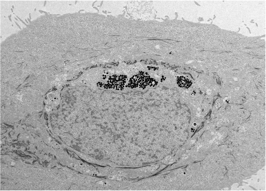
The scientists found that the synthetic nanoparticles were taken up in tissue culture by keratinocytes, the predominant cell type found in the epidermis, the outer layer of skin. Photo by Yuran Huang and Ying Jones/UC San Diego

Melanin particles are produced naturally in many different sizes and shapes by animals—for iridescent feathers in birds or the pigmented eyes and skin of some reptiles. But scientists have discovered that extracting melanins from natural sources is a difficult and potentially more complex process than producing them synthetically.

Gianneschi and his team discovered two years ago that synthetic melanin-like nanoparticles could be developed in a precisely controllable manner to mimic the performance of natural melanins used in bird feathers.

“We hypothesized that synthetic melanin-like nanoparticles would mimic naturally occurring melanosomes and be taken up by keratinocytes, the predominant cell type found in the epidermis, the outer layer of skin,” said Gianneschi.

In healthy humans, melanin is delivered to keratinocytes in the skin after being excreted as melanosomes from melanocytes.

The UC San Diego scientists prepared melanin-like nanoparticles through the spontaneous oxidation of dopamine—developing biocompatible, synthetic analogues of naturally occurring melanosomes. Then they studied their update, transport, distribution and ultraviolet radiation-protective capabilities in human keratinocytes in tissue culture.

The researchers found that these synthetic nanoparticles were not only taken up and distributed normally, like natural melanosomes, within the keratinocytes, they protected the skin cells from DNA damage due to ultraviolet radiation.

“Considering limitations in the treatment of melanin-defective related diseases and the biocompatibility of these synthetic melanin-like nanoparticles in terms of uptake and degradation, these systems have potential as artificial melanosomes for the development of novel therapies, possibly supplementing the biological functions of natural melanins,” the researchers said in their paper.

The other co-authors of the study were Yuran Huang and Ziying Hu of UC San Diego’s Materials Science and Engineering Program, Yiwen Li and Maria Proetto of the Department of Chemistry and Biochemistry; Xiujun Yue of the Department of Nanoengineering; and Ying Jones of the Electron Microscopy Core Facility.

The UC San Diego Office of Innovation and Commercialization has filed a patent application on the use of polydopamine-based artificial melanins as an intracellular UV-shield. Companies interested in commercializing this invention should contact Skip Cynar at invent@ucsd.edu

Here’s a link to and a citation fro the paper,

Mimicking Melanosomes: Polydopamine Nanoparticles as Artificial Microparasols by
Yuran Huang, Yiwen Li, Ziying Hu, Xiujun Yue, Maria T. Proetto, Ying Jones, and Nathan C. Gianneschi. ACS Cent. Sci., Article ASAP DOI: 10.1021/acscentsci.6b00230 Publication Date (Web): May 18, 2017

Copyright © 2017 American Chemical Society

This is an open access paper,

3D bioprinting: a conference about the latest trends (May 3 – 5, 2017 at the University of British Columbia, Vancouver)

The University of British Columbia’s (UBC) Peter Wall Institute for Advanced Studies (PWIAS) is hosting along with local biotech firm, Aspect Biosystems, a May 3 -5, 2017 international research roundtable known as ‘Printing the Future of Therapeutics in 3D‘.

A May 1, 2017 UBC news release (received via email) offers some insight into the field of bioprinting from one of the roundtable organizers,

This week, global experts will gather [4] at the University of British
Columbia to discuss the latest trends in 3D bioprinting—a technology
used to create living tissues and organs.

In this Q&A, UBC chemical and biological engineering professor
Vikramaditya Yadav [5], who is also with the Regenerative Medicine
Cluster Initiative in B.C., explains how bioprinting could potentially
transform healthcare and drug development, and highlights Canadian
innovations in this field.

WHY IS 3D BIOPRINTING SIGNIFICANT?

Bioprinted tissues or organs could allow scientists to predict
beforehand how a drug will interact within the body. For every
life-saving therapeutic drug that makes its way into our medicine
cabinets, Health Canada blocks the entry of nine drugs because they are
proven unsafe or ineffective. Eliminating poor-quality drug candidates
to reduce development costs—and therefore the cost to consumers—has
never been more urgent.

In Canada alone, nearly 4,500 individuals are waiting to be matched with
organ donors. If and when bioprinters evolve to the point where they can
manufacture implantable organs, the concept of an organ transplant
waiting list would cease to exist. And bioprinted tissues and organs
from a patient’s own healthy cells could potentially reduce the risk
of transplant rejection and related challenges.

HOW IS THIS TECHNOLOGY CURRENTLY BEING USED?

Skin, cartilage and bone, and blood vessels are some of the tissue types
that have been successfully constructed using bioprinting. Two of the
most active players are the Wake Forest Institute for Regenerative
Medicine in North Carolina, which reports that its bioprinters can make
enough replacement skin to cover a burn with 10 times less healthy
tissue than is usually needed, and California-based Organovo, which
makes its kidney and liver tissue commercially available to
pharmaceutical companies for drug testing.

Beyond medicine, bioprinting has already been commercialized to print
meat and artificial leather. It’s been estimated that the global
bioprinting market will hit $2 billion by 2021.

HOW IS CANADA INVOLVED IN THIS FIELD?

Canada is home to some of the most innovative research clusters and
start-up companies in the field. The UBC spin-off Aspect Biosystems [6]
has pioneered a bioprinting paradigm that rapidly prints on-demand
tissues. It has successfully generated tissues found in human lungs.

Many initiatives at Canadian universities are laying strong foundations
for the translation of bioprinting and tissue engineering into
mainstream medical technologies. These include the Regenerative Medicine
Cluster Initiative in B.C., which is headed by UBC, and the University
of Toronto’s Institute of Biomaterials and Biomedical Engineering.

WHAT ETHICAL ISSUES DOES BIOPRINTING CREATE?

There are concerns about the quality of the printed tissues. It’s
important to note that the U.S. Food and Drug Administration and Health
Canada are dedicating entire divisions to regulation of biomanufactured
products and biomedical devices, and the FDA also has a special division
that focuses on regulation of additive manufacturing – another name
for 3D printing.

These regulatory bodies have an impressive track record that should
assuage concerns about the marketing of substandard tissue. But cost and
pricing are arguably much more complex issues.

Some ethicists have also raised questions about whether society is not
too far away from creating Replicants, à la _Blade Runner_. The idea is
fascinating, scary and ethically grey. In theory, if one could replace
the extracellular matrix of bones and muscles with a stronger substitute
and use cells that are viable for longer, it is not too far-fetched to
create bones or muscles that are stronger and more durable than their
natural counterparts.

WILL DOCTORS BE PRINTING REPLACEMENT BODY PARTS IN 20 YEARS’ TIME?

This is still some way off. Optimistically, patients could see the
technology in certain clinical environments within the next decade.
However, some technical challenges must be addressed in order for this
to occur, beginning with faithful replication of the correct 3D
architecture and vascularity of tissues and organs. The bioprinters
themselves need to be improved in order to increase cell viability after
printing.

These developments are happening as we speak. Regulation, though, will
be the biggest challenge for the field in the coming years.

There are some events open to the public (from the international research roundtable homepage),

OPEN EVENTS

You’re invited to attend the open events associated with Printing the Future of Therapeutics in 3D.

Café Scientifique

Thursday, May 4, 2017
Telus World of Science
5:30 – 8:00pm [all tickets have been claimed as of May 2, 2017 at 3:15 pm PT]

3D Bioprinting: Shaping the Future of Health

Imagine a world where drugs are developed without the use of animals, where doctors know how a patient will react to a drug before prescribing it and where patients can have a replacement organ 3D-printed using their own cells, without dealing with long donor waiting lists or organ rejection. 3D bioprinting could enable this world. Join us for lively discussion and dessert as experts in the field discuss the exciting potential of 3D bioprinting and the ethical issues raised when you can print human tissues on demand. This is also a rare opportunity to see a bioprinter live in action!

Open Session

Friday, May 5, 2017
Peter Wall Institute for Advanced Studies
2:00 – 7:00pm

A Scientific Discussion on the Promise of 3D Bioprinting

The medical industry is struggling to keep our ageing population healthy. Developing effective and safe drugs is too expensive and time-consuming to continue unchanged. We cannot meet the current demand for transplant organs, and people are dying on the donor waiting list every day.  We invite you to join an open session where four of the most influential academic and industry professionals in the field discuss how 3D bioprinting is being used to shape the future of health and what ethical challenges may be involved if you are able to print your own organs.

ROUNDTABLE INFORMATION

The University of British Columbia and the award-winning bioprinting company Aspect Biosystems, are proud to be co-organizing the first “Printing the Future of Therapeutics in 3D” International Research Roundtable. This event will congregate global leaders in tissue engineering research and pharmaceutical industry experts to discuss the rapidly emerging and potentially game-changing technology of 3D-printing living human tissues (bioprinting). The goals are to:

Highlight the state-of-the-art in 3D bioprinting research
Ideate on disruptive innovations that will transform bioprinting from a novel research tool to a broadly adopted systematic practice
Formulate an actionable strategy for industry engagement, clinical translation and societal impact
Present in a public forum, key messages to educate and stimulate discussion on the promises of bioprinting technology

The Roundtable will bring together a unique collection of industry experts and academic leaders to define a guiding vision to efficiently deploy bioprinting technology for the discovery and development of new therapeutics. As the novel technology of 3D bioprinting is more broadly adopted, we envision this Roundtable will become a key annual meeting to help guide the development of the technology both in Canada and globally.

We thank you for your involvement in this ground-breaking event and look forward to you all joining us in Vancouver for this unique research roundtable.

Kind Regards,
The Organizing Committee
Christian Naus, Professor, Cellular & Physiological Sciences, UBC
Vikram Yadav, Assistant Professor, Chemical & Biological Engineering, UBC
Tamer Mohamed, CEO, Aspect Biosystems
Sam Wadsworth, CSO, Aspect Biosystems
Natalie Korenic, Business Coordinator, Aspect Biosystems

I’m glad to see this event is taking place—and with public events too! (Wish I’d seen the Café Scientifique announcement earlier when I first checked for tickets  yesterday. I was hoping there’d been some cancellations today.) Finally, for the interested, you can find Aspect Biosystems here.