Tag Archives: smart bandages

Imprinting fibres at the nanometric scale

Switzerland’s École Polytechnique Fédérale de Lausanne (EPFL) announces a discovery in a Jan. 24, 2017 press release (also on EurkeAlert),

Researchers at EPFL have come up with a way of imprinting nanometric patterns on the inside and outside of polymer fibers. These fibers could prove useful in guiding nerve regeneration and producing optical effects, for example, as well as in eventually creating artificial tissue and smart bandages.

Researchers at EPFL’s Laboratory of Photonic Materials and Fibre Devices, which is run by Fabien Sorin, have come up with a simple and innovative technique for drawing or imprinting complex, nanometric patterns on hollow polymer fibers. Their work has been published in Advanced Functional Materials.

The potential applications of this breakthrough are numerous. The imprinted designs could be used to impart certain optical effects on a fiber or make it water-resistant. They could also guide stem-cell growth in textured fiber channels or be used to break down the fiber at a specific location and point in time in order to release drugs as part of a smart bandage.

Stretching the fiber like molten plastic

To make their nanometric imprints, the researchers began with a technique called thermal drawing, which is the technique used to fabricate optical fibers. Thermal drawing involves engraving or imprinting millimeter-sized patterns on a preform, which is a macroscopic version of the target fiber. The imprinted preform is heated to change its viscosity, stretched like molten plastic into a long, thin fiber and then allowed to harden again. Stretching causes the pattern to shrink while maintaining its proportions and position. Yet this method has a major shortcoming: the pattern does not remain intact below the micrometer scale. “When the fiber is stretched, the surface tension of the structured polymer causes the pattern to deform and even disappear below a certain size, around several microns,” said Sorin.

To avoid this problem, the EPFL researchers came up with the idea of sandwiching the imprinted preform in a sacrificial polymer [emphasis mine]. This polymer protects the pattern during stretching by reducing the surface tension. It is discarded once the stretching is complete. Thanks to this trick, the researchers are able to apply tiny and highly complex patterns to various types of fibers. “We have achieved 300-nanometer patterns, but we could easily make them as small as several tens of nanometers,” said Sorin. This is the first time that such minute and highly complex patterns have been imprinted on flexible fiber on a very large scale. “This technique enables to achieve textures with feature sizes two order of magnitude smaller than previously reported,” said Sorin. “It could be applied to kilometers of fibers at a highly reasonable cost.”

To highlight potential applications of their achievement, the researchers teamed up with the Bertarelli Foundation Chair in Neuroprosthetic Technology, led by Stéphanie Lacour. Working in vitro, they were able to use their fibers to guide neurites from a spinal ganglion (on the spinal nerve). This was an encouraging step toward using these fibers to help nerves regenerate or to create artificial tissue.

This development could have implications in many other fields besides biology. “Fibers that are rendered water-resistant by the pattern could be used to make clothes. Or we could give the fibers special optical effects for design or detection purposes. There is also much to be done with the many new microfluidic systems out there,” said Sorin. The next step for the researchers will be to join forces with other EPFL labs on initiatives such as studying in vivo nerve regeneration. All this, thanks to the wonder of imprinted polymer fibers.

I like the term “sacrificial polymer.”

Here’s a link to and a citation for the paper,

Controlled Sub-Micrometer Hierarchical Textures Engineered in Polymeric Fibers and Microchannels via Thermal Drawing by Tung Nguyen-Dang, Alba C. de Luca, Wei Yan, Yunpeng Qu, Alexis G. Page, Marco Volpi, Tapajyoti Das Gupta, Stéphanie P. Lacour, and Fabien Sorin. Advanced Functional Materials DOI: 10.1002/adfm.201605935 Version of Record online: 24 JAN 2017

© 2017 WILEY-VCH Verlag GmbH & Co. KGaA, Weinheim

This paper is behind a paywall.
Sutures that can gather data wirelessly

Are sutures which gather data hackable? It’s a little early to start thinking about that issue as this seems to be brand new research. A July 18, 2016 news item on ScienceDaily tells more,

For the first time, researchers led by Tufts University engineers have integrated nano-scale sensors, electronics and microfluidics into threads — ranging from simple cotton to sophisticated synthetics — that can be sutured through multiple layers of tissue to gather diagnostic data wirelessly in real time, according to a paper published online July 18 [2016] in Microsystems & Nanoengineering. The research suggests that the thread-based diagnostic platform could be an effective substrate for a new generation of implantable diagnostic devices and smart wearable systems.

A July 18, 2016 Tufts University news release (also on EurekAlert), which originated the news item, provides more detail,

The researchers used a variety of conductive threads that were dipped in physical and chemical sensing compounds and connected to wireless electronic circuitry to create a flexible platform that they sutured into tissue in rats as well as in vitro. The threads collected data on tissue health (e.g. pressure, stress, strain and temperature), pH and glucose levels that can be used to determine such things as how a wound is healing, whether infection is emerging, or whether the body’s chemistry is out of balance. The results were transmitted wirelessly to a cell phone and computer.

The three-dimensional platform is able to conform to complex structures such as organs, wounds or orthopedic implants.

While more study is needed in a number of areas, including investigation of long-term biocompatibility, researchers said initial results raise the possibility of optimizing patient-specific treatments.

“The ability to suture a thread-based diagnostic device intimately in a tissue or organ environment in three dimensions adds a unique feature that is not available with other flexible diagnostic platforms,” said Sameer Sonkusale, Ph.D., corresponding author on the paper and director of the interdisciplinary Nano Lab in the Department of Electrical and Computer Engineering at Tufts School of Engineering. “We think thread-based devices could potentially be used as smart sutures for surgical implants, smart bandages to monitor wound healing, or integrated with textile or fabric as personalized health monitors and point-of-care diagnostics.”

Until now, the structure of substrates for implantable devices has essentially been two-dimensional, limiting their usefulness to flat tissue such as skin, according to the paper. Additionally, the materials in those substrates are expensive and require specialized processing.

Here’s a link to and a citation for the paper,

A toolkit of thread-based microfluidics, sensors, and electronics for 3D tissue embedding for medical diagnostics by Pooria Mostafalu, Mohsen Akbari, Kyle A. Alberti, Qiaobing Xu, Ali Khademhosseini, & Sameer R. Sonkusale. Microsystems & Nanoengineering 2, Article number: 16039 (2016) doi:10.1038/micronano.2016.39 Published online 18 July 2016

This paper is open access.

‘Glow in the dark’, paint-on bandage heals

Somewhat unexpectedly (to me), this research about a ‘smart’ paint-on bandage is being published by The Optical Society of America (OSA). Here’s more about the work from an Oct. 1, 2014 news item on Nanowerk,

Inspired by a desire to help wounded soldiers, an international, multidisciplinary team of researchers led by Assistant Professor Conor L. Evans at the Wellman Center for Photomedicine of Massachusetts General Hospital (MGH) and Harvard Medical School (HMS) has created a paint-on, see-through, “smart” bandage that glows to indicate a wound’s tissue oxygenation concentration. Because oxygen plays a critical role in healing, mapping these levels in severe wounds and burns can help to significantly improve the success of surgeries to restore limbs and physical functions.

An Oct. 1, 2014 OSA news release (also on EurekAlert), which originated the news item, describes the interest in oxygenation in more detail,

“Information about tissue oxygenation is clinically relevant but is often inaccessible due to a lack of accurate or noninvasive measurements,” explained lead author Zongxi Li, an HMS research fellow on Evans’ team.

Now, the “smart” bandage developed by the team provides direct, noninvasive measurement of tissue oxygenation by combining three simple, compact and inexpensive components: a bright sensor molecule with a long phosphorescence lifetime and appropriate dynamic range; a bandage material compatible with the sensor molecule that conforms to the skin’s surface to form an airtight seal; and an imaging device capable of capturing the oxygen-dependent signals from the bandage with high signal-to-noise ratio.

This work is part of the team’s long-term program “to develop a Sensing, Monitoring And Release of Therapeutics (SMART) bandage for improved care of patients with acute or chronic wounds,” says Evans …

The news release goes on to briefly explain the technology,

For starters, the bandage’s not-so-secret key ingredient is phosphors—molecules that absorb light and then emit it via a process known as phosphorescence.

Phosphorescence is encountered by many on a daily basis—ranging from glow-in-the-dark dials on watches to t-shirt lettering. “How brightly our phosphorescent molecules emit light depends on how much oxygen is present,” said Li. “As the concentration of oxygen is reduced, the phosphors glow both longer and more brightly.” To make the bandage simple to interpret, the team also incorporated a green oxygen-insensitive reference dye, so that changes in tissue oxygenation are displayed as a green-to-red colormap.

The bandage is applied by “painting” it onto the skin’s surface as a viscous liquid, which dries to a solid thin film within a minute. Once the first layer has dried, a transparent barrier layer is then applied atop it to protect the film and slow the rate of oxygen exchange between the bandage and room air—making the bandage sensitive to the oxygen within tissue.

The final piece involves a camera-based readout device, which performs two functions: it provides a burst of excitation light that triggers the emission of the phosphors inside the bandage, and then it records the phosphors’ emission. “Depending on the camera’s configuration, we can measure either the brightness or color of the emitted light across the bandage or the change in brightness over time,” Li said. “Both of these signals can be used to create an oxygenation map.”  The emitted light from the bandage is bright enough that it can be acquired using a regular camera or smartphone—opening the possibility to a portable, field-ready device.

There are some immediate applications, as well as, plans for research that will yield applications (from the news release),

Immediate applications for the oxygen-sensing bandage include monitoring patients with a risk of developing ischemic (restricted blood supply) conditions, postoperative monitoring of skin grafts or flaps, and burn-depth determination as a guide for surgical debridement—the removal of dead or damaged tissue from the body.

“The need for a reliable, accurate and easy-to-use method of rapid assessment of blood flow to the skin for patients remains a clinical necessity,” said co-author Samuel Lin, an HMS associate professor of surgery at Beth Israel Deaconess Medical Center. “Plastic surgeons continuously monitor the state of blood flow to the skin, so the liquid-bandage oxygenation sensor is an exciting step toward improving patient care within the realm of vascular blood flow examination of the skin.”

What’s the next step for the bandage? “We’re developing brighter sensor molecules to improve the bandage’s oxygen sensing efficiency,” said Emmanuel Roussakis, another research fellow in Evans’ laboratory and co-author, who is leading the sensor development effort.  The team’s laboratory research will also focus on expanding the sensing capability of the bandage to other treatment-related parameters—such as pH, bacterial load, oxidative states and specific disease markers—and incorporating an on-demand drug release capacity.

“In the future, our goal for the bandage is to incorporate therapeutic release capabilities that allow for on-demand drug administration at a desired location,” says Evans. “It allows for the visual assessment of the wound bed, so treatment-related wound parameters are readily accessible without the need for bandage removal—preventing unnecessary wound disruption and reducing the chance for bacterial infection.”

Should you be interested, the researchers are looking for industry partners,

Beyond the lab, the team’s aim is to move this technology from the bench to the bedside, so they are actively searching for industry partners. They acknowledge research support from the Military Medical Photonics Program from the U.S. Department of Defense, and National Institutes of Health.

Here’s a link to and a citation for the paper,

Non-invasive transdermal two-dimensional mapping of cutaneous oxygenation with a rapid-drying liquid bandage by Zongxi Li, Emmanuel Roussakis, Pieter G. L. Koolen, Ahmed M. S. Ibrahim, Kuylhee Kim, Lloyd F. Rose, Jesse Wu, Alexander J. Nichols, Yunjung Baek, Reginald Birngruber, Gabriela Apiou-Sbirlea, Robina Matyal, Thomas Huang, Rodney Chan, Samuel J. Lin, and Conor L. Evans. Biomedical Optics Express, Vol. 5, Issue 11, pp. 3748-3764 (2014) http://dx.doi.org/10.1364/BOE.5.003748

This article is open access.

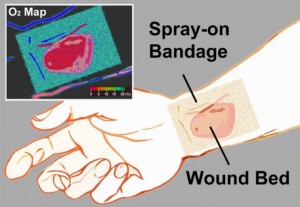
The researcher’s have provided an illustration of the bandage,

Caption: The transparent liquid bandage displays a quantitative, oxygenation-sensitive colormap that can be easily acquired using a simple camera or smartphone. Credit: Li/Wellman Center for Photomedicine.

Caption: The transparent liquid bandage displays a quantitative, oxygenation-sensitive colormap that can be easily acquired using a simple camera or smartphone. Credit: Li/Wellman Center for Photomedicine.