Tag Archives: Srikanth Ramaswamy

Guide for memristive hardware design

An August 15 ,2022 news item on ScienceDaily announces a type of guide for memristive hardware design,

They are many times faster than flash memory and require significantly less energy: memristive memory cells could revolutionize the energy efficiency of neuromorphic [brainlike] computers. In these computers, which are modeled on the way the human brain works, memristive cells function like artificial synapses. Numerous groups around the world are working on the use of corresponding neuromorphic circuits — but often with a lack of understanding of how they work and with faulty models. Jülich researchers have now summarized the physical principles and models in a comprehensive review article in the renowned journal Advances in Physics.

An August 15, 2022 Forschungszentrum Juelich press release (also on EurekAlert), which originated the news item, describes two papers designed to help researchers better understand and design memristive hardware,

Certain tasks – such as recognizing patterns and language – are performed highly efficiently by a human brain, requiring only about one ten-thousandth of the energy of a conventional, so-called “von Neumann” computer. One of the reasons lies in the structural differences: In a von Neumann architecture, there is a clear separation between memory and processor, which requires constant moving of large amounts of data. This is time and energy consuming – the so-called von Neumann bottleneck. In the brain, the computational operation takes place directly in the data memory and the biological synapses perform the tasks of memory and processor at the same time.

In Jülich, scientists have been working for more than 15 years on special data storage devices and components that can have similar properties to the synapses in the human brain. So-called memristive memory devices, also known as memristors, are considered to be extremely fast, energy-saving and can be miniaturized very well down to the nanometer range. The functioning of memristive cells is based on a very special effect: Their electrical resistance is not constant, but can be changed and reset again by applying an external voltage, theoretically continuously. The change in resistance is controlled by the movement of oxygen ions. If these move out of the semiconducting metal oxide layer, the material becomes more conductive and the electrical resistance drops. This change in resistance can be used to store information.

The processes that can occur in cells are very complex and vary depending on the material system. Three researchers from the Jülich Peter Grünberg Institute – Prof. Regina Dittmann, Dr. Stephan Menzel, and Prof. Rainer Waser – have therefore compiled their research results in a detailed review article, “Nanoionic memristive phenomena in metal oxides: the valence change mechanism”. They explain in detail the various physical and chemical effects in memristors and shed light on the influence of these effects on the switching properties of memristive cells and their reliability.

“If you look at current research activities in the field of neuromorphic memristor circuits, they are often based on empirical approaches to material optimization,” said Rainer Waser, director at the Peter Grünberg Institute. “Our goal with our review article is to give researchers something to work with in order to enable insight-driven material optimization.” The team of authors worked on the approximately 200-page article for ten years and naturally had to keep incorporating advances in knowledge.

“The analogous functioning of memristive cells required for their use as artificial synapses is not the normal case. Usually, there are sudden jumps in resistance, generated by the mutual amplification of ionic motion and Joule heat,” explains Regina Dittmann of the Peter Grünberg Institute. “In our review article, we provide researchers with the necessary understanding of how to change the dynamics of the cells to enable an analog operating mode.”

“You see time and again that groups simulate their memristor circuits with models that don’t take into account high dynamics of the cells at all. These circuits will never work.” said Stephan Menzel, who leads modeling activities at the Peter Grünberg Institute and has developed powerful compact models that are now in the public domain (www.emrl.de/jart.html). “In our review article, we provide the basics that are extremely helpful for a correct use of our compact models.”

Roadmap neuromorphic computing

The “Roadmap of Neuromorphic Computing and Engineering”, which was published in May 2022, shows how neuromorphic computing can help to reduce the enormous energy consumption of IT globally. In it, researchers from the Peter Grünberg Institute (PGI-7), together with leading experts in the field, have compiled the various technological possibilities, computational approaches, learning algorithms and fields of application. 

According to the study, applications in the field of artificial intelligence, such as pattern recognition or speech recognition, are likely to benefit in a special way from the use of neuromorphic hardware. This is because they are based – much more so than classical numerical computing operations – on the shifting of large amounts of data. Memristive cells make it possible to process these gigantic data sets directly in memory without transporting them back and forth between processor and memory. This could reduce the energy efficiency of artificial neural networks by orders of magnitude.

Memristive cells can also be interconnected to form high-density matrices that enable neural networks to learn locally. This so-called edge computing thus shifts computations from the data center to the factory floor, the vehicle, or the home of people in need of care. Thus, monitoring and controlling processes or initiating rescue measures can be done without sending data via a cloud. “This achieves two things at the same time: you save energy, and at the same time, personal data and data relevant to security remain on site,” says Prof. Dittmann, who played a key role in creating the roadmap as editor.

Here’s a link to and a citation for the ‘roadmap’,

2022 roadmap on neuromorphic computing and engineering by Dennis V Christensen, Regina Dittmann, Bernabe Linares-Barranco, Abu Sebastian, Manuel Le Gallo, Andrea Redaelli, Stefan Slesazeck, Thomas Mikolajick, Sabina Spiga, Stephan Menzel, Ilia Valov, Gianluca Milano, Carlo Ricciardi, Shi-Jun Liang, Feng Miao, Mario Lanza, Tyler J Quill, Scott T Keene, Alberto Salleo, Julie Grollier, Danijela Marković, Alice Mizrahi, Peng Yao, J Joshua Yang, Giacomo Indiveri, John Paul Strachan, Suman Datta, Elisa Vianello, Alexandre Valentian, Johannes Feldmann, Xuan Li, Wolfram H P Pernice, Harish Bhaskaran, Steve Furber, Emre Neftci, Franz Scherr, Wolfgang Maass, Srikanth Ramaswamy, Jonathan Tapson, Priyadarshini Panda, Youngeun Kim, Gouhei Tanaka, Simon Thorpe, Chiara Bartolozzi, Thomas A Cleland, Christoph Posch, ShihChii Liu, Gabriella Panuccio, Mufti Mahmud, Arnab Neelim Mazumder, Morteza Hosseini, Tinoosh Mohsenin, Elisa Donati, Silvia Tolu, Roberto Galeazzi, Martin Ejsing Christensen, Sune Holm, Daniele Ielmini and N Pryds. Neuromorphic Computing and Engineering , Volume 2, Number 2 DOI: 10.1088/2634-4386/ac4a83 20 May 2022 • © 2022 The Author(s)

This paper is open access.

Here’s the most recent paper,

Nanoionic memristive phenomena in metal oxides: the valence change mechanism by Regina Dittmann, Stephan Menzel & Rainer Waser. Advances in Physics
Volume 70, 2021 – Issue 2 Pages 155-349 DOI: https://doi.org/10.1080/00018732.2022.2084006 Published online: 06 Aug 2022

This paper is behind a paywall.
Blue Brain Project builds a digital piece of brain

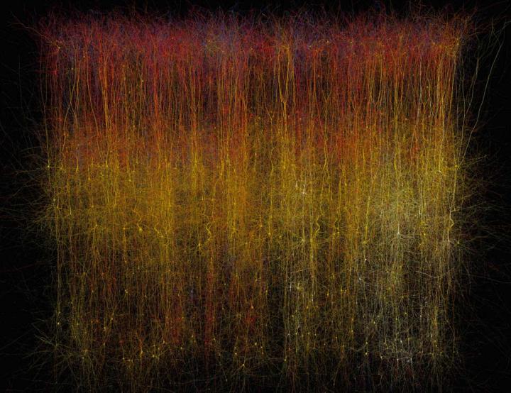
Caption: This is a photo of a virtual brain slice. Credit: Makram et al./Cell 2015

Caption: This is a photo of a virtual brain slice. Credit: Makram et al./Cell 2015

Here’s more *about this virtual brain slice* from an Oct. 8, 2015 Cell (magazine) news release on EurekAlert,

If you want to learn how something works, one strategy is to take it apart and put it back together again [also known as reverse engineering]. For 10 years, a global initiative called the Blue Brain Project–hosted at the Ecole Polytechnique Federale de Lausanne (EPFL)–has been attempting to do this digitally with a section of juvenile rat brain. The project presents a first draft of this reconstruction, which contains over 31,000 neurons, 55 layers of cells, and 207 different neuron subtypes, on October 8 [2015] in Cell.

Heroic efforts are currently being made to define all the different types of neurons in the brain, to measure their electrical firing properties, and to map out the circuits that connect them to one another. These painstaking efforts are giving us a glimpse into the building blocks and logic of brain wiring. However, getting a full, high-resolution picture of all the features and activity of the neurons within a brain region and the circuit-level behaviors of these neurons is a major challenge.

Henry Markram and colleagues have taken an engineering approach to this question by digitally reconstructing a slice of the neocortex, an area of the brain that has benefitted from extensive characterization. Using this wealth of data, they built a virtual brain slice representing the different neuron types present in this region and the key features controlling their firing and, most notably, modeling their connectivity, including nearly 40 million synapses and 2,000 connections between each brain cell type.

“The reconstruction required an enormous number of experiments,” says Markram, of the EPFL. “It paves the way for predicting the location, numbers, and even the amount of ion currents flowing through all 40 million synapses.”

Once the reconstruction was complete, the investigators used powerful supercomputers to simulate the behavior of neurons under different conditions. Remarkably, the researchers found that, by slightly adjusting just one parameter, the level of calcium ions, they could produce broader patterns of circuit-level activity that could not be predicted based on features of the individual neurons. For instance, slow synchronous waves of neuronal activity, which have been observed in the brain during sleep, were triggered in their simulations, suggesting that neural circuits may be able to switch into different “states” that could underlie important behaviors.

“An analogy would be a computer processor that can reconfigure to focus on certain tasks,” Markram says. “The experiments suggest the existence of a spectrum of states, so this raises new types of questions, such as ‘what if you’re stuck in the wrong state?'” For instance, Markram suggests that the findings may open up new avenues for explaining how initiating the fight-or-flight response through the adrenocorticotropic hormone yields tunnel vision and aggression.

The Blue Brain Project researchers plan to continue exploring the state-dependent computational theory while improving the model they’ve built. All of the results to date are now freely available to the scientific community at https://bbp.epfl.ch/nmc-portal.

An Oct. 8, 2015 Hebrew University of Jerusalem press release on the Canadian Friends of the Hebrew University of Jerusalem website provides more detail,

Published by the renowned journal Cell, the paper is the result of a massive effort by 82 scientists and engineers at EPFL and at institutions in Israel, Spain, Hungary, USA, China, Sweden, and the UK. It represents the culmination of 20 years of biological experimentation that generated the core dataset, and 10 years of computational science work that developed the algorithms and built the software ecosystem required to digitally reconstruct and simulate the tissue.

The Hebrew University of Jerusalem’s Prof. Idan Segev, a senior author of the research paper, said: “With the Blue Brain Project, we are creating a digital reconstruction of the brain and using supercomputer simulations of its electrical behavior to reveal a variety of brain states. This allows us to examine brain phenomena within a purely digital environment and conduct experiments previously only possible using biological tissue. The insights we gather from these experiments will help us to understand normal and abnormal brain states, and in the future may have the potential to help us develop new avenues for treating brain disorders.”

Segev, a member of the Hebrew University’s Edmond and Lily Safra Center for Brain Sciences and director of the university’s Department of Neurobiology, sees the paper as building on the pioneering work of the Spanish anatomist Ramon y Cajal from more than 100 years ago: “Ramon y Cajal began drawing every type of neuron in the brain by hand. He even drew in arrows to describe how he thought the information was flowing from one neuron to the next. Today, we are doing what Cajal would be doing with the tools of the day: building a digital representation of the neurons and synapses, and simulating the flow of information between neurons on supercomputers. Furthermore, the digitization of the tissue is open to the community and allows the data and the models to be preserved and reused for future generations.”

While a long way from digitizing the whole brain, the study demonstrates that it is feasible to digitally reconstruct and simulate brain tissue, and most importantly, to reveal novel insights into the brain’s functioning. Simulating the emergent electrical behavior of this virtual tissue on supercomputers reproduced a range of previous observations made in experiments on the brain, validating its biological accuracy and providing new insights into the functioning of the neocortex. This is a first step and a significant contribution to Europe’s Human Brain Project, which Henry Markram founded, and where EPFL is the coordinating partner.

Cell has made a video abstract available (it can be found with the Hebrew University of Jerusalem press release)

Here’s a link to and a citation for the paper,

Reconstruction and Simulation of Neocortical Microcircuitry by Henry Markram, Eilif Muller, Srikanth Ramaswamy, Michael W. Reimann, Marwan Abdellah, Carlos Aguado Sanchez, Anastasia Ailamaki, Lidia Alonso-Nanclares, Nicolas Antille, Selim Arsever, Guy Antoine Atenekeng Kahou, Thomas K. Berger, Ahmet Bilgili, Nenad Buncic, Athanassia Chalimourda, Giuseppe Chindemi, Jean-Denis Courcol, Fabien Delalondre, Vincent Delattre, Shaul Druckmann, Raphael Dumusc, James Dynes, Stefan Eilemann, Eyal Gal, Michael Emiel Gevaert, Jean-Pierre Ghobril, Albert Gidon, Joe W. Graham, Anirudh Gupta, Valentin Haenel, Etay Hay, Thomas Heinis, Juan B. Hernando, Michael Hines, Lida Kanari, Daniel Keller, John Kenyon, Georges Khazen, Yihwa Kim, James G. King, Zoltan Kisvarday, Pramod Kumbhar, Sébastien Lasserre, Jean-Vincent Le Bé, Bruno R.C. Magalhães, Angel Merchán-Pérez, Julie Meystre, Benjamin Roy Morrice, Jeffrey Muller, Alberto Muñoz-Céspedes, Shruti Muralidhar, Keerthan Muthurasa, Daniel Nachbaur, Taylor H. Newton, Max Nolte, Aleksandr Ovcharenko, Juan Palacios, Luis Pastor, Rodrigo Perin, Rajnish Ranjan, Imad Riachi, José-Rodrigo Rodríguez, Juan Luis Riquelme, Christian Rössert, Konstantinos Sfyrakis, Ying Shi, Julian C. Shillcock, Gilad Silberberg, Ricardo Silva, Farhan Tauheed, Martin Telefont, Maria Toledo-Rodriguez, Thomas Tränkler, Werner Van Geit, Jafet Villafranca Díaz, Richard Walker, Yun Wang, Stefano M. Zaninetta, Javier DeFelipe, Sean L. Hill, Idan Segev, Felix Schürmann. Cell, Volume 163, Issue 2, p456–492, 8 October 2015 DOI: http://dx.doi.org/10.1016/j.cell.2015.09.029

This paper appears to be open access.

My most substantive description of the Blue Brain Project , previous to this, was in a Jan. 29, 2013 posting featuring the European Union’s (EU) Human Brain project and involvement from countries that are not members.

* I edited a redundant lede (That’s a virtual slice of a rat brain.), moved the second sentence to the lede while adding this:  *about this virtual brain slice* on Oct. 16, 2015 at 0955 hours PST.