Tag Archives: Staphylococcus aureus

Immune to CRISPR?

I guess if you’re going to use bacteria as part of your gene editing technology (CRISPR [clustered regularly interspaced short palindromic repeats]/Cas9) then, you might half expect the body’s immune system may have developed some defenses. A Jan. 9, 2018 article by Sarah Zhang for The Atlantic provides some insight into what the new research suggests (Note: Links have been removed),

2018 is supposed to be the year of CRISPR in humans. The first U.S. and European clinical trials that test the gene-editing tool’s ability to treat diseases—such as sickle-cell anemia, beta thalassemia, and a type of inherited blindness—are slated to begin this year.

But the year has begun on a cautionary note. On Friday [January 5, 2018], Stanford researchers posted a preprint (which has not been peer reviewed) to the website biorXiv highlighting a potential obstacle to using CRISPR in humans: Many of us may already be immune to it. That’s because CRISPR actually comes from bacteria that often live on or infect humans, and we have built up immunity to the proteins from these bacteria over our lives.

Not all CRISPR therapies in humans will be doomed. “We don’t think this is the end of the story. This is the start of the story,” says Porteus [Matthew Porteus, a pediatrician and stem-cell researcher at Stanford]. There are likely ways around the problem of immunity to CRISPR proteins, and many of the early clinical trials appear to be designed around this problem.

Porteus and his colleagues focused on two versions of Cas9, the bacterial protein mostly commonly used in CRISPR gene editing. One comes from Staphylococcus aureus, which often harmlessly lives on skin but can sometimes causes staph infections, and another from Streptococcus pyogenes, which causes strep throat but can also become “flesh-eating bacteria” when it spreads to other parts of the body. So yeah, you want your immune system to be on guard against these bacteria.

The human immune system has a couple different ways of recognizing foreign proteins, and the team tested for both. First, they looked to see if people have molecules in their blood called antibodies that can specifically bind to Cas9. Among 34 people they tested, 79 percent had antibodies against the staph Cas9 and 65 percent against the strep Cas9.

The Stanford team only tested for preexisting immunity against Cas9, but anytime you inject a large bacterial protein into the human body, it can provoke an immune response. After all, that’s how the immune system learns to fight off bacteria it’s never seen before. (Preexisting immunity can make the response faster and more robust, though.)

The danger of the immune system turning on a patient’s body hangs over a lot of research into correcting genes. In the late 1990s and 2000s, research into gene therapy was derailed by the death of 18-year-old Jesse Gelsinger, who died from an immune reaction to the virus used to deliver the corrected gene. This is the worst-case scenario that the CRISPR world hopes to avoid.

Here’s a link to and a citation for the preprint,

Identification of Pre-Existing Adaptive Immunity to Cas9 Proteins in Humans by Carsten Trevor Charlesworth, Priyanka S Deshpande, Daniel P Dever, Beruh Dejene, Natalia Gomez-Ospina, Sruthi Mantri, Mara Pavel-Dinu, Joab Camarena, Kenneth I Weinberg, Matthew H Porteus. bioRxiv posted January 5, 2018 doi: https://doi.org/10.1101/243345

This article is a preprint and has not been peer-reviewed …

This preprint (not yet published paper) is open access and open for feedback.

Meanwhile, the year of CRISPR takes off (from a January 10, 2018 American Chemical Society news release on EurekAlert),

This year could be a defining one for CRISPR, the gene editing technique, which has been hailed as an important breakthrough in laboratory research. That’s because the first company-sponsored clinical studies will be conducted to see if it can help treat diseases in humans, according to an article in Chemical & Engineering News (C&EN), the weekly newsmagazine of the American Chemical Society.

C&EN Assistant Editor Ryan Cross reports that a big push is coming from industry, specifically from three companies that are each partly founded by one of the three inventors of the method. They are zeroing in on the blood diseases called sickle-cell anemia and β-thalassemia, mostly because their precise cause is known. In these diseases, hemoglobin doesn’t function properly, leading to severe health issues in some people. Crispr Therapeutics and Intellia Therapeutics plan to test the technique to boost levels of an alternative version of healthy hemoglobin. Editas Medicine, however, will also use CRISPR to correct mutations in the faulty hemoglobin gene. Labs led by university researchers are also joining the mix, starting or continuing clinical trials with the approach in 2018.

Because CRISPR is being used to cut a cell’s DNA and insert a new sequence, concerns have been raised about the potential for accidents. A cut in the wrong place could mean introducing a new mutation that could be benign — or cancerous. But according to proponents of the method, researchers are conducting extensive computer predictions and in vitro tests to help avoid this outcome.

The January 8, 2018 Chemical and Engineering News (C&EN) open access article by Ryan Cross is here.

Finally, if you are interested in how this affects research as it’s being developed, there’s University of British Columbia researcher Rosie Redfield’s January 16, 2018 posting on RRResearch blog,

Thursday’s [January 11, 2018] post described the hypothesis that bacteria might use gene transfer agent particles to inoculate other cells in the population with fragments of phage DNA, and outlined an experiment to test this.  Now I’m realizing that I need to know a lot more about the kind of immunity I should expect to see if this GTA-as-vaccine hypothesis is correct.

That should give you some idea of what I meant by “research as it’s being developed.” Redfield’s blog is not for the mildly interested.

Redfield is well-known internationally as being one of the first to refute research which suggested the existence of an ‘arsenic bacterium’ (see my Dec. 8, 2010 posting: My apologies for arsenic blooper. She’s first mentioned in the second excerpt, second paragraph.) The affair was known online as #arseniclife. There’s a May 27, 2011 essay by Carl Zimmer on Slate titled: The Discovery of Arsenic-Based Twitter: How #arseniclife changed science.
Nanofiber coating for artificial joints and implants

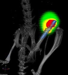
The researchers have a great image to accompany their research, which fit well with Hallowe’en and the Day of the Dead celebrations taking place around the same time as the research was published.

 A titanium implant (blue) without a nanofiber coating in the femur of a mouse. Bacteria are shown in red and responding immune cells in yellow. Credit: Lloyd Miller/Johns Hopkins Medicine

A titanium implant (blue) without a nanofiber coating in the femur of a mouse. Bacteria are shown in red and responding immune cells in yellow.
Credit: Lloyd Miller/Johns Hopkins Medicine

An Oct. 24, 2016 news item on ScienceDaily announces the research on nanofibers,

In a proof-of-concept study with mice, scientists at The Johns Hopkins University show that a novel coating they made with antibiotic-releasing nanofibers has the potential to better prevent at least some serious bacterial infections related to total joint replacement surgery.

An Oct. 24, 2016 Johns Hopkins Medicine news release (also on EurekAlert), provides further details (Note: Links have been removed),

A report on the study, published online the week of Oct. 24 [2016] in Proceedings of the National Academy of Sciences, was conducted on the rodents’ knee joints, but, the researchers say, the technology would have “broad applicability” in the use of orthopaedic prostheses, such as hip and knee total joint replacements, as well pacemakers, stents and other implantable medical devices. In contrast to other coatings in development, the researchers report the new material can release multiple antibiotics in a strategically timed way for an optimal effect.

“We can potentially coat any metallic implant that we put into patients, from prosthetic joints, rods, screws and plates to pacemakers, implantable defibrillators and dental hardware,” says co-senior study author Lloyd S. Miller, M.D., Ph.D., an associate professor of dermatology and orthopaedic surgery at the Johns Hopkins University School of Medicine.

Surgeons and biomedical engineers have for years looked for better ways —including antibiotic coatings — to reduce the risk of infections that are a known complication of implanting artificial hip, knee and shoulder joints.

Every year in the U.S., an estimated 1 to 2 percent of the more than 1 million hip and knee replacement surgeries are followed by infections linked to the formation of biofilms — layers of bacteria that adhere to a surface, forming a dense, impenetrable matrix of proteins, sugars and DNA. Immediately after surgery, an acute infection causes swelling and redness that can often be treated with intravenous antibiotics. But in some people, low-grade chronic infections can last for months, causing bone loss that leads to implant loosening and ultimately failure of the new prosthesis. These infections are very difficult to treat and, in many cases of chronic infection, prostheses must be removed and patients placed on long courses of antibiotics before a new prosthesis can be implanted. The cost per patient often exceeds $100,000 to treat a biofilm-associated prosthesis infection, Miller says.

Major downsides to existing options for local antibiotic delivery, such as antibiotic-loaded cement, beads, spacers or powder, during the implantation of medical devices are that they can typically only deliver one antibiotic at a time and the release rate is not well-controlled. To develop a better approach that addresses those problems, Miller teamed up with Hai-Quan Mao, Ph.D., a professor of materials science and engineering at the Johns Hopkins University Whiting School of Engineering, and a member of the Institute for NanoBioTechnology, Whitaker Biomedical Engineering Institute and Translational Tissue Engineering Center.

Over three years, the team focused on designing a thin, biodegradable plastic coating that could release multiple antibiotics at desired rates. This coating is composed of a nanofiber mesh embedded in a thin film; both components are made of polymers used for degradable sutures.

To test the technology’s ability to prevent infection, the researchers loaded the nanofiber coating with the antibiotic rifampin in combination with one of three other antibiotics: vancomycin, daptomycin or linezolid. “Rifampin has excellent anti-biofilm activity but cannot be used alone because bacteria would rapidly develop resistance,” says Miller. The coatings released vancomycin, daptomycin or linezolid for seven to 14 days and rifampin over three to five days. “We were able to deploy two antibiotics against potential infection while ensuring rifampin was never present as a single agent,” Miller says.

The team then used each combination to coat titanium Kirschner wires — a type of pin used in orthopaedic surgery to fix bone in place after wrist fractures — inserted them into the knee joints of anesthetized mice and introduced a strain of Staphylococcus aureus, a bacterium that commonly causes biofilm-associated infections in orthopaedic surgeries. The bacteria were engineered to give off light, allowing the researchers to noninvasively track infection over time.

Miller says that after 14 days of infection in mice that received an antibiotic-free coating on the pins, all of the mice had abundant bacteria in the infected tissue around the knee joint, and 80 percent had bacteria on the surface of the implant. In contrast, after the same time period in mice that received pins with either linezolid-rifampin or daptomycin-rifampin coating, none of the mice had detectable bacteria either on the implants or in the surrounding tissue.

“We were able to completely eradicate infection with this coating,” says Miller. “Most other approaches only decrease the number of bacteria but don’t generally or reliably prevent infections.”

After the two-week test, each of the rodents’ joints and adjacent bones were removed for further study. Miller and Mao found that not only had infection been prevented, but the bone loss often seen near infected joints — which creates the prosthetic loosening in patients — had also been completely avoided in animals that received pins with the antibiotic-loaded coating.

Miller emphasized that further research is needed to test the efficacy and safety of the coating in humans, and in sorting out which patients would best benefit from the coating — people with a previous prosthesis joint infection receiving a new replacement joint, for example.

The polymers they used to generate the nanofiber coating have already been used in many approved devices by the U.S. Food and Drug Administration, such as degradable sutures, bone plates and drug delivery systems.

Here’s a link to and a citation for the paper,

Polymeric nanofiber coating with tunable combinatorial antibiotic delivery prevents biofilm-associated infection in vivo by Alyssa G. Ashbaugh, Xuesong Jiang, Jesse Zheng, Andrew S. Tsai, Woo-Shin Kim, John M. Thompson, Robert J. Miller, Jonathan H. Shahbazian, Yu Wang, Carly A. Dillen, Alvaro A. Ordonez, Yong S. Chang, Sanjay K. Jain, Lynne C. Jones, Robert S. Sterling, Hai-Quan Mao, and Lloyd S. Miller. PNAS [Proceedings of the National Academy of Sciences] 2016 doi: 10.1073/pnas.1613722113 Published ahead of print October 24, 2016

This paper is behind a paywall.

A city of science in Japan: Kawasaki (Kanagawa)

Happily, I’m getting more nanotechnology (for the most part) information from Japan. Given Japan’s prominence in this field of endeavour I’ve long felt FrogHeart has not adequately represented Japanese contributions. Now that I’m receiving English language translations, I hope to better address the situation.

This morning (March 26, 2015), there were two news releases from Kawasaki INnovation Gateway at SKYFRONT (KING SKYFRONT), Coastal Area International Strategy Office, Kawasaki City, Japan in my mailbox. Before getting on to the news releases, here’s a little about  the city of Kawasaki and about its innovation gateway. From the Kawasaki, Kanagawa entry in Wikipedia (Note: Links have been removed),

Kawasaki (川崎市 Kawasaki-shi?) is a city in Kanagawa Prefecture, Japan, located between Tokyo and Yokohama. It is the 9th most populated city in Japan and one of the main cities forming the Greater Tokyo Area and Keihin Industrial Area.

Kawasaki occupies a belt of land stretching about 30 kilometres (19 mi) along the south bank of the Tama River, which divides it from Tokyo. The eastern end of the belt, centered on JR Kawasaki Station, is flat and largely consists of industrial zones and densely built working-class housing, the Western end mountainous and more suburban. The coastline of Tokyo Bay is occupied by vast heavy industrial complexes built on reclaimed land.

There is a 2014 video about Kawasaki’s innovation gateway, which despite its 14 mins. 39 secs. running time I am embedding here. (Caution: They highlight their animal testing facility at some length.)

Now on to the two news releases. The first concerns research on gold nanoparticles that was published in 2014. From a March 26, 2015 Kawasaki INnovation Gateway news release,

Gold nanoparticles size up to cancer treatment

Incorporating gold nanoparticles helps optimise treatment carrier size and stability to improve delivery of cancer treatment to cells.

Treatments that attack cancer cells through the targeted silencing of cancer genes could be developed using small interfering RNA molecules (siRNA). However delivering the siRNA into the cells intact is a challenge as it is readily degraded by enzymes in the blood and small enough to be eliminated from the blood stream by kidney filtration.  Now Kazunori Kataoka at the University of Tokyo and colleagues at Tokyo Institute of Technology have designed a protective treatment delivery vehicle with optimum stability and size for delivering siRNA to cells.

The researchers formed a polymer complex with a single siRNA molecule. The siRNA-loaded complex was then bonded to a 20 nm gold nanoparticle, which thanks to advances in synthesis techniques can be produced with a reliably low size distribution. The resulting nanoarchitecture had the optimum overall size – small enough to infiltrate cells while large enough to accumulate.

In an assay containing heparin – a biological anti-coagulant with a high negative charge density – the complex was found to release the siRNA due to electrostatic interactions. However when the gold nanoparticle was incorporated the complex remained stable. Instead, release of the siRNA from the complex with the gold nanoparticle could be triggered once inside the cell by the presence of glutathione, which is present in high concentrations in intracellular fluid. The glutathione bonded with the gold nanoparticles and the complex, detaching them from each other and leaving the siRNA prone to release.

The researchers further tested their carrier in a subcutaneous tumour model. The authors concluded that the complex bonded to the gold nanoparticle “enabled the efficient tumor accumulation of siRNA and significant in vivo gene silencing effect in the tumor, demonstrating the potential for siRNA-based cancer therapies.”

The news release provides links to the March 2015 newsletter which highlights this research and to the specific article and video,

March 2015 Issue of Kawasaki SkyFront iNewsletter: http://inewsletter-king-skyfront.jp/en/

Contents

Feature video on Professor Kataoka’s research : http://inewsletter-king-skyfront.jp/en/video_feature/vol_3/feature01/

Research highlights: http://inewsletter-king-skyfront.jp/en/research_highlights/vol_3/research01/

Here’s a link to and a citation for the paper,

Precise Engineering of siRNA Delivery Vehicles to Tumors Using Polyion Complexes and Gold Nanoparticles by Hyun Jin Kim, Hiroyasu Takemoto, Yu Yi, Meng Zheng, Yoshinori Maeda, Hiroyuki Chaya, Kotaro Hayashi, Peng Mi, Frederico Pittella, R. James Christie, Kazuko Toh, Yu Matsumoto, Nobuhiro Nishiyama, Kanjiro Miyata, and Kazunori Kataoka. ACS Nano, 2014, 8 (9), pp 8979–8991 DOI: 10.1021/nn502125h Publication Date (Web): August 18, 2014
Copyright © 2014 American Chemical Society

This article is behind a paywall.

The second March 26, 2015 Kawasaki INnovation Gateway news release concerns a DNA chip and food-borne pathogens,

Rapid and efficient DNA chip technology for testing 14 major types of food borne pathogens

Conventional methods for testing food-borne pathogens is based on the cultivation of pathogens, a process that is complicated and time consuming. So there is demand for alternative methods to test for food-borne pathogens that are simpler, quick and applicable to a wide range of potential applications.

Now Toshiba Ltd and Kawasaki City Institute for Public Health have collaborated in the development of a rapid and efficient automatic abbreviated DNA detection technology that can test for 14 major types of food borne pathogens. The so called ‘DNA chip card’ employs electrochemical DNA chips and overcomes the complicated procedures associated with genetic testing of conventional methods. The ‘DNA chip card’ is expected to find applications in hygiene management in food manufacture, pharmaceuticals, and cosmetics.

Details

The so-called automatic abbreviated DNA detection technology ‘DNA chip card’ was developed by Toshiba Ltd and in a collaboration with Kawasaki City Institute for Public Health, used to simultaneously detect 14 different types of food-borne pathogens in less than 90 minutes. The detection sensitivity depends on the target pathogen and has a range of 1E+01~05 cfu/mL.

Notably, such tests would usually take 4-5 days using conventional methods based on pathogen cultivation. Furthermore, in contrast to conventional DNA protocols that require high levels of skill and expertise, the ‘DNA chip card’ only requires the operator to inject nucleic acid, thereby making the procedure easier to use and without specialized operating skills.

Examples of pathogens associated with food poisoning that were tested with the “DNA chip card”

Enterohemorrhagic Escherichia coli

Salmonella

Campylobacter

Vibrio parahaemolyticus

Shigella

Staphylococcus aureus

Enterotoxigenic Escherichia coli

Enteroaggregative Escherichia coli

Enteropathogenic Escherichia coli

Clostridium perfringens

Bacillus cereus

Yersinia

Listeria

Vibrio cholerae

I think 14 is the highest number of tests I’ve seen for one of these chips. This chip is quite an achievement.

One final bit from the news release about the DNA chip provides a brief description of the gateway and something they call King SkyFront,

About KING SKYFRONT

The Kawasaki INnovation Gateway (KING) SKYFRONT is the flagship science and technology innovation hub of Kawasaki City. KING SKYFRONT is a 40 hectare area located in the Tonomachi area of the Keihin Industrial Region that spans Tokyo and Kanagawa Prefecture and Tokyo International Airport (also often referred to as Haneda Airport).

KING SKYFRONT was launched in 2013 as a base for scholars, industrialists and government administrators to work together to devise real life solutions to global issues in the life sciences and environment.

I find this emphasis on the city interesting. It seems that cities are becoming increasingly important and active where science research and development are concerned. Europe seems to have adopted a biannual event wherein a city is declared a European City of Science in conjunction with the EuroScience Open Forum (ESOF) conferences. The first such city was Dublin in 2012 (I believe the Irish came up with the concept themselves) and was later adopted by Copenhagen for 2014. The latest city to embrace the banner will be Manchester in 2016.

MMA (mixed martial arts) and nano silver wound dressings

I had never, ever expected to mention mixed martial arts (MMA) here but that’s one of the delightful aspects of writing about nanotechnology; you never know where it will take you. A March 9, 2015 news item on Azonano describes the wound situation for athletes and a new product,

..

As an MMA Champion athlete, Rich Franklin knows all too well about germs and how easily they spread. During training he dealt with them on a regular basis, but it wasn’t until the first time he had staph, did he realize these infections could cost him a victory. Now, working in a global setting, Franklin trains in locations around the world which leaves him exposed to a plethora of bacteria and fungi. So he teamed up with American Biotech Labs (ABL) to develop Armor Gel, nano silver-based, wound dressing gel that can stay active on the skin for up to seventy-two hours (3 days). Using patented nano silver technology, Armor Gel has been scientifically tested to reduce the levels of bacteria and other pathogens, while forming a protective barrier “armor” over the wound. By shielding the body from external bacterial, the body’s natural healing process can be expedited. Its use is recommended by doctors, trainers, coaches, and athletes alike.

A March 6, 2015 ABL news release on BusinessWire, which originated the news item, provides a little more detail about Armor Gel,

Engineered for today’s modern athletes, Armor Gel is safe, nontoxic and provides a personal first line of defense. Already proven to reduce the levels of MRSA, VRE, pseudomonas aeruginosa, E. coli, A. niger and Candida albicans, Armor Gel is formulated using a unique and patented 24 SilverSol Technology®.

American Biotech Labs (ABL) was started in 2002 as a nano silver biotech company with the goal of creating a more stable and powerful silver technology for consumer products. …

I am providing a link to the product website (neither the link nor this post are endorsements), you can find out more about Armor Gel here.

Armor Gel was announced previously in a Sept. 16, 2014 ABL news release on PR Newswire, At the time no mention was made of Rich Franklin, their MMA athlete,

American Biotech Labs, LLC, is pleased to announce the availability of three new silver hydrogel wound-dressing products.  The new products will allow American Biotech Labs (ABL) to market in the wound-care market focusing on ultimate sports and fitness, spa and health, and animal markets.

The new over-the-counter (OTC) products will have wound-dressing claims for minor cuts, lacerations, abrasions, 1st and 2nd degree burns, and skin irritations.  The products also have pathogen-inhibiting barrier claims against pathogens, such as Staphylococcus aureus, Pseudomonas aeruginosa, Escherichia coli, MRSA and VRE, as well as fungi, such as Candida albicans and Aspergillus niger.  These new gels can provide a barrier that will help protect wounds for 24 to 72 hours.

The new products will be found under the names of Armor Gel™ (for the ultimate sports and fitness market), ASAP OTC™ (for the spa and health markets), and ASAP Pet Shield® (for the animal market).

Along with the release of these new products, ABL has formed a strategic alliance with Stuart Evey, founder and former chairman of ESPN, and Gary Bernstein, marketing executive and professional photographer and film maker.  ABL will utilize these talented individuals to help introduce these revolutionary new products to high-profile organizations in sports, pet stores, fashion and beauty, medical, and direct-marketing areas, etc.

Said Keith Moeller, ABL Director, “We are very grateful to the numerous top scientists, labs and universities that have helped move this amazing, patented, silver technology forward.  We believe that these products have the ability to impact the future of wound management worldwide.”

Note: Any statements released by American Biotech Labs, LLC that are forward looking are made pursuant to the safe harbor provisions of the Private Securities Litigation Reform Act of 1995.  Editors and investors are cautioned that forward looking statements invoke risk and uncertainties that may affect the company’s business prospects and performance.

You can find out more about ABL and its entire product line here.