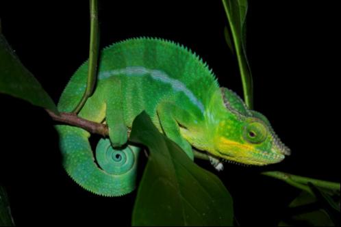
A March 12, 2015 news item on phys.org describes artificial skin inspired by chameleons,
Borrowing a trick from nature, engineers from the University of California at Berkeley have created an incredibly thin, chameleon-like material that can be made to change color—on demand—by simply applying a minute amount of force.
This new material-of-many-colors offers intriguing possibilities for an entirely new class of display technologies, color-shifting camouflage, and sensors that can detect otherwise imperceptible defects in buildings, bridges, and aircraft.
“This is the first time anybody has made a flexible chameleon-like skin that can change color simply by flexing it,” said Connie J. Chang-Hasnain, a member of the Berkeley team and co-author on a paper published today in Optica, The Optical Society’s (OSA) new journal.
A March 12, 2015 OSA news release (also on EurekAlert), which originated the news item, provides more information about this structural color project,
The colors we typically see in paints, fabrics, and other natural substances occur when white, broad spectrum light strikes their surfaces. The unique chemical composition of each surface then absorbs various bands, or wavelengths of light. Those that aren’t absorbed are reflected back, with shorter wavelengths giving objects a blue hue and longer wavelengths appearing redder and the entire rainbow of possible combinations in between. Changing the color of a surface, such as the leaves on the trees in autumn, requires a change in chemical make-up.
Recently, engineers and scientists have been exploring another approach, one that would create designer colors without the use of chemical dyes and pigments. Rather than controlling the chemical composition of a material, it’s possible to control the surface features on the tiniest of scales so they interact and reflect particular wavelengths of light. This type of “structural color” is much less common in nature, but is used by some butterflies and beetles to create a particularly iridescent display of color.
Controlling light with structures rather than traditional optics is not new. In astronomy, for example, evenly spaced slits known as diffraction gratings are routinely used to direct light and spread it into its component colors. Efforts to control color with this technique, however, have proved impractical because the optical losses are simply too great.
The authors of the Optica paper applied a similar principle, though with a radically different design, to achieve the color control they were looking for. In place of slits cut into a film they instead etched rows of ridges onto a single, thin layer of silicon. Rather than spreading the light into a complete rainbow, however, these ridges — or bars — reflect a very specific wavelength of light. By “tuning” the spaces between the bars, it’s possible to select the specific color to be reflected. Unlike the slits in a diffraction grating, however, the silicon bars were extremely efficient and readily reflected the frequency of light they were tuned to.
Fascinatingly, the reflected colors can be selected (from the news release),
Since the spacing, or period, of the bars is the key to controlling the color they reflect, the researchers realized it would be possible to subtly shift the period — and therefore the color — by flexing or bending the material.
“If you have a surface with very precise structures, spaced so they can interact with a specific wavelength of light, you can change its properties and how it interacts with light by changing its dimensions,” said Chang-Hasnain.
Earlier efforts to develop a flexible, color shifting surface fell short on a number of fronts. Metallic surfaces, which are easy to etch, were inefficient, reflecting only a portion of the light they received. Other surfaces were too thick, limiting their applications, or too rigid, preventing them from being flexed with sufficient control.
The Berkeley researchers were able to overcome both these hurdles by forming their grating bars using a semiconductor layer of silicon approximately 120 nanometers thick. Its flexibility was imparted by embedding the silicon bars into a flexible layer of silicone. As the silicone was bent or flexed, the period of the grating spacings responded in kind.
The semiconductor material also allowed the team to create a skin that was incredibly thin, perfectly flat, and easy to manufacture with the desired surface properties. This produces materials that reflect precise and very pure colors and that are highly efficient, reflecting up to 83 percent of the incoming light.
Their initial design, subjected to a change in period of a mere 25 nanometers, created brilliant colors that could be shifted from green to yellow, orange, and red – across a 39-nanometer range of wavelengths. Future designs, the researchers believe, could cover a wider range of colors and reflect light with even greater efficiency.
Here’s a link to and a citation for the paper,
Flexible photonic metastructures for tunable coloration by Li Zhu, Jonas Kapraun, James Ferrara, and Connie J. Chang-Hasnain. Optica, Vol. 2, Issue 3, pp. 255-258 (2015)
http://dx.doi.org/10.1364/OPTICA.2.000255
This paper is open access (for now at least).
Final note: I recently wrote about research into how real chameleons are able to effect colour changes in a March 16, 2015 post.
![AGELESS BRILLIANCE: Although the pigment-derived leaf color of this decades-old specimen of the African perennial Pollia condensata has faded, the fruit still maintains its intense metallic-blue iridescence.COURTESY OF P.J. RUDALL [downloaded from http://www.the-scientist.com/?articles.view/articleNo/34200/title/Color-from-Structure/]](http://www.frogheart.ca/wp-content/uploads/2014/06/AgelessColour_-Pollia_condensata_Herbarium2.jpg)
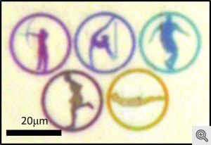
![This shows the diffusion of the neurotransmitter applied to squid skin at upper right, which induces a wave of iridescence traveling to the lower left and progressing from red to blue. Each object in the image is a living cell, 10 microns long; the dark object in the center of each cell is the cell nucleus. [downloaded from http://www.ia.ucsb.edu/pa/display.aspx?pkey=3076]](http://www.frogheart.ca/wp-content/uploads/2013/07/CephalopodCamuuflage.jpeg)
![Credit: Researchers at the University of Georgia [downloaded from http://pubs.acs.org.proxy.lib.sfu.ca/doi/full/10.1021/ja310587c#]](http://www.frogheart.ca/wp-content/uploads/2013/02/nanoEgyptianBlue.gif)
![AGELESS BRILLIANCE: Although the pigment-derived leaf color of this decades-old specimen of the African perennial Pollia condensata has faded, the fruit still maintains its intense metallic-blue iridescence.COURTESY OF P.J. RUDALL [downloaded from http://www.the-scientist.com/?articles.view/articleNo/34200/title/Color-from-Structure/]](http://www.frogheart.ca/wp-content/uploads/2013/02/AgelessColour_-Pollia_condensata_Herbarium2.jpg)