Tag Archives: structural colour

A structural colour solution for energy-saving paint (thank the butterflies)

The UCF-developed plasmonic paint uses nanoscale structural arrangement of colorless materials — aluminum and aluminum oxide — instead of pigments to create colors. Here the plasmonic paint is applied to the wings of metal butterflies, the insect that inspired the research. Credit: University of Central Florida

A March 9, 2023 news item on Nanowerk announces research into multicolour energy-saving coating/paint, so, this is a structural colour story, Note: Links have been removed,

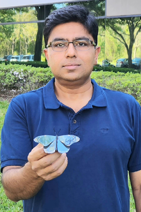
University of Central Florida researcher Debashis Chanda, a professor in UCF’s NanoScience Technology Center, has drawn inspiration from butterflies to create the first environmentally friendly, large-scale and multicolor alternative to pigment-based colorants, which can contribute to energy-saving efforts and help reduce global warming.

A March 8, 2023 University of Central Florida (UCF) news release (also on EurekAlert) by Katrina Cabansay, which originated the news item, provides more context and more details,

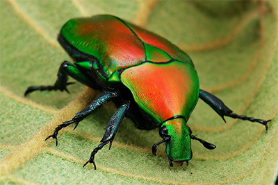
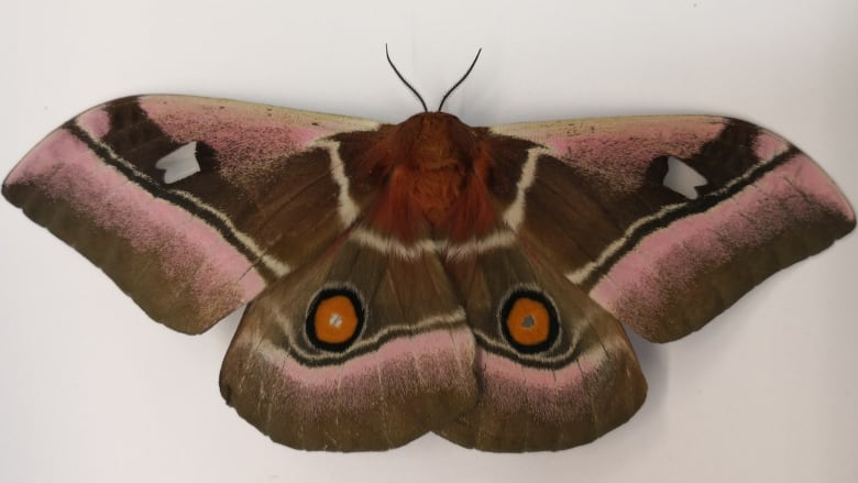
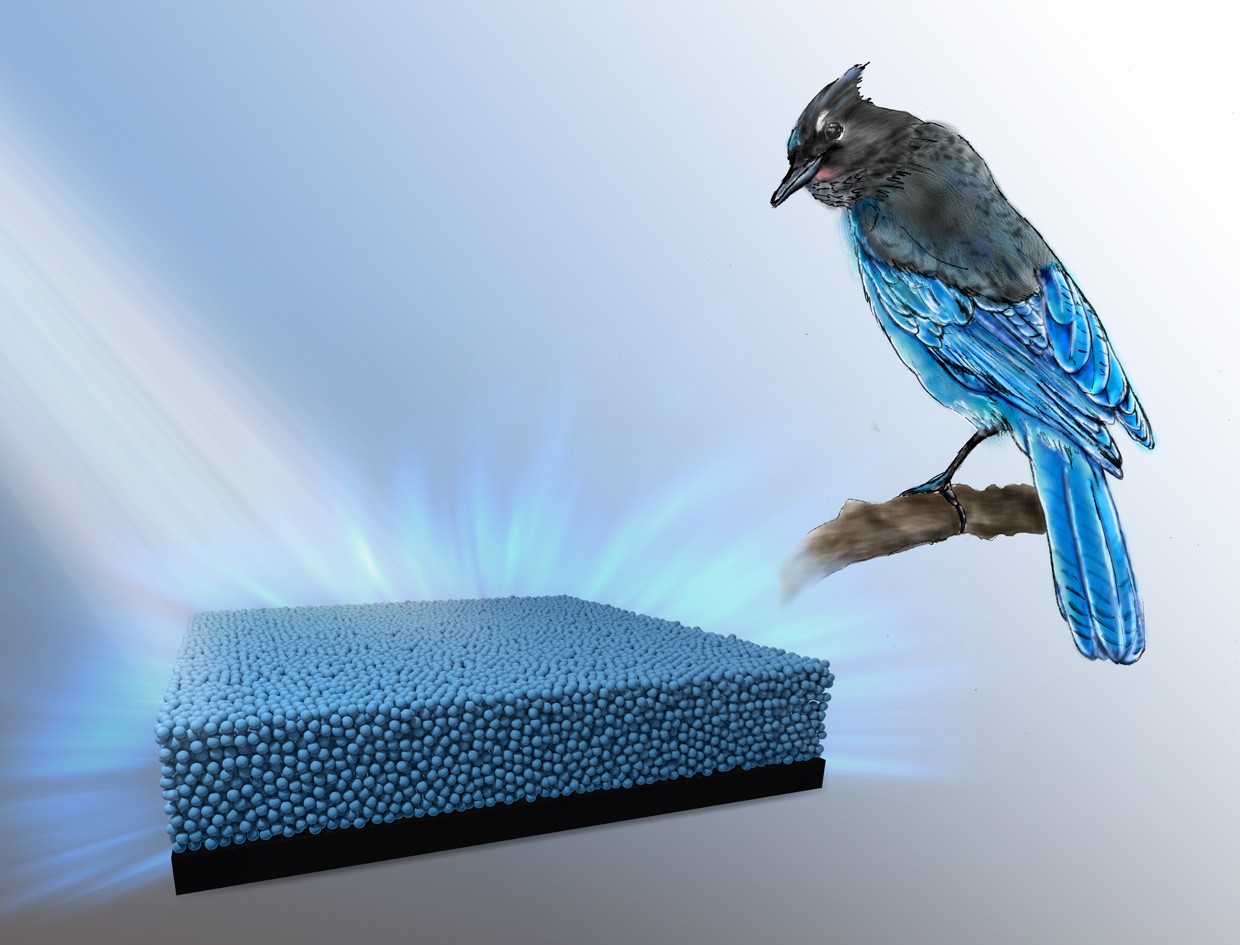
“The range of colors and hues in the natural world are astonishing — from colorful flowers, birds and butterflies to underwater creatures like fish and cephalopods,” Chanda says. “Structural color serves as the primary color-generating mechanism in several extremely vivid species where geometrical arrangement of typically two colorless materials produces all colors. On the other hand, with manmade pigment, new molecules are needed for every color present.”

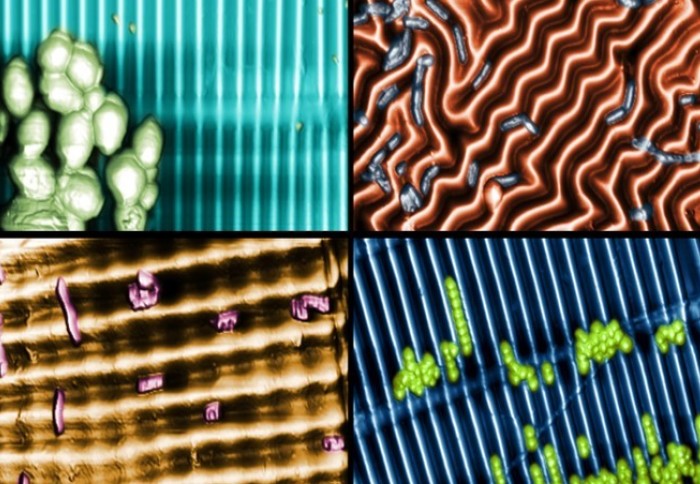
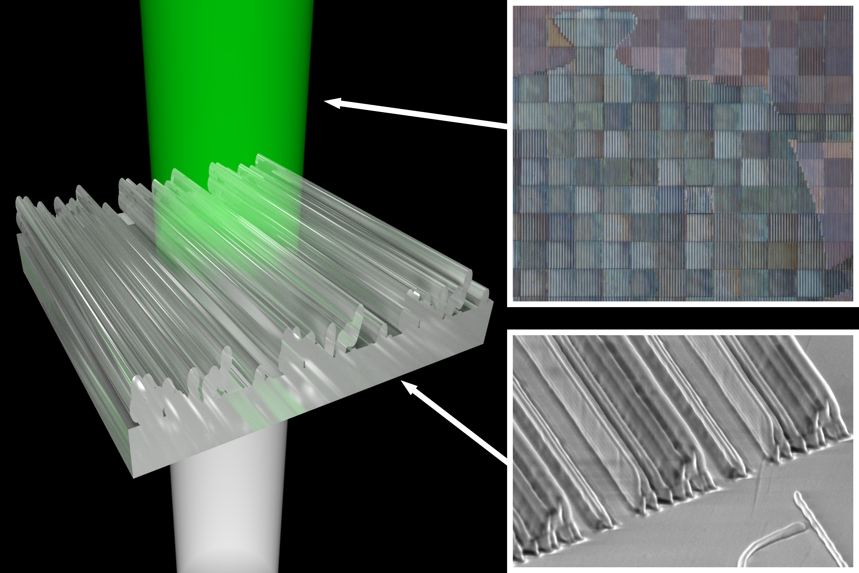
Based on such bio-inspirations, Chanda’s research group innovated a plasmonic paint, which utilizes nanoscale structural arrangement of colorless materials — aluminum and aluminum oxide — instead of pigments to create colors.

While pigment colorants control light absorption based on the electronic property of the pigment material and hence every color needs a new molecule, structural colorants control the way light is reflected, scattered or absorbed based purely on the geometrical arrangement of nanostructures.

Such structural colors are environmentally friendly as they only use metals and oxides, unlike present pigment-based colors that use artificially synthesized molecules.

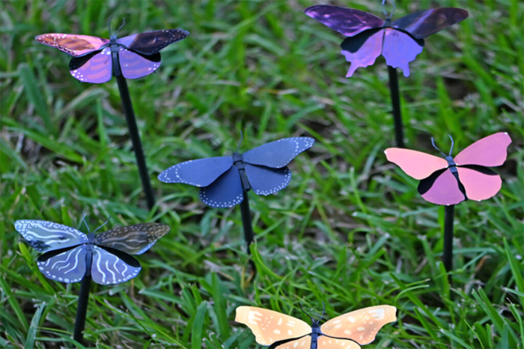
The researchers have combined their structural color flakes with a commercial binder to form long-lasting paints of all colors.

“Normal color fades because pigment loses its ability to absorb photons,” Chanda says. “Here, we’re not limited by that phenomenon. Once we paint something with structural color, it should stay for centuries.”

Additionally, because plasmonic paint reflects the entire infrared spectrum, less heat is absorbed by the paint, resulting in the underneath surface staying 25 to 30 degrees Fahrenheit cooler than it would if it were covered with standard commercial paint, the researcher says.

“Over 10% of total electricity in the U.S. goes toward air conditioner usage,” Chanda says. “The temperature difference plasmonic paint promises would lead to significant energy savings. Using less electricity for cooling would also cut down carbon dioxide emissions, lessening global warming.”

Plasmonic paint is also extremely lightweight, the researcher says.

This is due to the paint’s large area-to-thickness ratio, with full coloration achieved at a paint thickness of only 150 nanometers, making it the lightest paint in the world, Chanda says.

The paint is so lightweight that only about 3 pounds of plasmonic paint could cover a Boeing 747, which normally requires more than 1,000 pounds of conventional paint, he says.

Chanda says his interest in structural color stems from the vibrancy of butterflies.

“As a kid, I always wanted to build a butterfly,” he says. “Color draws my interest.”

Future Research

Chanda says the next steps of the project include further exploration of the paint’s energy-saving aspects to improve its viability as commercial paint.

“The conventional pigment paint is made in big facilities where they can make hundreds of gallons of paint,” he says. “At this moment, unless we go through the scale-up process, it is still expensive to produce at an academic lab.”

“We need to bring something different like, non-toxicity, cooling effect, ultralight weight, to the table that other conventional paints can’t.” Chanda says.

Licensing Opportunity

For more information about licensing this technology, please visit the Inorganic Paint Pigment for Vivid Plasmonic Color technology sheet.

Researcher’s Credentials

Chanda has joint appointments in UCF’s NanoScience Technology Center, Department of Physics and College of Optics and Photonics. He received his doctorate in photonics from the University of Toronto and worked as a postdoctoral fellow at the University of Illinois at Urbana-Champaign. He joined UCF in Fall 2012.

Here’s a link to and a citation for the paper,

Ultralight plasmonic structural color paint by Pablo Cencillo-Abad, Daniel Franklin, Pamela Mastranzo-Ortega, Javier Sanchez-Mondragon, and Debashis Chanda. Science Advances 8 Mar 2023 Vol 9, Issue 10 DOI: 10.1126/sciadv.adf7207

This paper is open access.

Here’s the researcher with one of ‘his butterflies’ (I may be reading a little too much into this but it looks like he’s uncomfortable having his photo taken but game to do it for work that he’s proud of),

Caption: Debashis Chanda, a professor in UCF’s NanoScience Technology Center, drew inspiration from butterflies to create the innovative new plasmonic paint, shown here applied to metal butterfly wings. Credit: University of Central Florida

The reddest red and Schrödinger’s red pixel

Caption: Schrödinger’s red pixel by quasi-bound-states in-the-continuum Credit: 123RF

Science keeps moving. First, there was the June 2022 news and, then, there was the August 2022 news.

A June 8, 2022 Agency for Science, Technology and Research (A*STAR) press release (also on EurekAlert but published June 7, 2022 as an ‘article highlight’) announces more research into structural colour along with some colour theory from Erwin Schrödinger,

The brilliant and often iridescent colours that we see in some species of birds, beetles and butterflies arise from a regular arrangement of nanostructures that scatter selective wavelengths of light more strongly to generate colour. These colours are called structural colours, which usually range from blues to greens, and even magenta. However, vibrant or saturated reds are elusive and notably absent from the structural colour range in both natural and synthetic realms.

To achieve highly saturated reds, the material needs to absorb light from all wavelengths shorter than ~600 nm and reflect the remaining longer wavelengths, doing both as completely as possible. This sharp transition from absorption to reflection was prescribed theoretically by none other than Erwin Schrödinger of quantum theory fame. However, the physics of resonators tell us that high-order optical resonances in blue will also occur as soon as we have a fundamental resonance in red. This combination of blue and red thus results in the magenta observed in nature. It is therefore challenging to achieve the Schrödinger’s red pixel, which would produce the most saturated red in the world. Current nanoantenna-based approaches are insufficient to simultaneously satisfy the above conditions.

Researchers from the Agency for Science, Technology and Research’s (A*STAR) Institute of Materials Research and Engineering (IMRE), National University of Singapore (NUS) and Singapore University of Technology and Design (SUTD) have collaborated to design and realise reds at the ultimate limit of saturation as predicted by theory, where the team worked together on conceptualisation methodology, fabrications, characterisations and simulations. This research was published in Science Advances on 23 February 2022.

The design consists of regularly arranged silicon nanoantennas in the shape of ellipses. These produce possibly the most saturated and brightest reds with ~80% reflectance, exceeding the reds in the standard red, green and blue gamut (sRGB) and other well-known red pigments, e.g. cadmium red .

The nanoantennas support two partially overlapping quasi bound-states-in-the-continuum modes, where the optimal dimensions of the silicon nanoantenna arrays are derived by using a gradient descent algorithm to enable the antennas to achieve sharp spectral edges at red wavelengths. At the same time, high-order modes at blue or green wavelengths are suppressed via engineering the substrate‑induced diffraction channels and the absorption of amorphous silicon.

Potential uses for Schrödinger’s red include developing a polarisation dependent encryption method, with plans to scale up the Schrödinger’s red pixel for applications like functional nanofabrication devices such as optical spectrometers and reflective displays with high colour saturation.

“With this new design that can achieve the most saturated and brightest reds, we can exploit its sensitivity to polarisation and illumination angle on potential applications for information encryption. This proposed concept and design methodology could also be generalised to other Schrödinger’s colour pixels. The highly-saturated red achieved could be potentially scaled up through methods such as deep ultraviolet and nano-imprint lithography, to reach the dimensions of reflective displays based on multilayer film configuration, which could lead to potential applications like compact red filters, highly saturated reflective displays, nonlocal metasurfaces, and miniaturised spectrometers”, said Dr. Dong Zhaogang, Deputy Department Head of Nanofabrication at A*STAR’s IMRE.

“The creation of the record-high saturation and brightness in red opens up possibilities for a plethora of applications related to anti-counterfeiting technologies, high-calibre colour display and more, which were previously perceived as unachievable with structural colour. It showcases a wonderful synergy between conceptual breakthrough, powerful algorithm and advanced nanofabrication”, said Prof. Cheng-Wei Qiu, Dean’s Chair Professor at NUS.

“This work in structural colours goes to show that we can sometimes outdo evolution through clever use of the tools in nanofabrication and accurate optical simulations”, said Prof. Joel Yang, Provost Chair Professor and Associate Professor in Engineering Product Development at SUTD.

Here’s a link to and a citation for the paper,

Schrödinger’s red pixel by quasi-bound-states-in-the-continuum by Zhaogang Dong, Lei Jin, Soroosh Daqiqeh Rezaei, Hao Wang, Yang Chen, Febiana Tjiptoharsono, Jinfa Ho, Sergey Gorelik, Ray Jia Hong Ng, Qifeng Ruan, Cheng-Wei Qiu and Joel K. W. Yang. Science Advances Vol 8, Issue 8 DOI: 10.1126/sciadv.abm4512 Published 23 Feb 2022

This paper is open access.

Math error, colour theory, and perception

An August 10, 2022 news item on phys.org announced a math error made by Erwin Schrödinger and others,

A new study corrects an important error in the 3D mathematical space developed by the Nobel Prize-winning physicist Erwin Schrödinger and others, and used by scientists and industry for more than 100 years to describe how your eye distinguishes one color from another. The research has the potential to boost scientific data visualizations, improve TVs and recalibrate the textile and paint industries.

“The assumed shape of color space requires a paradigm shift,” said Roxana Bujack, a computer scientist with a background in mathematics who creates scientific visualizations at Los Alamos National Laboratory. Bujack is lead author of the paper by a Los Alamos team in the Proceedings of the National Academy of Sciences on the mathematics of color perception.

“Our research shows that the current mathematical model of how the eye perceives color differences is incorrect. That model was suggested by Bernhard Riemann and developed by Hermann von Helmholtz and Erwin Schrödinger—all giants in mathematics and physics—and proving one of them wrong is pretty much the dream of a scientist,” said Bujack.

While the Los Alamos National Laboratory work was published in April 2022 (online) and May 2022 (in print), their news announcement doesn’t seem to have been made until August. I can’t be certain but I believe this should have an impact on the work from A*STAR as that team’s paper cites: E. Schrödinger, Theorie der Pigmente von größter Leuchtkraft. Ann. Phys. 367, 603–622 (1920).

An August 10, 2022 Los Alamos National Laboratory (LANL) news release (also on EurekAlert) provides more information about the discovery,

Modeling human color perception enables automation of image processing, computer graphics and visualization tasks.

“Our original idea was to develop algorithms to automatically improve color maps for data visualization, to make them easier to understand and interpret,” Bujack said. So the team was surprised when they discovered they were the first to determine that the longstanding application of Riemannian geometry, which allows generalizing straight lines to curved surfaces, didn’t work.

To create industry standards, a precise mathematical model of perceived color space is needed. First attempts used Euclidean spaces—the familiar geometry taught in many high schools; more advanced models used Riemannian geometry. The models plot red, green and blue in the 3D space. Those are the colors registered most strongly by light-detecting cones on our retinas, and—not surprisingly—the colors that blend to create all the images on your RGB computer screen.

In the study, which blends psychology, biology and mathematics, Bujack and her colleagues discovered that using Riemannian geometry overestimates the perception of large color differences. That’s because people perceive a big difference in color to be less than the sum you would get if you added up small differences in color that lie between two widely separated shades.

Riemannian geometry cannot account for this effect.

“We didn’t expect this, and we don’t know the exact geometry of this new color space yet,” Bujack said. “We might be able to think of it normally but with an added dampening or weighing function that pulls long distances in, making them shorter. But we can’t prove it yet.”

Here’s a link to and a citation for the paper,

The non-Riemannian nature of perceptual color space by Roxana Bujack, Emily Teti, Jonah Miller, Elektra Caffrey, and Terece L. Turton. Proceedings of the National Academy of Sciences (PNAS) 119 (18) e2119753119 DOI: https://doi.org/10.1073/pnas.2119753119 Published: April 29, 2022

This paper is behind a paywall.

Spiky materials that can pop bacteria?

Bacteria interacting with four different topographies Courtesy: Imperial College London

A February 9, 2022 news item on phys.org describes some bioinspired research that could help cut down on the use of disinfectants,

Researchers have created intricately patterned materials that mimic antimicrobial, adhesive and drag reducing properties found in natural surfaces.

The team from Imperial College London found inspiration in the wavy and spiky surfaces found in insects, including on cicada and dragonfly wings, which ward off bacteria.

They hope the new materials could be used to create self-disinfecting surfaces and offer an alternative to chemically functionalized surfaces and cleaners, which can promote the growth of antibiotic-resistant bacteria.

A February 9, 2022 Imperial College London (ICL) press release by Caroline Brogan, which originated the news item, describes the work in more technical detail,

The tiny waves, which overlap at defined angles to create spikes and ripples, could also help to reduce drag on marine transport by mimicking shark skin, and to enhance the vibrancy of color without needing pigment, by mimicking insects.

Senior author Professor Joao Cabral, of Imperial’s Department of Chemical Engineering, said, “It’s inspiring to see in miniscule detail how the wings and skins of animals help them master their environments. Animals evolved wavy surfaces to kill bacteria, enhance color, and reduce drag while moving through water. We’re borrowing these natural tricks for the very same purposes, using a trick reminiscent of a Fourier wave superposition.”

Spiky structures

Researchers created the new materials by stretching and compressing a thin, soft, sustainable plastic resembling clingfilm to create three-dimensional nano- and microscale wavy patterns, compatible with sustainable and biodegradable polymers. 

The spiky structure was inspired by the way insects and fish have evolved to interact with their environments. The corrugated ripple effect is seen in the wings of cicadas and dragonflies, whose surfaces are made of tiny spikes which pop bacterial cells to keep the insects clean.  

The structure could also be applied to ships to reduce drag and boost efficiency – an application inspired by shark skin, which contains nanoscale horizontal ridges to reduce friction and drag.

Another application is in producing vibrant colours like those seen in the wings of morpho blue butterflies, whose cells are arranged to reflect and bend light into a brilliant blue without using pigment. Known as structural colour, other examples include the blue in peacock feathers, the shells of iridescent beetles, and blue human eyes.

Scaling up waves

To conduct the research, which is published in Physical Review Letters, the researchers studied specimens of cicadas and dragonflies from the Natural History Museum, and sedimentary deposits and rock formations documented by Trinity College Dublin.

They discovered that they could recreate these naturally occurring surface waves by stretching and then relaxing thin polymer skins in precise directions at the nanoscale.

While complex patterns can be fabricated by lithography and other methods, for instance in silicon microchip production, these are generally prohibitively expensive to use over large areas. This new technique, on the other hand, is ready to be scaled up relatively inexpensively if confirmed to be effective and robust. 

Potential applications include self-disinfecting surfaces in hospitals, schools, public transport, and food manufacturing. They could even help keep medical implants clean, which is important as these can host networks of bacterial matter known as biofilms that are notoriously difficult to kill. 

Naturally occurring wave patterns are also seen in the wrinkling of the human brain and fingertips as well as the ripples in sand beds. First author Dr Luca Pellegrino from the Department of Chemical Engineering, said: “The idea is compelling because it is simple: by mimicking the surface waves found in nature, we can create a palette of patterns with important applications. Through this work we can also learn more about the possible origins of these natural forms – a field called morphogenesis.” 

he next focus for the team is to test the effectiveness and robustness of the material in real-world settings, like on bus surfaces. The researchers hope it can contribute to solutions to surface cleanliness that are not reliant on chemical cleaners. To this end, they have been awarded a €5.4million EU HORIZON grant with collaborators ranging from geneticists at KU Leuven to a bus manufacturer to develop sustainable and robust antimicrobial surfaces for high traffic contexts. 

Here’s a link (the press release also has a link) to and a citation for the paper,

Ripple Patterns Spontaneously Emerge through Sequential Wrinkling Interference in Polymer Bilayers by Luca Pellegrino, Annabelle Tan, and João T. Cabral. Phys. Rev. Lett. 128, 058001 Vol. 128, Issue 5 — 4 February 2022 Published online 2 February 2022

This paper is behind a paywall.

This work reminds me of Sharklet, a company that was going to produce materials that mimicked the structure of sharkskin. Apparently, sharks have nanostructures on their skin which prevents bacteria and more from finding a home there.

Tiny Matters: podcast from the American Chemical Society (ACS)

I was expecting a news release mentioning some of the smaller scales at which scientists work, e.g., micro, nano, pico, femto, etc. That was not the case.

From a January 26, 2022 American Chemical Society (ACS) news release on EurekAlert,

The American Chemical Society (ACS) is producing a new, biweekly science podcast called Tiny Matters, which is available wherever you listen to podcasts. Head to ACS’ website or your favorite platform and subscribe.

The first episode drops today. Hosts Sam Jones, Ph.D., and Deboki Chakravarti, Ph.D., chat with experts about the ancient beasts that went extinct 65 million years ago, but whose remains still captivate us today — dinosaurs. Scientists around the world regularly discover new fossils, and that helps piece together the mystery of what dinosaurs and other extinct creatures were like. That information doesn’t just inspire movies like “Jurassic Park”; it also helps researchers predict Earth’s future and could even lead to more sustainable technology.

Tiny Matters is a science podcast about things small in size but big in impact. Every other Wednesday, the hosts will uncover little stuff that makes big stuff possible. Upcoming episodes will find them answering questions such as “How does our brain form memories?”, “Why haven’t we terraformed Mars yet?” and “Why isn’t there a vaccine for HIV?” Tune in!

The American Chemical Society (ACS) is a nonprofit organization chartered by the U.S. Congress. ACS’ mission is to advance the broader chemistry enterprise and its practitioners for the benefit of Earth and all its people. The Society is a global leader in promoting excellence in science education and providing access to chemistry-related information and research through its multiple research solutions, peer-reviewed journals, scientific conferences, eBooks and weekly news periodical Chemical & Engineering News. ACS journals are among the most cited, most trusted and most read within the scientific literature; however, ACS itself does not conduct chemical research. As a leader in scientific information solutions, its CAS division partners with global innovators to accelerate breakthroughs by curating, connecting and analyzing the world’s scientific knowledge. ACS’ main offices are in Washington, D.C., and Columbus, Ohio.

I was not expecting dinosaurs and fossils. So, I listened.

First, it’s not that easy to define what a fossil is. (I had no idea this was a problem.) And, the hosts interview a scientist who studies what happens to fossils at the molecular level, which in this case means DNA (deoxyribonucleic acid) and proteins. it;s a field known as molecular taphonomy.

I found the programme fascinating (scientists think dinosaurs were feathered; they mention evolutionary photonics and structural colour). This despite the fact I’m not very interested in dinosaurs or fossils. Bravo to the hosts for keeping it interesting and light while providing lots of technical information.

(I imagine that the excessive perkiness and multiple declarations that something or other is cool are a consequence of nerves when recording the first episode in a brand new podcast series.)

Getting back to the strengths, the hosts (Jones and Chakravarti) have taken some very technical material and found a way to describe it without patronizing the listener or making it impossible to understand.

For people who prefer to read, there’s a transcript of the first episode here. The scientists interviewed in the “Dinosaur Fossils: Inspiring Jurassic Park and helping us predict Earth’s future” episode were Caitlin Colleary, a paleontologist at the Cleveland Museum of Natural History (Ohio), Emma Dunne, a paleobiologist at University of Birmingham (England), and Vinod Saranathan, a physicist and evolutionary biologist at Yale-NUS [National University of Singapore] College in Singapore.

Tough colour and the flower beetle

The flower beetle Torynorrhina flammea. [downloaded from https://www.nanowerk.com/nanotechnology-news2/newsid=58269.php]

That is one gorgeous beetle and a June 17, 2021 news item on Nanowerk reveals that it features in a structural colour story (i.e, how structures rather than pigments create colour),

The unique mechanical and optical properties found in the exoskeleton of a humble Asian beetle has the potential to offer a fascinating new insight into how to develop new, effective bio-inspired technologies.

Pioneering new research by a team of international scientists, including Professor Pete Vukusic from the University of Exeter, has revealed a distinctive, and previously unknown property within the carapace of the flower beetle – a member of the scarab beetle family.

The study showed that the beetle has small micropillars within the carapace – or the upper section of the exoskeleton – that give the insect both strength and flexibility to withstand damage very effectively.

Crucially, these micropillars are incorporated into highly regular layering in the exoskeleton that concurrently give the beetle an intensely bright metallic colour appearance.

A June 18, 2021 University of Exeter press release (also on EurekAlert but published June 17, 2021), delves further into the researchers’ new insights,

For this new study, the scientists used sophisticated modelling techniques to determine which of the two functions – very high mechanical strength or conspicuously bright colour – were more important to the survival of the beetle.

They found that although these micropillars do create a highly enhanced toughness of the beetle shell, they were most beneficial for optimising the scattering of coloured light that generates its conspicuous appearance.

The research is published this week in the leading journal, Proceedings of the National Academy of Sciences, PNAS.

Professor Vukusic, one of three leads of the research along with Professor Li at Virginia Tech and Professor Kolle at MIT [Massachusetts Institute of Technology], said: “The astonishing insights generated by this research have only been possible through close collaborative work between Virginia Tech, MIT, Harvard and Exeter, in labs that trailblaze the fields of materials, mechanics and optics. Our follow-up venture to make use of these bio-inspired principles will be an even more exciting journey.”.

The seeds of the pioneering research were sown more than 16 years ago as part of a short project created by Professor Vukusic in the Exeter undergraduate Physics labs. Those early tests and measurements, made by enthusiastic undergraduate students, revealed the possibility of intriguing multifunctionality.

The original students examined the form and structure of beetles’ carapce to try to understand the simple origin of their colour. They noticed for the first time, however, the presence of strength-inducing micropillars.

Professor Vukusic ultimately carried these initial findings to collaborators Professor Ling Li at Virginia Tech and Professor Mathias Kolle at Harvard and then MIT who specialise in the materials sciences and applied optics. Using much more sophisticated measurement and modelling techniques, the combined research team were also to confirm the unique role played by the micropillars in enhancing the beetles’ strength and toughness without compromising its intense metallic colour.

The results from the study could also help inspire a new generation of bio-inspired materials, as well as the more traditional evolutionary research.

By understanding which of the functions provides the greater benefit to these beetles, scientists can develop new techniques to replicate and reproduce the exoskeleton structure, while ensuring that it has brilliant colour appearance with highly effective strength and toughness.

Professor Vukusic added: “Such natural systems as these never fail to impress with the way in which they perform, be it optical, mechanical or in another area of function. The way in which their optical or mechanical properties appear highly tolerant of all manner of imperfections too, continues to offer lessons to us about scientific and technological avenues we absolutely should explore. There is exciting science ahead of us on this journey.”

Here’s a link to and a citation for the paper,

Microstructural design for mechanical–optical multifunctionality in the exoskeleton of the flower beetle Torynorrhina flammea by Zian Jia, Matheus C. Fernandes, Zhifei Deng, Ting Yang, Qiuting Zhang, Alfie Lethbridge, Jie Yin, Jae-Hwang Lee, Lin Han, James C. Weaver, Katia Bertoldi, Joanna Aizenberg, Mathias Kolle, Pete Vukusic, and Ling Li. PNAS June 22, 2021 118 (25) e2101017118; DOI: https://doi.org/10.1073/pnas.2101017118

This paper is behind a paywall.

Structural colo(u)r from transparent 3D printed nanostructures

Caption: Light hits the 3-D printed nanostructures from below. After it is transmitted through, the viewer sees only green light — the remaining colors are redirected. Credit: Thomas Auzinger [downloaded from http://visualcomputing.ist.ac.at/publications/2018/StructCol/]

An August 17, 2018 news item on ScienceDaily announces the work illustrated by the image above,

Most of the objects we see are colored by pigments, but using pigments has disadvantages: such colors can fade, industrial pigments are often toxic, and certain color effects are impossible to achieve. The natural world, however, also exhibits structural coloration, where the microstructure of an object causes various colors to appear. Peacock feathers, for instance, are pigmented brown, but — because of long hollows within the feathers — reflect the gorgeous, iridescent blues and greens we see and admire. Recent advances in technology have made it practical to fabricate the kind of nanostructures that result in structural coloration, and computer scientists from the Institute of Science and Technology Austria (IST Austria) and the King Abdullah University of Science and Technology (KAUST) have now created a computational tool that automatically creates 3D-print templates for nanostructures that correspond to user-defined colors. Their work demonstrates the great potential for structural coloring in industry, and opens up possibilities for non-experts to create their own designs. This project will be presented at this year’s top computer graphics conference, SIGGRAPH 2018, by first author and IST Austria postdoc Thomas Auzinger. This is one of five IST Austria presentations at the conference this year.

SIGGRAPH 2018, now ended, was mentioned in my Aug. 9, 2018 posting.but since this presentation is accompanied by a paper, it rates its own posting. For one more excuse, there’s my fascination with structural colour.

An August 17, 2018 Institute of Science and Technology Austria press release (also on EurekAlert), which originated the news item, delves into the work,

The changing colors of a chameleon and the iridescent blues and greens of the morpho butterfly, among many others in nature, are the result of structural coloration, where nanostructures cause interference effects in light, resulting in a variety of colors when viewed macroscopically. Structural coloration has certain advantages over coloring with pigments (where particular wavelengths are absorbed), but until recently, the limits of technology meant fabricating such nanostructures required highly specialized methods. New “direct laser writing” set-ups, however, cost about as much as a high-quality industrial 3D printer, and allow for printing at the scale of hundreds of nanometers (hundred to thousand time thinner than a human hair), opening up possibilities for scientists to experiment with structural coloration.

So far, scientists have primarily experimented with nanostructures that they had observed in nature, or with simple, regular nanostructural designs (e.g. row after row of pillars). Thomas Auzinger and Bernd Bickel of IST Austria, together with Wolfgang Heidrich of KAUST, however, took an innovative new approach that differs in several key ways. First, they solve the inverse design task: the user enters the color they want to replicate, and then the computer creates a nanostructure pattern that gives that color, rather than attempting to reproduce structures found in nature. Moreover, “our design tool is completely automatic,” says Thomas Auzinger. “No extra effort is required on the part of the user.”

Second, the nanostructures in the template do not follow a particular pattern or have a regular structure; they appear to be randomly composed—a radical break from previous methods, but one with many advantages. “When looking at the template produced by the computer I cannot tell by the structure alone, if I see a pattern for blue or red or green,” explains Auzinger. “But that means the computer is finding solutions that we, as humans, could not. This free-form structure is extremely powerful: it allows for greater flexibility and opens up possibilities for additional coloring effects.” For instance, their design tool can be used to print a square that appears red from one angle, and blue from another (known as directional coloring).

Finally, previous efforts have also stumbled when it came to actual fabrication: the designs were often impossible to print. The new design tool, however, guarantees that the user will end up with a printable template, which makes it extremely useful for the future development of structural coloration in industry. “The design tool can be used to prototype new colors and other tools, as well as to find interesting structures that could be produced industrially,” adds Auzinger. Initial tests of the design tool have already yielded successful results. “It’s amazing to see something composed entirely of clear materials appear colored, simply because of structures invisible to the human eye,” says Bernd Bickel, professor at IST Austria, “we’re eager to experiment with additional materials, to expand the range of effects we can achieve.”

“It’s particularly exciting to witness the growing role of computational tools in fabrication,” concludes Auzinger, “and even more exciting to see the expansion of ‘computer graphics’ to encompass physical as well as virtual images.”

Here’s a link to and a citation for the paper,

Computational Design of Nanostructural Color for Additive Manufacturing by Thomas Auzinger, Wolfgang Heidrich, and Bernd Bickel. ACM Trans. Graph. 37, 4, Article 159 (August 2018). 16 pages. doi.org/10.1145/3197517.3201376

This appears to be open access.

There is also a project page bearing the same title as the paper, Computational Design of Nanostructural Color for Additive Manufacturing.

Moths with sound absorption stealth technology

The cabbage tree emperor moth (Thomas Neil) [downloaded from https://www.cbc.ca/radio/quirks/nov-17-2018-greenland-asteroid-impact-short-people-in-the-rain-forest-reef-islands-and-sea-level-and-more-1.4906857/how-moths-evolved-a-kind-of-stealth-jet-technology-to-sneak-past-bats-1.4906866]

I don’t think I’ve ever seen a more gorgeous moth and it seems a perfect way to enter 2019, from a November 16, 2018 news item on CBC (Canadian Broadcasting Corporation),

A species of silk moth has evolved special sound absorbing scales on its wings to absorb the sonar pulses from hunting bats. This is analogous to the special coatings on stealth aircraft that allow them to be nearly invisible to radar.

“It’s a battle out there every night, insects flying for their lives trying to avoid becoming a bat’s next dinner,” said Dr. Marc Holderied, the senior author on the paper and an associate professor in the School of Biological Sciences at the University of Bristol.

“If you manage to absorb some of these sound energies, it would make you look smaller and let you be detectable over a shorter distance because echoe isn’t strong enough outside the detection bubble.”

Many moths have ears that warn them when a bat is nearby. But not the big and juicy cabbage tree emperor moths which would ordinarily make the perfect meal for bats.

The researchers prepared a brief animated feature illustrating the research,

Prior to publication of the study, the scientists made a presentation at the Acoustical Society of America’s 176th Meeting, held in conjunction with the Canadian Acoustical Association’s 2018 Acoustics Week, Nov. 5-9 at the Victoria Conference Centre in Victoria, Canada according to a November 7, 2018 University of Bristol press release (also on EurekAlert but submitted by the Acoustical Society of America on November 6, 2018),

Moths are a mainstay food source for bats, which use echolocation (biological sonar) to hunt their prey. Scientists such as Thomas Neil, from the University of Bristol in the U.K., are studying how moths have evolved passive defenses over millions of years to resist their primary predators.

While some moths have evolved ears that detect the ultrasonic calls of bats, many types of moths remain deaf. In those moths, Neil has found that the insects developed types of “stealth coating” that serve as acoustic camouflage to evade hungry bats.

Neil will describe his work during the Acoustical Society of America’s 176th Meeting, held in conjunction with the Canadian Acoustical Association’s 2018 Acoustics Week, Nov. 5-9 at the Victoria Conference Centre in Victoria, Canada.

In his presentation, Neil will focus on how fur on a moth’s thorax and wing joints provide acoustic stealth by reducing the echoes of these body parts from bat calls.

“Thoracic fur provides substantial acoustic stealth at all ecologically relevant ultrasonic frequencies,” said Neil, a researcher at Bristol University. “The thorax fur of moths acts as a lightweight porous sound absorber, facilitating acoustic camouflage and offering a significant survival advantage against bats.” Removing the fur from the moth’s thorax increased its detection risk by as much as 38 percent.

Neil used acoustic tomography to quantify echo strength in the spatial and frequency domains of two deaf moth species that are subject to bat predation and two butterfly species that are not.

In comparing the effects of removing thorax fur from insects that serve as food for bats to those that don’t, Neil’s research team found that thoracic fur determines acoustic camouflage of moths but not butterflies.

“We found that the fur on moths was both thicker and denser than that of the butterflies, and these parameters seem to be linked with the absorptive performance of their respective furs,” Neil said. “The thorax fur of the moths was able to absorb up to 85 percent of the impinging sound energy. The maximum absorption we found in butterflies was just 20 percent.”

Neil’s research could contribute to the development of biomimetic materials for ultrathin sound absorbers and other noise-control devices.

“Moth fur is thin and lightweight,” said Neil, “and acts as a broadband and multidirectional ultrasound absorber that is on par with the performance of current porous sound-absorbing foams.”

Moth fur? This has changed my view of moths although I reserve the right to get cranky when local moths chew through my wool sweaters. Here’s a link to and a citation for the paper,

Biomechanics of a moth scale at ultrasonic frequencies by Zhiyuan Shen, Thomas R. Neil, Daniel Robert, Bruce W. Drinkwater, and Marc W. Holderied. PNAS [Proccedings of the National Academy of Sciences of the United States of America] November 27, 2018 115 (48) 12200-12205; published ahead of print November 12, 2018 https://doi.org/10.1073/pnas.1810025115

This paper is behind a paywall.

Unusually I’m going to include the paper’s abstract here,

The wings of moths and butterflies are densely covered in scales that exhibit intricate shapes and sculptured nanostructures. While certain butterfly scales create nanoscale photonic effects [emphasis mine], moth scales show different nanostructures suggesting different functionality. Here we investigate moth-scale vibrodynamics to understand their role in creating acoustic camouflage against bat echolocation, where scales on wings provide ultrasound absorber functionality. For this, individual scales can be considered as building blocks with adapted biomechanical properties at ultrasonic frequencies. The 3D nanostructure of a full Bunaea alcinoe moth forewing scale was characterized using confocal microscopy. Structurally, this scale is double layered and endowed with different perforation rates on the upper and lower laminae, which are interconnected by trabeculae pillars. From these observations a parameterized model of the scale’s nanostructure was formed and its effective elastic stiffness matrix extracted. Macroscale numerical modeling of scale vibrodynamics showed close qualitative and quantitative agreement with scanning laser Doppler vibrometry measurement of this scale’s oscillations, suggesting that the governing biomechanics have been captured accurately. Importantly, this scale of B. alcinoe exhibits its first three resonances in the typical echolocation frequency range of bats, suggesting it has evolved as a resonant absorber. Damping coefficients of the moth-scale resonator and ultrasonic absorption of a scaled wing were estimated using numerical modeling. The calculated absorption coefficient of 0.50 agrees with the published maximum acoustic effect of wing scaling. Understanding scale vibroacoustic behavior helps create macroscopic structures with the capacity for broadband acoustic camouflage.

Those nanoscale photonic effects caused by butterfly scales are something I’d usually describe as optical effects due to the nanoscale structures on some butterfly wings, notably those of the Blue Morpho butterfly. In fact there’s a whole field of study on what’s known as structural colo(u)r. Strictly speaking I’m not sure you could describe the nanostructures on Glasswing butterflies as an example of structure colour since those structures make that butterfly’s wings transparent but they are definitely an optical effect. For the curious, you can use ‘blue morpho butterfly’, ‘glasswing butterfly’ or ‘structural colo(u)r’ to search for more on this blog or pursue bigger fish with an internet search.

Colo(u)r-changing bandage for better compression

This is a structural colo(u)r story, from a May 29, 2018 news item on Nanowerk,

Compression therapy is a standard form of treatment for patients who suffer from venous ulcers and other conditions in which veins struggle to return blood from the lower extremities. Compression stockings and bandages, wrapped tightly around the affected limb, can help to stimulate blood flow. But there is currently no clear way to gauge whether a bandage is applying an optimal pressure for a given condition.

Now engineers at MIT {Massachusetts Institute of Technology] have developed pressure-sensing photonic fibers that they have woven into a typical compression bandage. As the bandage is stretched, the fibers change color. Using a color chart, a caregiver can stretch a bandage until it matches the color for a desired pressure, before, say, wrapping it around a patient’s leg.

The photonic fibers can then serve as a continuous pressure sensor — if their color changes, caregivers or patients can use the color chart to determine whether and to what degree the bandage needs loosening or tightening.

A May 29, 2018 MIT news release (also on EurekAlert), which originated the news item, provides more detail,

“Getting the pressure right is critical in treating many medical conditions including venous ulcers, which affect several hundred thousand patients in the U.S. each year,” says Mathias Kolle, assistant professor of mechanical engineering at MIT. “These fibers can provide information about the pressure that the bandage exerts. We can design them so that for a specific desired pressure, the fibers reflect an easily distinguished color.”

Kolle and his colleagues have published their results in the journal Advanced Healthcare Materials. Co-authors from MIT include first author Joseph Sandt, Marie Moudio, and Christian Argenti, along with J. Kenji Clark of the Univeristy of Tokyo, James Hardin of the United States Air Force Research Laboratory, Matthew Carty of Brigham and Women’s Hospital-Harvard Medical School, and Jennifer Lewis of Harvard University.

Natural inspiration

The color of the photonic fibers arises not from any intrinsic pigmentation, but from their carefully designed structural configuration. Each fiber is about 10 times the diameter of a human hair. The researchers fabricated the fiber from ultrathin layers of transparent rubber materials, which they rolled up to create a jelly-roll-type structure. Each layer within the roll is only a few hundred nanometers thick.

In this rolled-up configuration, light reflects off each interface between individual layers. With enough layers of consistent thickness, these reflections interact to strengthen some colors in the visible spectrum, for instance red, while diminishing the brightness of other colors. This makes the fiber appear a certain color, depending on the thickness of the layers within the fiber.

“Structural color is really neat, because you can get brighter, stronger colors than with inks or dyes just by using particular arrangements of transparent materials,” Sandt says. “These colors persist as long as the structure is maintained.”

The fibers’ design relies upon an optical phenomenon known as “interference,” in which light, reflected from a periodic stack of thin, transparent layers, can produce vibrant colors that depend on the stack’s geometric parameters and material composition. Optical interference is what produces colorful swirls in oily puddles and soap bubbles. It’s also what gives peacocks and butterflies their dazzling, shifting shades, as their feathers and wings are made from similarly periodic structures.

“My interest has always been in taking interesting structural elements that lie at the origin of nature’s most dazzling light manipulation strategies, to try recreating and employing them in useful applications,” Kolle says.

A multilayered approach

The team’s approach combines known optical design concepts with soft materials, to create dynamic photonic materials.

While a postdoc at Harvard in the group of Professor Joanna Aizenberg, Kolle was inspired by the work of Pete Vukusic, professor of biophotonics at the University of Exeter in the U.K., on Margaritaria nobilis, a tropical plant that produces extremely shiny blue berries. The fruits’ skin is made up of cells with a periodic cellulose structure, through which light can reflect to give the fruit its signature metallic blue color.

Together, Kolle and Vukusic sought ways to translate the fruit’s photonic architecture into a useful synthetic material. Ultimately, they fashioned multilayered fibers from stretchable materials, and assumed that stretching the fibers would change the individual layers’ thicknesses, enabling them to tune the fibers’ color. The results of these first efforts were published in Advanced Materials in 2013.

When Kolle joined the MIT faculty in the same year, he and his group, including Sandt, improved on the photonic fiber’s design and fabrication. In their current form, the fibers are made from layers of commonly used and widely available transparent rubbers, wrapped around highly stretchable fiber cores. Sandt fabricated each layer using spin-coating, a technique in which a rubber, dissolved into solution, is poured onto a spinning wheel. Excess material is flung off the wheel, leaving a thin, uniform coating, the thickness of which can be determined by the wheel’s speed.

For fiber fabrication, Sandt formed these two layers on top of a water-soluble film on a silicon wafer. He then submerged the wafer, with all three layers, in water to dissolve the water-soluble layer, leaving the two rubbery layers floating on the water’s surface. Finally, he carefully rolled the two transparent layers around a black rubber fiber, to produce the final colorful photonic fiber.

Reflecting pressure

The team can tune the thickness of the fibers’ layers to produce any desired color tuning, using standard optical modeling approaches customized for their fiber design.

“If you want a fiber to go from yellow to green, or blue, we can say, ‘This is how we have to lay out the fiber to give us this kind of [color] trajectory,'” Kolle says. “This is powerful because you might want to have something that reflects red to show a dangerously high strain, or green for ‘ok.’ We have that capacity.”

The team fabricated color-changing fibers with a tailored, strain-dependent color variation using the theoretical model, and then stitched them along the length of a conventional compression bandage, which they previously characterized to determine the pressure that the bandage generates when it’s stretched by a certain amount.

The team used the relationship between bandage stretch and pressure, and the correlation between fiber color and strain, to draw up a color chart, matching a fiber’s color (produced by a certain amount of stretching) to the pressure that is generated by the bandage.

To test the bandage’s effectiveness, Sandt and Moudio enlisted over a dozen student volunteers, who worked in pairs to apply three different compression bandages to each other’s legs: a plain bandage, a bandage threaded with photonic fibers, and a commercially-available bandage printed with rectangular patterns. This bandage is designed so that when it is applying an optimal pressure, users should see that the rectangles become squares.

Overall, the bandage woven with photonic fibers gave the clearest pressure feedback. Students were able to interpret the color of the fibers, and based on the color chart, apply a corresponding optimal pressure more accurately than either of the other bandages.

The researchers are now looking for ways to scale up the fiber fabrication process. Currently, they are able to make fibers that are several inches long. Ideally, they would like to produce meters or even kilometers of such fibers at a time.

“Currently, the fibers are costly, mostly because of the labor that goes into making them,” Kolle says. “The materials themselves are not worth much. If we could reel out kilometers of these fibers with relatively little work, then they would be dirt cheap.”

Then, such fibers could be threaded into bandages, along with textiles such as athletic apparel and shoes as color indicators for, say, muscle strain during workouts. Kolle envisions that they may also be used as remotely readable strain gauges for infrastructure and machinery.

“Of course, they could also be a scientific tool that could be used in a broader context, which we want to explore,” Kolle says.

Here’s what the bandage looks like,

Caption: Engineers at MIT have developed pressure-sensing photonic fibers that they have woven into a typical compression bandage. Credit Courtesy of the researchers

Here’s a link to and a citation for the paper,

Stretchable Optomechanical Fiber Sensors for Pressure Determination in Compressive Medical Textiles by Joseph D. Sandt, Marie Moudio, J. Kenji Clark, James Hardin, Christian Argenti, Matthew Carty, Jennifer A. Lewis, Mathias Kolle. Advanced Healthcare Materials https://doi.org/10.1002/adhm.201800293 First published: 29 May 2018

This paper is behind a paywall.

Stellar’s jay gives structural colo(u)r a new look

The structural colo(u)r stories I’ve posted previously identify nanostructures as the reason for why certain animals and plants display a particular set of optical properties, colours that can’t be obtained by pigment or dye. However, the Stellar’s jay structural colour story is a little different.

Caption: Bio-inspired bright structurally colored colloidal amorphous array enhanced by controlling thickness and black background. ©Yukikazu Takeoka

From a May 8, 2017 news item on ScienceDaily,

A Nagoya University-led [Japan] research team mimics the rich color of bird plumage and demonstrates new ways to control how light interacts with materials.

Bright colors in the natural world often result from tiny structures in feathers or wings that change the way light behaves when it’s reflected. So-called “structural color” is responsible for the vivid hues of birds and butterflies. Artificially harnessing this effect could allow us to engineer new materials for applications such as solar cells and chameleon-like adaptive camouflage.

Inspired by the deep blue coloration of a native North American bird, Stellar’s jay, a team at Nagoya University reproduced the color in their lab, giving rise to a new type of artificial pigment. This development was reported in Advanced Materials.

“The Stellar’s jay’s feathers provide an excellent example of angle-independent structural color,” says last author Yukikazu Takeoka, “This color is enhanced by dark materials, which in this case can be attributed to black melanin particles in the feathers.

A May 8, 2017 Nagoya University press release (also on EurekAlert), which originated the news item, expands on the theme of what makes the structural colour of a Stellar’s jay feather different,

In most cases, structural colors appear to change when viewed from different perspectives. For example, imagine the way that the colors on the underside of a CD appear to shift when the disc is viewed from a different angle. The difference in Stellar’s jay’s blue is that the structures, which interfere with light, sit on top of black particles that can absorb a part of this light. This means that at all angles, however you look at it, the color of the Stellar’s Jay does not change.

The team used a “layer-by-layer” approach to build up films of fine particles that recreated the microscopic sponge-like texture and black backing particles of the bird’s feathers.

To mimic the feathers, the researchers covered microscopic black core particles with layers of even smaller transparent particles, to make raspberry-like particles. The size of the core and the thickness of the layers controlled the color and saturation of the resulting pigments. Importantly, the color of these particles did not change with viewing angle.

“Our work represents a much more efficient way to design artificially produced angle-independent structural colors,” Takeoka adds. “We still have much to learn from biological systems, but if we can understand and successfully apply these phenomena, a whole range of new metamaterials will be accessible for all kinds of advanced applications where interactions with light are important.”

Here’s a link to and a citation for the paper,

Bio-Inspired Bright Structurally Colored Colloidal Amorphous Array Enhanced by Controlling Thickness and Black Background by Masanori Iwata, Midori Teshima, Takahiro Seki, Shinya Yoshioka, and Yukikazu Takeoka. Advanced Materials DOI: 10.1002/adma.201605050 Version of Record online: 26 APR 2017

© 2017 WILEY-VCH Verlag GmbH & Co. KGaA, Weinheim

This paper is behind a paywall.

Ordinarily, I’d expect to see the term ‘nano’ somewhere in the press release or in the abstract but that’s not the case here. The best I could find was a reference to ‘submicrometer-sized .. particles” in the abstract. I suppose that could refer to the nanoscale but given that a Japanese researcher (Norio Taniguchi in 1974) coined the phrase ‘nanotechnology’ to describe research at that scale it seems unlikely that Japanese researchers some forty years later wouldn’t use that term when appropriate.

More on the blue tarantula noniridescent photonics

Covered in an Oct. 19, 2016 posting here, some new details have been released about noniridescent photonics and blue tarantulas, this time from the Karlsruhe Institute of Technology (KIT) in a Nov. 17, 2016 (?) press release (also on EurekAlert; h/t Nanowerk Nov. 17, 2016 news item) ,

Colors are produced in a variety of ways. The best known colors are pigments. However, the very bright colors of the blue tarantula or peacock feathers do not result from pigments, but from nanostructures that cause the reflected light waves to overlap. This produces extraordinarily dynamic color effects. Scientists from Karlsruhe Institute of Technology (KIT), in cooperation with international colleagues, have now succeeded in replicating nanostructures that generate the same color irrespective of the viewing angle. DOI: 10.1002/adom.201600599

In contrast to pigments, structural colors are non-toxic, more vibrant and durable. In industrial production, however, they have the drawback of being strongly iridescent, which means that the color perceived depends on the viewing angle. An example is the rear side of a CD. Hence, such colors cannot be used for all applications. Bright colors of animals, by contrast, are often independent of the angle of view. Feathers of the kingfisher always appear blue, no matter from which angle we look. The reason lies in the nanostructures: While regular structures are iridescent, amorphous or irregular structures always produce the same color. Yet, industry can only produce regular nanostructures in an economically efficient way.

Radwanul Hasan Siddique, researcher at KIT in collaboration with scientists from USA and Belgium has now discovered that the blue tarantula does not exhibit iridescence in spite of periodic structures on its hairs. First, their study revealed that the hairs are multi-layered, flower-like structure. Then, the researchers analyzed its reflection behavior with the help of computer simulations. In parallel, they built models of these structures using nano-3D printers and optimized the models with the help of the simulations. In the end, they produced a flower-like structure that generates the same color over a viewing angle of 160 degrees. This is the largest viewing angle of any synthetic structural color reached so far.


Flower-shaped nanostructures generate the color of the blue tarantula. (Graphics: Bill Hsiung, University of Akron)


The 3D print of the optimized flower structure is only 15 µm in dimension. A human hair is about three times as thick. (Photo: Bill Hsiung, Universtiy of Akron)

Apart from the multi-layered structure and rotational symmetry, it is the hierarchical structure from micro to nano that ensures homogeneous reflection intensity and prevents color changes.

Via the size of the “flower,” the resulting color can be adjusted, which makes this coloring method interesting for industry. “This could be a key first step towards a future where structural colorants replace the toxic pigments currently used in textile, packaging, and cosmetic industries,” says Radwanul Hasan Siddique of KIT’s Institute of Microstructure Technology, who now works at the California Institute of Technology. He considers short-term application in textile industry feasible.


The synthetically generated flower structure inspired by the blue tarantula reflects light in the same color over a viewing angle of 160 degrees. (Graphics: Derek Miller)  

Dr. Hendrik Hölscher thinks that the scalability of nano-3D printing is the biggest challenge on the way towards industrial use. Only few companies in the world are able to produce such prints. In his opinion, however, rapid development in this field will certainly solve this problem in the near future.

Once again, here’s a link to and a citation for the paper,

Tarantula-Inspired Noniridescent Photonics with Long-Range Order by Bor-Kai Hsiung, Radwanul Hasan Siddique, Lijia Jiang, Ying Liu, Yongfeng Lu, Matthew D. Shawkey, and Todd A. Blackledge. Advanced Materials DOI: 10.1002/adom.201600599 Version of Record online: 11 OCT 2016

© 2016 WILEY-VCH Verlag GmbH & Co. KGaA, Weinheim

The paper is behind a paywall. You can see the original Oct. 19, 2016 posting for my comments and some excerpts from the paper.