Reassuringly, you won’t be waking up in the hospital to see your sutures glowing in the dark. On that note, here’s more about the innovation in a February 1, 2023 news item on ScienceDaily,
A new antimicrobial suture material that glows in medical imaging could provide a promising alternative for mesh implants and internal stitches.
Surgical site infections are one of the most common medical infections, occurring in 2 to 4% of patients post-surgery. For some procedures, such as vaginal mesh implants to treat prolapse, infection rates can be higher.
Study lead author and Vice Chancellor’s Senior Research Fellow [RMIT University, Australia], Dr Shadi Houshyar, said their suture was being developed in partnership with clinicians specifically for this type of procedure.
…
A February 1, 2023 RMIT University press release (also on EurekAlert but published January 31, 2023), which originated the news item, provides more context and technical detail about the research,
“Our smart surgical sutures can play an important role in preventing infection and monitoring patient recovery and the proof-of-concept material we’ve developed has several important properties that make it an exciting candidate for this,” said Houshyar, from the School of Engineering at RMIT University, Australia.
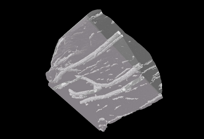
Lab tests on the surgical filament, published in OpenNano, showed it was easily visible in CT scans when threaded through samples of chicken meat, even after three weeks. It also showed strong antimicrobial properties, killing 99% of highly drug-resistant bacteria after six hours at body temperature.
Houshyar said the team was not aware of any commercially available suture products that combined these properties.
How they did it
The multidisciplinary team led by RMIT – included nano-engineering, biomedical and textile experts working in partnership with a practicing surgeon – used the university’s cutting-edge textile manufacturing facility to develop their proof-of-concept material.
The suture’s properties come from the combination of iodine and tiny nanoparticles, called carbon dots, throughout the material.
Carbon dots are inherently fluorescent, due to their particular wavelength, but they can also be tuned to various levels of luminosity that easily stand out from surrounding tissue in medical imaging.
Attaching iodine to these carbon dots, meanwhile, provides them with their strong antimicrobial properties and greater X-ray visibility.
Houshyar said carbon nano dots were safe, cheap and easy to produce in the lab from natural ingredients.
“They can be tailored to create biodegradable stitches or a permanent suture, or even to be adhesive on one side only, where required,” she said.
“This project opens up a lot of practical solutions for surgeons, which has been our aim from the start and the reason we have involved clinicians in the study.”
Clinical possibilities
Consultant colorectal surgeon and Professor of Surgery at the University of Melbourne, Justin Yeung, was involved in the study. He said it addressed a real challengefaced by surgeons in trying to identify the precise anatomical location of internal meshes on CT scans.
“This mesh will enable us to help with improved identification of the causes of symptoms, reduce the incidence of mesh infections and will help with precise preoperative planning, if there is a need to surgically remove this mesh,” he said.
“It has the potential to improve surgery outcomes and improve quality of life for a huge proportion of women, if used as vaginal mesh for example, by reducing the need for infected mesh removal.”
“It may also significantly reduce surgery duration and increase surgical accuracy in general through the ability to visualise mesh location accurately on preoperative imaging.”
Next steps
Study co-author from RMIT’s School of Health and Biomedical Sciences, Professor Elisa Hill-Yardin, said the next steps were pre-clinical trials.
“While this research is at an early stage, we believe we are onto something very promising that could help a lot of people and are really keen to speak with industry partners who are interested in working with us to take it further,” she said.
“We see potential especially in vaginal mesh implants and similar procedures.”
The research team used Australia’s leading university-based textile manufacturing facilities at RMIT’s Centre for Materials Innovation and Future Fashion to produce the proof-of-concept material. They will soon be producing larger suture samples to use in pre-clinical trials, which they have just received seed funding for from RMIT.
Here’s a link to and a citation for the paper,
Smart suture with iodine contrasting nanoparticles for computed tomography by Shadi Houshyar, Hong Yin, Leon Pope, Rumbidzai Zizhou, Chaitali Dekiwadia, Elisa L. Hill-Yardin, Justin MC Yeung, Sabu John, Kate Fox, Nhiem Tran, Ivan Cole, Aaron Elbourne, Vi Khanh Truong, and Adam Truskewycz. OpenNano Volume 9, January 2023, 100120 DOI: https://doi.org/10.1016/j.onano.2022.100120
This paper is open access.