Tag Archives: teeth

Oxygen-deficient nanotitania (titanium dioxide nanoparticles) for whiter teeth without the damage

A September 8, 2021 news item on phys.org announces research that could make the process of whitening teeth safer,

Most people would like to flash a smile of pearly whites, but over time teeth can become stained by foods, beverages and some medications. Unfortunately, the high levels of hydrogen peroxide in dentists’ bleaching treatments can damage enamel and cause tooth sensitivity and gum irritation. Now, researchers reporting in ACS Applied Materials & Interfaces have developed a gel that, when exposed to near infrared (NIR) light, safely whitens teeth without the burn.

Caption: A new bleaching gel whitened tooth samples by six shades, using a low level of hydrogen peroxide (12%). Credit: Adapted from ACS Applied Materials & Interfaces 2021, DOI: 10.1021/acsami.1c06774

A September 8, 2021Amercian Chemical Society (ACS) news release (also on EurekAlert), which originated the news item, provides more detail,

The growing demand for selfie-ready smiles has made tooth whitening one of the most popular dental procedures. Treatments at a dentist’s office are effective, but they use high-concentration hydrogen peroxide (30–40%). Home bleaching products contain less peroxide (6–12%), but they usually require weeks of treatment and don’t work as well. When a bleaching gel is applied to teeth, hydrogen peroxide and peroxide-derived reactive oxygen species (mainly the hydroxyl radical) degrade pigments in stains. The hydroxyl radical is much better at doing this than hydrogen peroxide itself, so researchers have tried to improve the bleaching capacity of low-concentration hydrogen peroxide by boosting the generation of powerful hydroxyl radicals. Because previous approaches have had limitations, Xingyu Hu, Li Xie, Weidong Tian and colleagues wanted to develop a safe, effective whitening gel containing a catalyst that, when exposed to NIR light, would convert low levels of hydrogen peroxide into abundant hydroxyl radicals.

The researchers made oxygen-deficient titania nanoparticles that catalyzed hydroxyl radical production from hydrogen peroxide. Exposing the nanoparticles to NIR light increased their catalytic activity, allowing them to completely bleach tooth samples stained with orange dye, tea or red dye within 2 hours. Then, the researchers made a gel containing the nanoparticles, a carbomer gel and 12% hydrogen peroxide. They applied it to naturally stained tooth samples and treated them with NIR light for an hour. The gel bleached teeth just as well as a popular tooth whitening gel containing 40% hydrogen peroxide, with less damage to enamel. The nanoparticle system is highly promising for tooth bleaching and could also be extended to other biomedical applications, such as developing antibacterial materials, the researchers say.

The authors acknowledge funding from the National Natural Science Foundation of China, the National Key R&D Program of China and the Key Technologies R&D Program of Sichuan Province.

Here’s a link to and a citation for the paper,

Photothermal-Enhanced Fenton-like Catalytic Activity of Oxygen-Deficient Nanotitania for Efficient and Safe Tooth Whitening by Xingyu Hu, Li Xie, Zhaoyu Xu, Suru Liu, Xinzhi Tan, Ruojing Qian, Ruitao Zhang, Mingyan Jiang, Wenjia Xie, and Weidong Tian. ACS Appl. Mater. Interfaces 2021, 13, 30, 35315–35327 Publication Date: July 22, 2021 DOI: https://doi.org/10.1021/acsami.1c06774 Copyright © 2021 American Chemical Society

This paper is behind a paywall.

Regenerating dental enamel

For anyone who’s concerned about their dental enamel, this research might prove encouraging. From a June 1, 2018 news item on Nanowerk,

Researchers at Queen Mary University of London [UK][ have developed a new way to grow mineralised materials which could regenerate hard tissues such as dental enamel and bone.

Enamel, located on the outer part of our teeth, is the hardest tissue in the body and enables our teeth to function for a large part of our lifetime despite biting forces, exposure to acidic foods and drinks and extreme temperatures. This remarkable performance results from its highly organised structure.

However, unlike other tissues of the body, enamel cannot regenerate once it is lost, which can lead to pain and tooth loss. These problems affect more than 50 per cent of the world’s population and so finding ways to recreate enamel has long been a major need in dentistry.

A June 1, 2018 Queen Mary University of London press release, which originated the news item, provides more detail,

The study, published in Nature Communications, shows that this new approach can create materials with remarkable precision and order that look and behave like dental enamel.

The materials could be used for a wide variety of dental complications such as the prevention and treatment of tooth decay or tooth sensitivity – also known as dentin hypersensitivity.

Simple and versatile

Dr Sherif Elsharkawy, a dentist and first author of the study from Queen Mary’s School of Engineering and Materials Science, said: “This is exciting because the simplicity and versatility of the mineralisation platform opens up opportunities to treat and regenerate dental tissues. For example, we could develop acid resistant bandages that can infiltrate, mineralise, and shield exposed dentinal tubules of human teeth for the treatment of dentin hypersensitivity.”

The mechanism that has been developed is based on a specific protein material that is able to trigger and guide the growth of apatite nanocrystals at multiple scales – similarly to how these crystals grow when dental enamel develops in our body. This structural organisation is critical for the outstanding physical properties exhibited by natural dental enamel.

Lead author Professor Alvaro Mata, also from Queen Mary’s School of Engineering and Materials Science, said: “A major goal in materials science is to learn from nature to develop useful materials based on the precise control of molecular building-blocks. The key discovery has been the possibility to exploit disordered proteins to control and guide the process of mineralisation at multiple scales. Through this, we have developed a technique to easily grow synthetic materials that emulate such hierarchically organised architecture over large areas and with the capacity to tune their properties.”

Mimic other hard tissues

Enabling control of the mineralisation process opens the possibility to create materials with properties that mimic different hard tissues beyond enamel such as bone and dentin. As such, the work has the potential to be used in a variety of applications in regenerative medicine. In addition, the study also provides insights into the role of protein disorder in human physiology and pathology.

Here’s a link to and a citation for the paper,

Protein disorder–order interplay to guide the growth of hierarchical mineralized structures by Sherif Elsharkawy, Maisoon Al-Jawad, Maria F. Pantano, Esther Tejeda-Montes, Khushbu Mehta, Hasan Jamal, Shweta Agarwal, Kseniya Shuturminska, Alistair Rice, Nadezda V. Tarakina, Rory M. Wilson, Andy J. Bushby, Matilde Alonso, Jose C. Rodriguez-Cabello, Ettore Barbieri, Armando del Río Hernández, Molly M. Stevens, Nicola M. Pugno, Paul Anderson, & Alvaro Mata. Nature Communicationsvolume 9, Article number: 2145 (2018) Published 01 June 2018 DOI: https://doi.org/10.1038/s41467-018-04319-0

This paper is open access.

One final comment, this work is at the ‘in vitro’ stage. More colloquially, this is being done in a petri dish or glass vial or some other container and it’s going to be a long time before there are going to be any human clinical trials, assuming the work gets that far.

Nano with green tea for sensitive teeth

The future will be beautiful if scientists are successful with a new DNA (deoxyribonucleic acid) sunscreen (my Aug. 3, 2017 posting) and a new dental material for people with sensitive teeth. From an Aug. 2, 2017 news item on phys.org,

An ice cold drink is refreshing in the summer, but for people with sensitive teeth, it can cause a painful jolt in the mouth. This condition can be treated, but many current approaches don’t last long. Now researchers report in the journal ACS [American Chemical Society] Applied Materials & Interfaces the development of a new material with an extract from green tea that could fix this problem—and help prevent cavities in these susceptible patients.

An Aug. 2, 2017 ACS news release, which originated the news item, describes the problem and the work in more detail,

Tooth sensitivity commonly occurs when the protective layers of teeth are worn away, revealing a bony tissue called dentin. This tissue contains microscopic hollow tubes that, when exposed, allow hot and cold liquids and food to contact the underlying nerve endings in the teeth, causing pain. Unprotected dentin is also vulnerable to cavity formation. Plugging these tubes with a mineral called nanohydroxyapatite is a long-standing approach to treating sensitivity. But the material doesn’t stand up well to regular brushing, grinding, erosion or acid produced by cavity-causing bacteria. Cui Huang and colleagues wanted to tackle sensitivity and beat the bacteria at the same time.

The researchers encapsulated nanohydroxyapatite and a green tea polyphenol — epigallocatechin-3-gallate, or EGCG — in silica nanoparticles, which can stand up to acid and wear and tear. EGCG has been shown in previous studies to fight Streptococcus mutans, which forms biofilms that cause cavities. Testing on extracted wisdom teeth showed that the material plugged the dentin tubules, released EGCG for at least 96 hours, stood up to tooth erosion and brushing and prevented biofilm formation. It also showed low toxicity. Based on these findings, the researchers say the material could indeed be a good candidate for combating tooth sensitivity and cavities.

The authors acknowledge funding from the National Natural Science Foundation of China, the Natural Science Foundation of Hubei Province of China and the Fundamental Research Funds for the Central Universities.

Here’s a link to and a citation for the paper,

Development of Epigallocatechin-3-gallate-Encapsulated Nanohydroxyapatite/Mesoporous Silica for Therapeutic Management of Dentin Surface by Jian Yu, Hongye Yang, Kang Li, Hongyu Ren, Jinmei Lei, and Cui Huang. ACS Appl. Mater. Interfaces, Article ASAP DOI: 10.1021/acsami.7b06597 Publication Date (Web): July 13, 2017

Copyright © 2017 American Chemical Society

This paper is behind a paywall.

Nanoscale elements that govern the behaviour of our teeth

Are we going to be adopting atomically correct dental hygiene practices in the future? It’s certainly a possibility given the latest Australian research announced in a Sept. 7, 2016 news item on Nanowerk (Note: A link has been removed),

With one in two Australian children reported to have tooth decay in their permanent teeth by age 12, researchers from the University of Sydney believe they have identified some nanoscale elements that govern the behaviour of our teeth.

Material and structures engineers worked with dentists and bioengineers to map the exact composition and structure of tooth enamel at the atomic scale.

Using a relatively new microscopy technique called atom probe tomography, their work produced the first-ever three-dimensional maps showing the positions of atoms critical in the decay process.

The new knowledge on atom composition at the nanolevel has the potential to aid oral health hygiene and caries prevention, and has been published today in the journal Science Advances(“Atomic-scale compositional mapping reveals Mg-rich amorphous calcium phosphate in human dental enamel”).

A Sept. 8, 2016 University of Sydney press release, which originated the news item, expands on the theme (Note: A link has been removed),

Professor Julie Cairney, Material and Structures Engineer in the Faculty of Engineering and Information Technologies, said:

“The dental professionals have known that certain trace ions are important in the tough structure of tooth enamel but until now it had been impossible to map the ions in detail.

“The structure of human tooth enamel is extremely intricate and while we have known that magnesium, carbonate and fluoride ions influence enamel properties scientists have never been able to capture its structure at a high enough resolution or definition.”

“What we have found are the magnesium-rich regions between the hydroxyapatite nanorods that make up the enamel.”

“This means we have the first direct evidence of the existence of a proposed amorphous magnesium-rich calcium phosphate phase that plays an essential role in governing the behaviour of teeth. “

Co-lead researcher on the study, Dr Alexandre La Fontaine from the University’s Australian Centre for Microscopy and Microanalysis, said:

“We were also able to see nanoscale ‘clumps’ of organic material, which indicates that proteins and peptides are heterogeneously distributed within the enamel rather than present along all the nanorod interfaces, which was what was previously suggested.

“The mapping has the potential for new treatments designed around protecting against the dissolution of this specific amorphous phase.

“The new understanding of how enamel forms will also help in tooth remineralisation research.”

Here’s a link to and a citation for the paper,

Atomic-scale compositional mapping reveals Mg-rich amorphous calcium phosphate in human dental enamel by Alexandre La Fontaine, Alexander Zavgorodniy, Howgwei Liu, Rongkun Zheng, Michael Swain, and Julie Cairney. Science Advances  07 Sep 2016: Vol. 2, no. 9, e1601145 DOI: 10.1126/sciadv.1601145

This paper is open access.

Nanoparticles in baby formula

Needle-like particles of hydroxyapatite found in infant formula by ASU researchers. Westerhoff and Schoepf/ASU, CC BY-ND

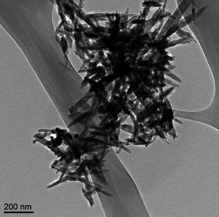
Needle-like particles of hydroxyapatite found in infant formula by ASU [Arizona State University] researchers. Westerhoff and Schoepf/ASU, CC BY-ND

Nanowerk is featuring an essay about hydroxyapatite nanoparticles in baby formula written by Dr. Andrew Maynard in a May 17, 2016 news item (Note: A link has been removed),

There’s a lot of stuff you’d expect to find in baby formula: proteins, carbs, vitamins, essential minerals. But parents probably wouldn’t anticipate finding extremely small, needle-like particles. Yet this is exactly what a team of scientists here at Arizona State University [ASU] recently discovered.

The research, commissioned and published by Friends of the Earth (FoE) – an environmental advocacy group – analyzed six commonly available off-the-shelf baby formulas (liquid and powder) and found nanometer-scale needle-like particles in three of them. The particles were made of hydroxyapatite – a poorly soluble calcium-rich mineral. Manufacturers use it to regulate acidity in some foods, and it’s also available as a dietary supplement.

Andrew’s May 17, 2016 essay first appeared on The Conversation website,

Looking at these particles at super-high magnification, it’s hard not to feel a little anxious about feeding them to a baby. They appear sharp and dangerous – not the sort of thing that has any place around infants. …

… questions like “should infants be ingesting them?” make a lot of sense. However, as is so often the case, the answers are not quite so straightforward.

Andrew begins by explaining about calcium and hydroxyapatite (from The Conversation),

Calcium is an essential part of a growing infant’s diet, and is a legally required component in formula. But not necessarily in the form of hydroxyapatite nanoparticles.

Hydroxyapatite is a tough, durable mineral. It’s naturally made in our bodies as an essential part of bones and teeth – it’s what makes them so strong. So it’s tempting to assume the substance is safe to eat. But just because our bones and teeth are made of the mineral doesn’t automatically make it safe to ingest outright.

The issue here is what the hydroxyapatite in formula might do before it’s digested, dissolved and reconstituted inside babies’ bodies. The size and shape of the particles ingested has a lot to do with how they behave within a living system.

He then discusses size and shape, which are important at the nanoscale,

Size and shape can make a difference between safe and unsafe when it comes to particles in our food. Small particles aren’t necessarily bad. But they can potentially get to parts of our body that larger ones can’t reach. Think through the gut wall, into the bloodstream, and into organs and cells. Ingested nanoscale particles may be able to interfere with cells – even beneficial gut microbes – in ways that larger particles don’t.

These possibilities don’t necessarily make nanoparticles harmful. Our bodies are pretty well adapted to handling naturally occurring nanoscale particles – you probably ate some last time you had burnt toast (carbon nanoparticles), or poorly washed vegetables (clay nanoparticles from the soil). And of course, how much of a material we’re exposed to is at least as important as how potentially hazardous it is.

Yet there’s a lot we still don’t know about the safety of intentionally engineered nanoparticles in food. Toxicologists have started paying close attention to such particles, just in case their tiny size makes them more harmful than otherwise expected.

Currently, hydroxyapatite is considered safe at the macroscale by the US Food and Drug Administration (FDA). However, the agency has indicated that nanoscale versions of safe materials such as hydroxyapatite may not be safe food additives. From Andrew’s May 17, 2016 essay,

Hydroxyapatite is a tough, durable mineral. It’s naturally made in our bodies as an essential part of bones and teeth – it’s what makes them so strong. So it’s tempting to assume the substance is safe to eat. But just because our bones and teeth are made of the mineral doesn’t automatically make it safe to ingest outright.

The issue here is what the hydroxyapatite in formula might do before it’s digested, dissolved and reconstituted inside babies’ bodies. The size and shape of the particles ingested has a lot to do with how they behave within a living system. Size and shape can make a difference between safe and unsafe when it comes to particles in our food. Small particles aren’t necessarily bad. But they can potentially get to parts of our body that larger ones can’t reach. Think through the gut wall, into the bloodstream, and into organs and cells. Ingested nanoscale particles may be able to interfere with cells – even beneficial gut microbes – in ways that larger particles don’t.These possibilities don’t necessarily make nanoparticles harmful. Our bodies are pretty well adapted to handling naturally occurring nanoscale particles – you probably ate some last time you had burnt toast (carbon nanoparticles), or poorly washed vegetables (clay nanoparticles from the soil). And of course, how much of a material we’re exposed to is at least as important as how potentially hazardous it is.Yet there’s a lot we still don’t know about the safety of intentionally engineered nanoparticles in food. Toxicologists have started paying close attention to such particles, just in case their tiny size makes them more harmful than otherwise expected.

Putting particle size to one side for a moment, hydroxyapatite is classified by the US Food and Drug Administration (FDA) as “Generally Regarded As Safe.” That means it considers the material safe for use in food products – at least in a non-nano form. However, the agency has raised concerns that nanoscale versions of food ingredients may not be as safe as their larger counterparts.Some manufacturers may be interested in the potential benefits of “nanosizing” – such as increasing the uptake of vitamins and minerals, or altering the physical, textural and sensory properties of foods. But because decreasing particle size may also affect product safety, the FDA indicates that intentionally nanosizing already regulated food ingredients could require regulatory reevaluation.In other words, even though non-nanoscale hydroxyapatite is “Generally Regarded As Safe,” according to the FDA, the safety of any nanoscale form of the substance would need to be reevaluated before being added to food products.Despite this size-safety relationship, the FDA confirmed to me that the agency is unaware of any food substance intentionally engineered at the nanoscale that has enough generally available safety data to determine it should be “Generally Regarded As Safe.”Casting further uncertainty on the use of nanoscale hydroxyapatite in food, a 2015 report from the European Scientific Committee on Consumer Safety (SCCS) suggests there may be some cause for concern when it comes to this particular nanomaterial.Prompted by the use of nanoscale hydroxyapatite in dental products to strengthen teeth (which they consider “cosmetic products”), the SCCS reviewed published research on the material’s potential to cause harm. Their conclusion?

The available information indicates that nano-hydroxyapatite in needle-shaped form is of concern in relation to potential toxicity. Therefore, needle-shaped nano-hydroxyapatite should not be used in cosmetic products.

This recommendation was based on a handful of studies, none of which involved exposing people to the substance. Researchers injected hydroxyapatite needles directly into the bloodstream of rats. Others exposed cells outside the body to the material and observed the effects. In each case, there were tantalizing hints that the small particles interfered in some way with normal biological functions. But the results were insufficient to indicate whether the effects were meaningful in people.

As Andrew also notes in his essay, none of the studies examined by the SCCS OEuropean Scientific Committee on Consumer Safety) looked at what happens to nano-hydroxyapatite once it enters your gut and that is what the researchers at Arizona State University were considering (from the May 17, 2016 essay),

The good news is that, according to preliminary studies from ASU researchers, hydroxyapatite needles don’t last long in the digestive system.

This research is still being reviewed for publication. But early indications are that as soon as the needle-like nanoparticles hit the highly acidic fluid in the stomach, they begin to dissolve. So fast in fact, that by the time they leave the stomach – an exceedingly hostile environment – they are no longer the nanoparticles they started out as.

These findings make sense since we know hydroxyapatite dissolves in acids, and small particles typically dissolve faster than larger ones. So maybe nanoscale hydroxyapatite needles in food are safer than they sound.

This doesn’t mean that the nano-needles are completely off the hook, as some of them may get past the stomach intact and reach more vulnerable parts of the gut. But the findings do suggest these ultra-small needle-like particles could be an effective source of dietary calcium – possibly more so than larger or less needle-like particles that may not dissolve as quickly.

Intriguingly, recent research has indicated that calcium phosphate nanoparticles form naturally in our stomachs and go on to be an important part of our immune system. It’s possible that rapidly dissolving hydroxyapatite nano-needles are actually a boon, providing raw material for these natural and essential nanoparticles.

While it’s comforting to know that preliminary research suggests that the hydroxyapatite nanoparticles are likely safe for use in food products, Andrew points out that more needs to be done to insure safety (from the May 17, 2016 essay),

And yet, even if these needle-like hydroxyapatite nanoparticles in infant formula are ultimately a good thing, the FoE report raises a number of unresolved questions. Did the manufacturers knowingly add the nanoparticles to their products? How are they and the FDA ensuring the products’ safety? Do consumers have a right to know when they’re feeding their babies nanoparticles?

Whether the manufacturers knowingly added these particles to their formula is not clear. At this point, it’s not even clear why they might have been added, as hydroxyapatite does not appear to be a substantial source of calcium in most formula. …

And regardless of the benefits and risks of nanoparticles in infant formula, parents have a right to know what’s in the products they’re feeding their children. In Europe, food ingredients must be legally labeled if they are nanoscale. In the U.S., there is no such requirement, leaving American parents to feel somewhat left in the dark by producers, the FDA and policy makers.

As far as I’m aware, the Canadian situation is much the same as the US. If the material is considered safe at the macroscale, there is no requirement to indicate that a nanoscale version of the material is in the product.

I encourage you to read Andrew’s essay in its entirety. As for the FoE report (Nanoparticles in baby formula: Tiny new ingredients are a big concern), that is here.

Discovering why your teeth aren’t perfectly crack-resistant

This helps make your teeth crack-resistant?

Caption: Illustration shows complex biostructure of dentin: the dental tubuli (yellow hollow cylinders, diameters appr. 1 micrometer) are surrounded by layers of mineralized collagen fibers (brown rods). The tiny mineral nanoparticles are embedded in the mesh of collagen fibers and not visible here. Credit: JB Forien @Charité

Caption: Illustration shows complex biostructure of dentin: the dental tubuli (yellow hollow cylinders, diameters appr. 1 micrometer) are surrounded by layers of mineralized collagen fibers (brown rods). The tiny mineral nanoparticles are embedded in the mesh of collagen fibers and not visible here. Credit: JB Forien @Charité

A June 10, 2015 Helmholtz Zentrum Berlin (HZB) press release (also on EurekAlert) explains how the illustration above relates to the research,

Human teeth have to serve for a lifetime, despite being subjected to huge forces. But the high failure resistance of dentin in teeth is not fully understood. An interdisciplinary team led by scientists of Charite Universitaetsmedizin Berlin has now analyzed the complex structure of dentin. At the synchrotron sources BESSY II at HZB, Berlin, Germany, and the European Synchrotron Radiation Facility ESRF, Grenoble, France, they could reveal that the mineral particles are precompressed.

The internal stress works against crack propagation and increases resistance of the biostructure.

Engineers use internal stresses to strengthen materials for specific technical purposes. Now it seems that evolution has long ‘known’ about this trick, and has put it to use in our natural teeth. Unlike bones, which are made partly of living cells, human teeth are not able to repair damage. Their bulk is made of dentin, a bonelike material consisting of mineral nanoparticles. These mineral nanoparticles are embedded in collagen protein fibres, with which they are tightly connected. In every tooth, such fibers can be found, and they lie in layers, making teeth tough and damage resistant. Still, it was not well understood, how crack propagation in teeth can be stopped.

The press release goes on to describe the new research and the teams which investigated the role of the mineral nanoparticles with regard to compression and cracking,

Now researchers from Charite Julius-Wolff-Institute, Berlin have been working with partners from Materials Engineering Department of Technische Universitaets Berlin, MPI of Colloids and Interfaces, Potsdam and Technion – Israel Institute of Technology, Haifa, to examine these biostructures more closely. They performed Micro-beam in-situ stress experiments in the mySpot BESSY facility of HZB, Berlin, Germany and analyzed the local orientation of the mineral nanoparticles using the nano-imaging facility of the European Synchrotron Radiation Facility (ESRF) in Grenoble, France.

When the tiny collagen fibers shrink, the attached mineral particles become increasingly compressed, the science team found out. “Our group was able to use changes in humidity to demonstrate how stress appears in the mineral in the collagen fibers, Dr. Paul Zaslansky from Julius Wolff-Institute of Charite Berlin explains. “The compressed state helps to prevents cracks from developing and we found that compression takes place in such a way that cracks cannot easily reach the tooth inner parts, which could damage the sensitive pulp. In this manner, compression stress helps to prevent cracks from rushing through the tooth.

The scientists also examined what happens if the tight mineral-protein link is destroyed by heating: In that case, dentin in teeth becomes much weaker. We therefore believe that the balance of stresses between the particles and the protein is important for the extended survival of teeth in the mouth, Charite scientist Jean-Baptiste Forien says. Their results may explain why artificial tooth replacements usually do not work as well as healthy teeth do: they are simply too passive, lacking the mechanisms found in the natural tooth structures, and consequently fillings cannot sustain the stresses in the mouth as well as teeth do. “Our results might inspire the development of tougher ceramic structures for tooth repair or replacement, Zaslansky hopes.

Experiments took place as part of the DFG project “Biomimetic Materials Research: Functionality by Hierarchical Structuring of Materials (SPP1420).

Here’s a link to and a citation for the paper,

Compressive Residual Strains in Mineral Nanoparticles as a Possible Origin of Enhanced Crack Resistance in Human Tooth Dentin by Jean-Baptiste Forien, Claudia Fleck, Peter Cloetens, Georg Duda, Peter Fratzl, Emil Zolotoyabko, and Paul Zaslansky. Nano Lett., 2015, 15 (6), pp 3729–3734 DOI: 10.1021/acs.nanolett.5b00143 Publication Date (Web): May 26, 2015

Copyright © 2015 American Chemical Society

This paper is behind a paywall.

Mesenchymal condensation (a process embryos use to begin forming a variety of organs, including teeth, cartilage, bone, muscle, tendon, and kidney) for complex 3D tissue engineering

It seems that there are three strategies for creating complex 3D tissues and until now scientists have used only two of the three. From a March 5, 2014 news item on ScienceDaily,

A bit of pressure from a new shrinking, sponge-like gel is all it takes to turn transplanted unspecialized cells into cells that lay down minerals and begin to form teeth.

The bioinspired gel material could one day help repair or replace damaged organs, such as teeth and bone, and possibly other organs as well, scientists from the Wyss Institute for Biologically Inspired Engineering at Harvard University, Harvard School of Engineering and Applied Sciences (SEAS), and Boston Children’s Hospital report recently in Advanced Materials.

“Tissue engineers have long raised the idea of using synthetic materials to mimic the inductive power of the embryo,” said Don Ingber, M.D., Ph.D., Founding Director of the Wyss Institute, …, Professor of Bioengineering at SEAS, and senior author of the study. “We’re excited about this work because it shows that it really is possible.”

The March 5, 2014 Wyss Institute news release, which originated the news item, delves into the nature of the research,

Embryonic tissues have the power to drive cells and tissues to specialize and form organs. To do that, they employ biomolecules called growth factors to stimulate growth; gene-activating chemicals that cause the cells to specialize, and mechanical forces that modulate cell responses to these other factors.

But so far tissue engineers who want to build organs in the laboratory have employed only two of the three strategies — growth factors and gene-activating chemicals. Perhaps as a result, they have not yet succeeded in producing complex three-dimensional tissues.

A few years ago, Ingber and Tadanori Mammoto, M.D., Ph.D., Instructor in Surgery at Boston Children’s Hospital and Harvard Medical School, investigated a process called mesenchymal condensation that embryos use to begin forming a variety of organs, including teeth, cartilage, bone, muscle, tendon, and kidney.

In mesenchymal condensation, two adjacent tissue layers — loosely packed connective-tissue cells called mesenchyme and sheet-like tissue called an epithelium that covers it — exchange biochemical signals. This exchange causes the mesenchymal cells to squeeze themselves tightly into a small knot directly below where the new organ will form.

Here’s a video from the Wyss Institute illustrating the squeezing process,

When the temperature rises to just below body temperature, this biocompatible gel shrinks dramatically within minutes, bringing tooth-precursor cells (green) closer together. Credit: Basma Hashmi

Getting back to the research (from the news release),

By examining tissues isolated from the jaws of embryonic mice, Mammoto and Ingber showed that when the compressed mesenchymal cells turn on genes that stimulate them to generate whole teeth composed of mineralized tissues, including dentin and enamel.

Inspired by this embryonic induction mechanism, Ingber and Basma Hashmi, a Ph.D. candidate at SEAS who is the lead author of the current paper, set out to develop a way to engineer artificial teeth by creating a tissue-friendly material that accomplishes the same goal. Specifically, they wanted a porous sponge-like gel that could be impregnated with mesenchymal cells, then, when implanted into the body, induced to shrink in 3D to physically compact the cells inside it.

To develop such a material, Ingber and Hashmi teamed up with researchers led by Joanna Aizenberg, Ph.D., a Wyss Institute Core Faculty member who leads the Institute’s Adaptive Materials Technologies platform. Aizenberg is the Amy Smith Berylson Professor of Materials Science at SEAS and Professor of Chemistry and Chemical Biology at Harvard University.

They chemically modified a special gel-forming polymer called PNIPAAm that scientists have used to deliver drugs to the body’s tissues. PNIPAAm gels have an unusual property: they contract abruptly when they warm.

But they do this at a lukewarm temperature, whereas the researchers wanted them to shrink specifically at 37°C — body temperature — so that they’d squeeze their contents as soon as they were injected into the body. Hashmi worked with Lauren Zarzar, Ph.D., a former SEAS graduate student who’s now a postdoctoral associate at Massachusetts Institute of Technology, for more than a year, modifying PNIPAAm and testing the resulting materials. Ultimately, they developed a polymer that forms a tissue-friendly gel with two key properties: cells stick to it, and it compresses abruptly when warmed to body temperature.

As an initial test, Hashmi implanted mesenchymal cells in the gel and warmed it in the lab. Sure enough, when the temperature reached 37°C, the gel shrank within 15 minutes, causing the cells inside the gel to round up, shrink, and pack tightly together.

“The reason that’s cool is that the cells are alive,” Hashmi said. “Usually when this happens, cells are dead or dying.”

Not only were they alive — they activated three genes that drive tooth formation.

To see if the shrinking gel also worked its magic in the body, Hashmi worked with Mammoto to load mesenchymal cells into the gel, then implant the gel beneath the mouse kidney capsule — a tissue that is well supplied with blood and often used for transplantation experiments.

The implanted cells not only expressed tooth-development genes — they laid down calcium and minerals, just as mesenchymal cells do in the body as they begin to form teeth.

“They were in full-throttle tooth-development mode,” Hashmi said.

The researchers have future plans (from the news release),

In the embryo, mesenchymal cells can’t build teeth alone — they need to be combined with cells that form the epithelium. In the future, the scientists plan to test whether the shrinking gel can stimulate both tissues to generate an entire functional tooth.

Here’s a link to and a citation for the paper about the successful attempt to stimulate mesenchymal cells into the beginnings of tooth formation,

Developmentally-Inspired Shrink-Wrap Polymers for Mechanical Induction of Tissue Differentiation by Basma Hashmi, Lauren D. Zarzar, Tadanori Mammoto, Akiko Mammoto, Amanda Jiang, Joanna Aizenberg, and Donald E. Ingber. Advanced Materials Article first published online: 18 FEB 2014 DOI: 10.1002/adma.201304995

© 2014 WILEY-VCH Verlag GmbH & Co. KGaA, Weinheim

This paper is behind a paywall.

Growing a tooth—as an adult

These days it seems that teeth are the most erogenous zone of all. Actors on screens of all types flash pearly whites that are increasingly blinding while the rest of us are enjoined to buy teeth whiteners in toothpastes, mouthwashes, whitening strips, and/or find dental professionals to assist us in our quest for the brightest and whitest teeth. It would all be so much easier if we could just grow new teeth and discard the old ones.

Coincidentally or not, it seems researchers at King’s College London have also been thinking about how we might grow new teeth. Ben Schiller in a Mar. 14, 2013 article for Fast Company highlights the work,

Researchers from the U.K. have successfully bioengineered teeth from gum tissue and cells taken from mice. By combining and transplanting two groups of cells, they were able to grow full teeth, complete with roots, dentine, and enamel.

This King’s College London Mar. 11, 2013 news release provides more details,

New research published in the Journal of Dental Research describes an advance in efforts to develop a method to replace missing teeth with new bioengineered teeth generated from a person’s own gum cells. …

Current implant-based methods of whole tooth replacement fail to reproduce a natural root structure and as a consequence of the friction from eating and other jaw movement, loss of jaw bone can occur around the implant.

Research towards achieving the aim of producing bioengineered teeth (bioteeth) has largely focused on the generation of immature teeth (teeth primordia) that mimic those in the embryo that can be transplanted as small cell ‘pellets’ into the adult jaw to develop into functional teeth. Remarkably, despite the very different environments, embryonic teeth primordia can develop normally in the adult mouth and thus if suitable cells can be identified that can be combined in such a way to produce an immature tooth, there is a realistic prospect bioteeth can become a clinical reality. Subsequent studies have largely focussed on the use of embryonic cells and although it is clear that embryonic tooth primordia cells can readily form immature teeth following dissociation into single cell populations and subsequent recombination, such cell sources are impractical to use in a general therapy.

Professor Sharpe [Paul Sharpe, an expert in craniofacial development and stem cell biology at King’s College London’s Dental Institute] said: ‘What is required is the identification of adult sources of human epithelial and mesenchymal cells that can be obtained in sufficient numbers to make biotooth formation a viable alternative to dental implants.’

In this new work, the researchers isolated adult human gum (gingival) tissue from patients at the Dental Institute at King’s College London, grew more of it in the lab, and then combined it with the cells of mice that form teeth (mesenchyme cells). By transplanting this combination of cells into mice the researchers were able to grow hybrid human/mouse teeth containing dentine and enamel, as well as viable roots.

Professor Sharpe concluded: ‘Epithelial cells derived from adult human gum tissue are capable of responding to tooth inducing signals from embryonic tooth mesenchyme in an appropriate way to contribute to tooth crown and root formation and give rise to relevant differentiated cell types, following in vitro culture. These easily accessible epithelial cells are thus a realistic source for consideration in human biotooth formation. The next major challenge is to identify a way to culture adult human mesenchymal cells to be tooth-inducing, as at the moment we can only make embryonic mesenchymal cells do this.’

If I read this rightly, researchers are several years away from actually growing a new tooth in an adult human mouth but this work suggests they might be on the right research track.

A ‘wandering meatloaf’ with teeth inspires nanomaterials for solar cells and Li-ion batteries

The ‘wandering meatloaf’ is a species of marine snail (or chiton) that has extraordinary teeth according to the Jan. 16, 2013 news item on ScienceDaily,

An assistant professor [David Kisailus] at the University of California, Riverside’s Bourns College of Engineering is using the teeth of a marine snail found off the coast of California to create less costly and more efficient nanoscale materials to improve solar cells and lithium-ion batteries.

The paper is focused on the gumboot chiton, the largest type of chiton, which can be up to a foot-long. They are found along the shores of the Pacific Ocean from central California to Alaska. They have a leathery upper skin, which is usually reddish-brown and occasionally orange, leading some to give it the nickname “wandering meatloaf.”

Over time, chitons have evolved to eat algae growing on and within rocks using a specialized rasping organ called a radula, a conveyer belt-like structure in the mouth that contains 70 to 80 parallel rows of teeth. During the feeding process, the first few rows of the teeth are used to grind rock to get to the algae. They become worn, but new teeth are continuously produced and enter the “wear zone” at the same rate as teeth are shed.

The University of California Riverside Jan. 15, 2013 news release by Sean Nealon, which originated the news item, describes the chiton’s teeth and the specifics of Kisailus’ inspiration (Note: A link has been removed),

Over time, chitons have evolved to eat algae growing on and within rocks using a specialized rasping organ called a radula, a conveyer belt-like structure in the mouth that contains 70 to 80 parallel rows of teeth. During the feeding process, the first few rows of the teeth are used to grind rock to get to the algae. They become worn, but new teeth are continuously produced and enter the “wear zone” at the same rate as teeth are shed.

Kisailus, who uses nature as inspiration to design next generation engineering products and materials, started studying chitons five years ago because he was interested in abrasion and impact-resistant materials. He has previously determined that the chiton teeth contain the hardest biomineral known on Earth, magnetite, which is the key mineral that not only makes the tooth hard, but also magnetic.

Kisailus is using the lessons learned from this biomineralization pathway as inspiration in his lab to guide the growth of minerals used in solar cells and lithium-ion [li-ion] batteries. By controlling the crystal size, shape and orientation of engineering nanomaterials, he believes he can build materials that will allow the solar cells and lithium-ion batteries to operate more efficiently. In other words, the solar cells will be able to capture a greater percentage of sunlight and convert it to electricity more efficiently and the lithium-ion batteries could need significantly less time to recharge.

Using the chiton teeth model has another advantage: engineering nanocrystals can be grown at significantly lower temperatures, which means significantly lower production costs.

While Kisailus is focused on solar cells and lithium-ion batteries, the same techniques could be used to develop everything from materials for car and airplane frames to abrasion resistant clothing. In addition, understanding the formation and properties of the chiton teeth could help to create better design parameters for better oil drills and dental drill bits.

Here’s a representation of the teeth from the University of California Riverside,

A series of images that show the teeth of the gumboot chiton (aka, snail, aka, wandering meatloaf)

A series of images that show the teeth of the gumboot chiton (aka, snail, aka, wandering meatloaf)

You can find other images and media materials in the ScienceDaily news item or the University of California Riverside news release. This citation and link for the research paper is from the ScienceDaily news item,

Qianqian Wang, Michiko Nemoto, Dongsheng Li, James C. Weaver, Brian Weden, John Stegemeier, Krassimir N. Bozhilov, Leslie R. Wood, Garrett W. Milliron, Christopher S. Kim, Elaine DiMasi, David Kisailus. Phase Transformations and Structural Developments in the Radular Teeth ofCryptochiton Stelleri. Advanced Functional Materials, 2013; DOI: 10.1002/adfm.201202894

This article is behind a paywall.