Tag Archives: Tina Hilding

Nanoflowers for better drug delivery; researchers looking for commercial partners

Caption: Schematic representation of the movement of the flower-like particle as it makes its way through a cellular trap to deliver therapeutic genes. Credit: WSU [Washington State University]

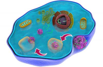
It looks more like a swimming pool with pool toys to me but I imagine that nobody wants to say that they’re sending ‘pool toys’ through your bloodstream. Nanoflowers or flower-shaped nanoparticles sounds nicer.

From a January 10, 2019 news item on Nanowerk,

Washington State University [WSU] researchers have developed a novel way to deliver drugs and therapies into cells at the nanoscale without causing toxic effects that have stymied other such efforts.

The work could someday lead to more effective therapies and diagnostics for cancer and other illnesses.

Led by Yuehe Lin, professor in WSU’s School of Mechanical and Materials Engineering, and Chunlong Chen, senior scientist at the Department of Energy’s Pacific Northwest National Laboratory (PNNL), the research team developed biologically inspired materials at the nanoscale that were able to effectively deliver model therapeutic genes into tumor cells. …

A January 10, 2019 WSU news release (also on EurekAlert) by Tina Hilding, which originated the news item, describes the work in greater detail,

Researchers have been working to develop nanomaterials that can effectively carry therapeutic genes directly into the cells for the treatment of diseases such as cancer. The key issues for gene delivery using nanomaterials are their low delivery efficiency of medicine and potential toxicity.

“To develop nanotechnology for medical purposes, the first thing to consider is toxicity — That is the first concern for doctors,” said Lin.

The flower-like particle the WSU and PNNL team developed is about 150 nanometers in size, or about one thousand times smaller than the width of a piece of paper. It is made of sheets of peptoids, which are similar to natural peptides that make up proteins. The peptoids make for a good drug delivery particle because they’re fairly easy to synthesize and, because they’re similar to natural biological materials, work well in biological systems.

The researchers added fluorescent probes in their peptoid nanoflowers, so they could trace them as they made their way through cells, and they added the element fluorine, which helped the nanoflowers more easily escape from tricky cellular traps that often impede drug delivery.

The flower-like particles loaded with therapeutic genes were able to make their way smoothly out of the predicted cellular trap, enter the heart of the cell, and release their drug there.

“The nanoflowers successfully and rapidly escaped (the cell trap) and exhibited minimal cytotoxicity,” said Lin.

After their initial testing with model drug molecules, the researchers hope to conduct further studies using real medicines.

“This paves a new way for us to develop nanocargoes that can efficiently deliver drug molecules into the cell and offers new opportunities for targeted gene therapies,” he said.

The WSU and PNNL team have filed a patent application for the new technology, and they are seeking industrial partners for further development.

Should you and your company be interested in partnering with the researchers, contact:

  • Yuehe Lin, professor, School of Mechanical and Materials Engineering, 509‑335‑8523, yuehe.lin@wsu.edu
  • Tina Hilding, communications director, Voiland College of Engineering and Architecture, 509‑335‑5095, thilding@wsu.edu

For those who’d like more information, here’s a link to and a citation for the paper,

Efficient Cytosolic Delivery Using Crystalline Nanoflowers Assembled from Fluorinated Peptoids by Yang Song, Mingming Wang, Suiqiong Li, Haibao Jin, Xiaoli Cai, Dan Du, He Li, Chun‐Long Chen, Yuehe Lin. Small DOI: https://doi.org/10.1002/smll.201803544 First published: 22 November 2018

This paper is behind a paywall.

Bone regeneration with a mix of 21st century techniques and an age-old natural cure

Curry was how I was introduced to turmeric. My father who came from Mauritius loved curry and we had it at least once a week. Nobody mentioned healing properties, which I was to discover them only after I started this blog. Usually, turmeric is mentioned in cancer cures but not this time.

Turmeric Courtesy: Washington State University

From a May 2, 2018 Washington State University news release by Tina Hilding (also on EurekAlert but dated May 3, 2018),

A WSU research team is bringing together natural medical cures with modern biomedical devices in hopes of bringing about better health outcomes for people with bone diseases.

In this first-ever effort, the team improved bone-growing capabilities on 3D-printed, ceramic bone scaffolds by 30-45 percent when coated with curcumin, a compound found in the spice, turmeric. They have published their work in the journal, Materials Today Chemistry.

The work could be important for the millions of Americans who suffer from injuries or bone diseases like osteoporosis.

Human bone includes bone forming and resorbing cells that constantly remodel throughout our lives. As people age, the bone cell cycling process often doesn’t work as well. Bones become weaker and likely to fracture. Many of the medicines used for osteoporosis work by slowing down or stopping the destruction of old bone or by forming new bone. While they may increase bone density, they also create an imbalance in the natural bone remodeling cycle and may create poorer quality bone.

Turmeric has been used as medicine for centuries in Asian countries, and curcumin has been shown to have antioxidant, anti-inflammatory and bone-building capabilities. It can also prevent various forms of cancers. However, when taken orally as medicine, the compound can’t be absorbed well in the body. It is metabolized and eliminated too quickly.

Led by Susmita Bose, Herman and Brita Lindholm Endowed Chair Professor in the School of Mechanical and Materials Engineering, the researchers encased the curcumin within a water-loving polymer, a large molecule, so that it could be gradually released from their ceramic scaffolds. The curcumin increased the viability and proliferation of new bone cells and blood vessels in surrounding tissue as well as accelerated the healing process.

Bose hopes that the work will lead to medicines that naturally create healthier bone without affecting the bone remodeling cycle.

“In the end, it’s the bone quality that matters,” she said.

The researchers are continuing the studies, looking at the protein and cellular level to gain better understanding of exactly how the natural compound works. They are also working to improve the process’ efficiency and control. The challenge with the natural compounds, said Bose, is that they are often large organic molecules.

“You have to use the right vehicle for delivery,” she said. “We need to load and get it released in a controlled and sustained way. The chemistry of vehicle delivery is very important.”

In addition to curcumin, the researchers are studying other natural remedies, including compounds from aloe vera, saffron, Vitamin D, garlic, oregano and ginger. Bose is focused on compounds that might help with bone disorders, including those that encourage bone growth or that have anti-inflammatory, infection control, or anti-cancer properties.

Starting with her own health issues, Bose has had a longtime interest in bridging natural medicinal compounds with modern medicine. That interest increased after she had her children.

“As a mother and having a chemistry background, I realized I didn’t want my children to be exposed to so many chemicals for every illness,” Bose said. “I started looking at home remedies.”

To her students, she always emphasizes healthy living as the best way to guarantee the best health outcomes, including healthy eating, proper sleep, interesting hobbies, and exercise.

Courtesy Washington State University

Here’s a link to and a citation for the paper,

Effects of PCL, PEG and PLGA polymers on curcumin release from calcium phosphate matrix for in vitro and in vivo bone regeneration by Susmita Bose, Naboneeta Sarkar, Dishary Banerjee. Materials Today Chemistry Vol. 8 June 2018, pp. 110-130 [Published online May 2, 2018] https://doi.org/10.1016/j.mtchem.2018.03.005

This paper is behind a paywall.

Mimicking the architecture of materials like wood and bone

Caption: Microstructures like this one developed at Washington State University could be used in batteries, lightweight ultrastrong materials, catalytic converters, supercapacitors and biological scaffolds. Credit: Washington State University

A March 3, 2017 news item on Nanowerk features a new 3D manufacturing technique for creating biolike materials, (Note: A link has been removed)

Washington State University nanotechnology researchers have developed a unique, 3-D manufacturing method that for the first time rapidly creates and precisely controls a material’s architecture from the nanoscale to centimeters. The results closely mimic the intricate architecture of natural materials like wood and bone.

They report on their work in the journal Science Advances (“Three-dimensional microarchitected materials and devices using nanoparticle assembly by pointwise spatial printing”) and have filed for a patent.

A March 3, 2017 Washington State University news release by Tina Hilding (also on EurekAlert), which originated the news item, expands on the theme,

“This is a groundbreaking advance in the 3-D architecturing of materials at nano- to macroscales with applications in batteries, lightweight ultrastrong materials, catalytic converters, supercapacitors and biological scaffolds,” said Rahul Panat, associate professor in the School of Mechanical and Materials Engineering, who led the research. “This technique can fill a lot of critical gaps for the realization of these technologies.”

The WSU research team used a 3-D printing method to create foglike microdroplets that contain nanoparticles of silver and to deposit them at specific locations. As the liquid in the fog evaporated, the nanoparticles remained, creating delicate structures. The tiny structures, which look similar to Tinkertoy constructions, are porous, have an extremely large surface area and are very strong.

Silver was used because it is easy to work with. However, Panat said, the method can be extended to any other material that can be crushed into nanoparticles – and almost all materials can be.

The researchers created several intricate and beautiful structures, including microscaffolds that contain solid truss members like a bridge, spirals, electronic connections that resemble accordion bellows or doughnut-shaped pillars.

The manufacturing method itself is similar to a rare, natural process in which tiny fog droplets that contain sulfur evaporate over the hot western Africa deserts and give rise to crystalline flower-like structures called “desert roses.”

Because it uses 3-D printing technology, the new method is highly efficient, creates minimal waste and allows for fast and large-scale manufacturing.

The researchers would like to use such nanoscale and porous metal structures for a number of industrial applications; for instance, the team is developing finely detailed, porous anodes and cathodes for batteries rather than the solid structures that are now used. This advance could transform the industry by significantly increasing battery speed and capacity and allowing the use of new and higher energy materials.

Here’s a link to and a citation for the paper,

Three-dimensional microarchitected materials and devices using nanoparticle assembly by pointwise spatial printing by Mohammad Sadeq Saleh, Chunshan Hu, and Rahul Panat. Science Advances  03 Mar 2017: Vol. 3, no. 3, e1601986 DOI: 10.1126/sciadv.1601986

This paper appears to be open access.

Finally, there is a video,