Tag Archives: tissue eningeering

Transplanting healthy neurons could be possible with walking molecules and 3D printing

A February 23, 2021 news item on ScienceDaily announces work which may lead to healing brain injuries and diseases,

Imagine if surgeons could transplant healthy neurons into patients living with neurodegenerative diseases or brain and spinal cord injuries. And imagine if they could “grow” these neurons in the laboratory from a patient’s own cells using a synthetic, highly bioactive material that is suitable for 3D printing.

By discovering a new printable biomaterial that can mimic properties of brain tissue, Northwestern University researchers are now closer to developing a platform capable of treating these conditions using regenerative medicine.

A February 22, 2021 Northwestern University news release (also received by email and available on EurekAlert) by Lila Reynolds, which originated the news item, delves further into self-assembling ‘walking’ molecules and the nanofibers resulting in a new material designed to promote the growth of healthy neurons,

A key ingredient to the discovery is the ability to control the self-assembly processes of molecules within the material, enabling the researchers to modify the structure and functions of the systems from the nanoscale to the scale of visible features. The laboratory of Samuel I. Stupp published a 2018 paper in the journal Science which showed that materials can be designed with highly dynamic molecules programmed to migrate over long distances and self-organize to form larger, “superstructured” bundles of nanofibers.

Now, a research group led by Stupp has demonstrated that these superstructures can enhance neuron growth, an important finding that could have implications for cell transplantation strategies for neurodegenerative diseases such as Parkinson’s and Alzheimer’s disease, as well as spinal cord injury.

“This is the first example where we’ve been able to take the phenomenon of molecular reshuffling we reported in 2018 and harness it for an application in regenerative medicine,” said Stupp, the lead author on the study and the director of Northwestern’s Simpson Querrey Institute. “We can also use constructs of the new biomaterial to help discover therapies and understand pathologies.

Walking molecules and 3D printing

The new material is created by mixing two liquids that quickly become rigid as a result of interactions known in chemistry as host-guest complexes that mimic key-lock interactions among proteins, and also as the result of the concentration of these interactions in micron-scale regions through a long scale migration of “walking molecules.”

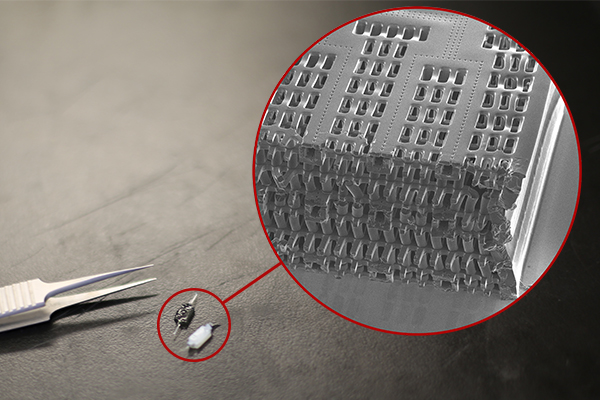
The agile molecules cover a distance thousands of times larger than themselves in order to band together into large superstructures. At the microscopic scale, this migration causes a transformation in structure from what looks like an uncooked chunk of ramen noodles into ropelike bundles.

“Typical biomaterials used in medicine like polymer hydrogels don’t have the capabilities to allow molecules to self-assemble and move around within these assemblies,” said Tristan Clemons, a research associate in the Stupp lab and co-first author of the paper with Alexandra Edelbrock, a former graduate student in the group. “This phenomenon is unique to the systems we have developed here.”

Furthermore, as the dynamic molecules move to form superstructures, large pores open that allow cells to penetrate and interact with bioactive signals that can be integrated into the biomaterials.

Interestingly, the mechanical forces of 3D printing disrupt the host-guest interactions in the superstructures and cause the material to flow, but it can rapidly solidify into any macroscopic shape because the interactions are restored spontaneously by self-assembly. This also enables the 3D printing of structures with distinct layers that harbor different types of neural cells in order to study their interactions.

Signaling neuronal growth

The superstructure and bioactive properties of the material could have vast implications for tissue regeneration. Neurons are stimulated by a protein in the central nervous system known as brain-derived neurotrophic factor (BDNF), which helps neurons survive by promoting synaptic connections and allowing neurons to be more plastic. BDNF could be a valuable therapy for patients with neurodegenerative diseases and injuries in the spinal cord but these proteins degrade quickly in the body and are expensive to produce.

One of the molecules in the new material integrates a mimic of this protein that activates its receptor known as Trkb, and the team found that neurons actively penetrate the large pores and populate the new biomaterial when the mimetic signal is present. This could also create an environment in which neurons differentiated from patient-derived stem cells mature before transplantation.

Now that the team has applied a proof of concept to neurons, Stupp believes he could now break into other areas of regenerative medicine by applying different chemical sequences to the material. Simple chemical changes in the biomaterials would allow them to provide signals for a wide range of tissues.

“Cartilage and heart tissue are very difficult to regenerate after injury or heart attacks, and the platform could be used to prepare these tissues in vitro from patient-derived cells,” Stupp said. “These tissues could then be transplanted to help restore lost functions. Beyond these interventions, the materials could be used to build organoids to discover therapies or even directly implanted into tissues for regeneration since they are biodegradable.”

Here’s a link to and a citation for the paper,

Superstructured Biomaterials Formed by Exchange Dynamics and Host–Guest Interactions in Supramolecular Polymers by Alexandra N. Edelbrock, Tristan D. Clemons, Stacey M. Chin, Joshua J. W. Roan, Eric P. Bruckner, Zaida Álvarez, Jack F. Edelbrock, Kristen S. Wek, Samuel I. Stupp. Advanced Science DOI: https://doi.org/10.1002/advs.202004042 First published: 22 February 2021

This paper is open access.

University of Toronto (Canada) researchers and lab-grown heart and liver tissue (person-on-a-chip)

Usually called ‘human-on-a-chip’, a team at the University of Toronto have developed a two-organ ‘person on a chip’ according to a March 7, 2016 news item on phys.org (Note: Links have been removed),

Researchers at U of T [University of Toronto] Engineering have developed a new way of growing realistic human tissues outside the body. Their “person-on-a-chip” technology, called AngioChip, is a powerful platform for discovering and testing new drugs, and could eventually be used to repair or replace damaged organs.

Professor Milica Radisic (IBBME, ChemE), graduate student Boyang Zhang and the rest of the team are among those research groups around the world racing to find ways to grow human tissues in the lab, under conditions that mimic a real person’s body. They have developed unique methods for manufacturing small, intricate scaffolds for individual cells to grow on. These artificial environments produce cells and tissues that resemble the real thing more closely than those grown lying flat in a petri dish.

The team’s recent creations have included BiowireTM—an innovative method of growing heart cells around a silk suture—as well as a scaffold for heart cells that snaps together like sheets of Velcro. But AngioChip takes tissue engineering to a whole new level. “It’s a fully three-dimensional structure complete with internal blood vessels,” says Radisic. “It behaves just like vasculature, and around it there is a lattice for other cells to attach and grow.” …

A March 7, 2016 University of Toronto news release (also on EurekAlert), which originated the news item, provides more detail about the AngioChip,

Zhang built the scaffold out of POMaC, a polymer that is both biodegradable and biocompatible. The scaffold is built out of a series of thin layers, stamped with a pattern of channels that are each about 50 to 100 micrometres wide. The layers, which resemble the computer microchips, are then stacked into a 3D structure of synthetic blood vessels. As each layer is added, UV light is used to cross-link the polymer and bond it to the layer below.

When the structure is finished, it is bathed in a liquid containing living cells. The cells quickly attach to the inside and outside of the channels and begin growing just as they would in the human body.

“Previously, people could only do this using devices that squish the cells between sheets of silicone and glass,” says Radisic. “You needed several pumps and vacuum lines to run just one chip. Our system runs in a normal cell culture dish, and there are no pumps; we use pressure heads to perfuse media through the vasculature. The wells are open, so you can easily access the tissue.”

Using the platform, the team has built model versions of both heart and liver tissues that function like the real thing. “Our liver actually produced urea and metabolized drugs,” says Radisic. They can connect the blood vessels of the two artificial organs, thereby modelling not just the organs themselves, but the interactions between them. They’ve even injected white blood cells into the vessels and watched as they squeezed through gaps in the vessel wall to reach the tissue on the other side, just as they do in the human body.

The news release also mentions potential markets and the work that needs to be accomplished before AngioChip is available for purchase,

AngioChip has great potential in the field of pharmaceutical testing. Current drug-testing methods, such as animal testing and controlled clinical trials, are costly and fraught with ethical concerns. Testing on lab-grown human tissues would provide a realistic model at a fraction of the cost, but this area of research is still in its infancy. “In the last few years, it has become possible to order cultures of human cells for testing, but they’re grown on a plate, a two-dimensional environment,” says Radisic. “They don’t capture all the functional hallmarks of a real heart muscle, for example.”

A more realistic platform like AngioChip could enable drug companies to detect dangerous side effects and interactions between organ compartments long before their products reach the market, saving countless lives. It could also be used to understand and validate the effectiveness of current drugs and even to screen libraries of chemical compounds to discover new drugs. Through TARA Biosystems Inc., a spin-off company co-founded by Radisic, the team is already working on commercializing the technology.

In future, Radisic envisions her lab-grown tissues being implanted into the body to repair organs damaged by disease. Because the cells used to seed the platform can come from anyone, the new tissues could be genetically identical to the intended host, reducing the risk of organ rejection. Even in its current form, the team has shown that the AngioChip can be implanted into a living animal, its artificial blood vessels connected to a real circulatory system. The polymer scaffolding itself simply biodegrades after several months.

The team still has much work to do. Each AngioChip is currently made by hand; if the platform is to be used industrially, the team will need to develop high-throughput manufacturing methods to create many copies at once. Still, the potential is obvious. “It really is multifunctional, and solves many problems in the tissue engineering space,” says Radisic. “It’s truly next-generation.”

Here’s a link to and a citation for the paper,

Biodegradable scaffold with built-in vasculature for organ-on-a-chip engineering and direct surgical anastomosis by Boyang Zhang, Miles Montgomery, M. Dean Chamberlain, Shinichiro Ogawa, Anastasia Korolj, Aric Pahnke, Laura A. Wells, Stéphane Massé, Jihye Kim, Lewis Reis, Abdul Momen, Sara S. Nunes, Aaron R. Wheeler, Kumaraswamy Nanthakumar, Gordon Keller, Michael V. Sefton, & Milica Radisic. Nature Materials (2016) doi:10.1038/nmat4570 Published online 07 March 2016

This paper is behind a paywall.

The researchers have made two images illustrating their work available. There’s this still image,

These tiny polymer scaffolds contain channels that are about 100 micrometres wide, about the same diameter as a human hair. When seeded with cells, the channels act as artificial blood vessels. By mimicking tissues in the human heart and other organs, these scaffolds provide a new way to test drugs for potentially dangerous side effects. (Image: Tyler Irving/Boyang Zhang/Kevin Soobrian)

These tiny polymer scaffolds contain channels that are about 100 micrometres wide, about the same diameter as a human hair. When seeded with cells, the channels act as artificial blood vessels. By mimicking tissues in the human heart and other organs, these scaffolds provide a new way to test drugs for potentially dangerous side effects. (Image: Tyler Irving/Boyang Zhang/Kevin Soobrian)

Perhaps more intriguing is this one,

UofT_AngioChipMoving

When seeded with heart cells, the flexible polymer scaffold contracts with a regular rhythm, just like real heart tissue. (Image: Boyang Zhang)

I have mentioned ‘human-on-a-chip’ projects many times here and as the news release writer notes, there is an international race. My July 1, 2015 posting (cross-posted from the June 30, 2015 posting [Testing times: the future of animal alternatives] on the International Innovation blog [a CORDIS-listed project dissemination partner for FP7 and H2020 projects]) notes a couple of those projects,

Organ-on-a-chip projects use stem cells to create human tissues that replicate the functions of human organs. Discussions about human-on-a-chip activities – a phrase used to describe 10 interlinked organ chips – were a highlight of the 9th World Congress on Alternatives to Animal Testing held in Prague, Czech Republic, last year. One project highlighted at the event was a joint US National Institutes of Health (NIH), US Food and Drug Administration (FDA) and US Defense Advanced Research Projects Agency (DARPA) project led by Dan Tagle that claimed it would develop functioning human-on-a-chip by 2017. However, he and his team were surprisingly close-mouthed and provided few details making it difficult to assess how close they are to achieving their goal.

By contrast, Uwe Marx – Leader of the ‘Multi-Organ-Chip’ programme in the Institute of Biotechnology at the Technical University of Berlin and Scientific Founder of TissUse, a human-on-a-chip start-up company – claims to have sold two-organ chips. He also claims to have successfully developed a four-organ chip and that he is on his way to building a human-on-a-chip. Though these chips remain to be seen, if they are, they will integrate microfluidics, cultured cells and materials patterned at the nanoscale to mimic various organs, and will allow chemical testing in an environment that somewhat mirrors a human.

As for where the University of Toronto efforts fit into the race, I don’t know for sure. It’s the first time I’ve come across a reference to liver tissue producing urea but I believe there’s at least one other team in China which has achieved a three-dimensional, more lifelike aspect for liver tissue in my Jan. 29, 2016 posting ‘Constructing a liver’.