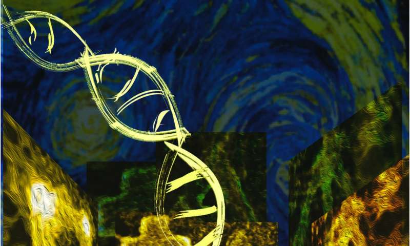
Tag Archives: transmission electron microscopy (TEM)

Canadian research into nanomaterial workplace exposure in the air and on surfaces

An August 30, 2018 news item on Nanowerk announces the report,

The monitoring of air contamination by engineered nanomaterials (ENM) is a complex process with many uncertainties and limitations owing to the presence of particles of nanometric size that are not ENMs, the lack of validated instruments for breathing zone measurements and the many indicators to be considered.

In addition, some organizations, France’s Institut national de recherche et de sécurité (INRS) and Québec’s Institut de recherche Robert-Sauvé en santé et en sécurité du travail (IRSST) among them, stress the need to also sample surfaces for ENM deposits.

In other words, to get a better picture of the risks of worker exposure, we need to fine-tune the existing methods of sampling and characterizing ENMs and develop new one. Accordingly, the main goal of this project was to develop innovative methodological approaches for detailed qualitative as well as quantitative characterization of workplace exposure to ENMs.

A PDF of the 88-page report is available in English or in French.

An August 30, 2018 (?) abstract of the IRSST report titled An Assessment of Methods of Sampling and Characterizing Engineered Nanomaterials in the Air and on Surfaces in the Workplace (2nd edition) by Maximilien Debia, Gilles L’Espérance, Cyril Catto, Philippe Plamondon, André Dufresne, Claude Ostiguy, which originated the news item, outlines what you can expect from the report,

This research project has two complementary parts: a laboratory investigation and a fieldwork component. The laboratory investigation involved generating titanium dioxide (TiO2) nanoparticles under controlled laboratory conditions and studying different sampling and analysis devices. The fieldwork comprised a series of nine interventions adapted to different workplaces and designed to test a variety of sampling devices and analytical procedures and to measure ENM exposure levels among Québec workers.

The methods for characterizing aerosols and surface deposits that were investigated include: i) measurement by direct-reading instruments (DRI), such as condensation particle counters (CPC), optical particle counters (OPC), laser photometers, aerodynamic diameter spectrometers and electric mobility spectrometer; ii) transmission electron microscopy (TEM) or scanning transmission electron microscopy (STEM) with a variety of sampling devices, including the Mini Particle Sampler® (MPS); iii) measurement of elemental carbon (EC); iv) inductively coupled plasma mass spectrometry (ICP-MS) and (v) Raman spectroscopy.

The workplace investigations covered a variety of industries (e.g., electronics, manufacturing, printing, construction, energy, research and development) and included producers as well as users or integrators of ENMs. In the workplaces investigated, we found nanometals or metal oxides (TiO2, SiO2, zinc oxides, lithium iron phosphate, titanate, copper oxides), nanoclays, nanocellulose and carbonaceous materials, including carbon nanofibers (CNF) and carbon nanotubes (CNT)—single-walled (SWCNT) as well as multiwalled (MWCNT).

The project helped to advance our knowledge of workplace assessments of ENMs by documenting specific tasks and industrial processes (e.g., printing and varnishing) as well as certain as yet little investigated ENMs (nanocellulose, for example).

Based on our investigations, we propose a strategy for more accurate assessment of ENM exposure using methods that require a minimum of preanalytical handling. The recommended strategy is a systematic two-step assessment of workplaces that produce and use ENMs. The first step involves testing with different DRIs (such as a CPC and a laser photometer) as well as sample collection and subsequent microscopic analysis (MPS + TEM/STEM) to clearly identify the work tasks that generate ENMs. The second step, once work exposure is confirmed, is specific quantification of the ENMs detected. The following findings are particularly helpful for detailed characterization of ENM exposure:

  1. The first conclusive tests of a technique using ICP-MS to quantify the metal oxide content of samples collected in the workplace
  2. The possibility of combining different sampling methods recommended by the National Institute for Occupational Safety and Health (NIOSH) to measure elemental carbon as an indicator of NTC/NFC, as well as demonstration of the limitation of this method stemming from observed interference with the black carbon particles required to synthesis carbon materials (for example, Raman spectroscopy showed that less than 6% of the particles deposited on the electron microscopy grid at one site were SWCNTs)
  3. The clear advantages of using an MPS (instead of the standard 37-mm cassettes used as sampling media for electron microscopy), which allows quantification of materials
  4. The major impact of sampling time: a long sampling time overloads electron microscopy grids and can lead to overestimation of average particle agglomerate size and underestimation of particle concentrations
  5. The feasibility and utility of surface sampling, either with sampling pumps or passively by diffusion onto the electron microscopy grids, to assess ENM dispersion in the workplace

These original findings suggest promising avenues for assessing ENM exposure, while also showing their limitations. Improvements to our sampling and analysis methods give us a better understanding of ENM exposure and help in adapting and implementing control measures that can minimize occupational exposure.

You can download the full report in either or both English and French from the ‘Nanomaterials – A Guide to Good Practices Facilitating Risk Management in the Workplace, 2nd Edition‘ webpage.

Calligraphy ink and cancer treatment

Courtesy of ACS Omega and the researchers

Nice illustration! I wish I could credit the artist. For anyone who needs a little text to make sense of it, there’s a Sept. 27, 2017 news item on Nanowerk (Note: A link has been removed),

For hundreds of years, Chinese calligraphers have used a plant-based ink to create beautiful messages and art. Now, one group reports in ACS Omega (“New Application of Old Material: Chinese Traditional Ink for Photothermal Therapy of Metastatic Lymph Nodes”) that this ink could noninvasively and effectively treat cancer cells that spread, or metastasize, to lymph nodes.

A Sept. 27, 2017 American Chemical Society (ACS) news release, which originated the news item, reveals more about the research,

As cancer cells leave a tumor, they frequently make their way to lymph nodes, which are part of the immune system. In this case, the main treatment option is surgery, but this can result in complications. Photothermal therapy (PTT) is an emerging noninvasive treatment option in which nanomaterials are injected and accumulate in cancer cells. A laser heats up the nanomaterials, and this heat kills the cells. Many of these nanomaterials are expensive, difficult-to-make and toxic. However, a traditional Chinese ink called Hu-Kaiwen ink (Hu-ink) has similar properties to the nanomaterials used in PTT. For example, they are the same color, and are both carbon-based and stable in water. So Wuli Yang and colleagues wanted to see if Hu-ink could be a good alternative material for PTT.

The researchers analyzed Hu-ink and found that it consists of nanoparticles and thin layers of carbon. When Hu-ink was heated with a laser, its temperature rose by 131 degrees Fahrenheit, much higher than current nanomaterials. Under PPT conditions, the Hu-ink killed cancer cells in a laboratory dish, but under normal conditions, the ink was non-toxic. This was also the scenario observed in mice with tumors. The researchers also noted that Hu-ink could act as a probe to locate tumors and metastases because it absorbs near-infrared light, which goes through skin.

Being a little curious about Hu-ink’s similarity to nanomaterial, I looked for more detail in the the paper (Note: Links have been removed), From the: Introduction,

Photothermal therapy (PTT) is an emerging tumor treatment strategy, which utilizes hyperthermia generated from absorbed near-infrared (NIR) light energy by photoabsorbing agents to kill tumor cells.(7-13) Different from chemotherapy, surgical treatment, and radiotherapy, PTT is noninvasive and more efficient.(7, 14, 15) In the past decade, PTT with diverse nanomaterials to eliminate cancer metastases lymph nodes has attracted extensive attention by several groups, including our group.(3, 16-20) For instance, Liu and his co-workers developed a treatment method based on PEGylated single-walled carbon nanotubes for PTT of tumor sentinel lymph nodes and achieved remarkably improved treatment effect in an animal tumor model.(21) To meet the clinical practice, the potential metastasis of deeper lymph nodes was further ablated in our previous work, using magnetic graphene oxide as a theranostic agent.(22) However, preparation of these artificial nanomaterials usually requires high cost, complicated synthetic process, and unavoidably toxic catalyst or chemicals,(23, 24) which impede their future clinical application. For the clinical application, exploring an environment-friendly material with simple preparation procedure, good biocompatibility, and excellent therapeutic efficiency is still highly desired. [emphases mine]

From the: Preparation and Characterization of Hu-Ink

To obtain an applicable sample, the condensed Hu-ink was first diluted into aqueous dispersion with a lower concentration. The obtained Hu-ink dispersion without any further treatment was black in color and stable in physiological environment, including water, phosphate-buffered saline (PBS), and Roswell Park Memorial Institute (RPMI) 1640; furthermore, no aggregation was observed even after keeping undisturbed for 3 days (Figure 2a). The nanoscaled morphology of Hu-ink was examined by transmission electron microscopy (TEM) (Figure 2b), which demonstrates that Hu-ink mainly exist in the form of small aggregates. These small aggregates consist of a few nanoparticles with diameter of about 20–50 nm. Dynamic light scattering (DLS) measurement (Figure 2c) further shows that Hu-ink aqueous dispersion possesses a hydrodynamic diameter of about 186 nm (polydispersity index: 0.18), which was a crucial prerequisite for biomedical applications.(29) In the X-ray diffraction (XRD) pattern, no other characteristic peaks are found except carbon peak (Figure S1, Supporting Information), which confirms that the main component of Hu-ink is carbon.(25) Raman spectroscopy was a common tool to characterize graphene-related materials.(30) D band (∼1300 cm–1, corresponding to the defects) and G band (∼1600 cm–1, related to the sp2 carbon sites) peaks could be observed in Figure 2d with the ratio ID/IG = 0.96, which confirms the existence of graphene sheetlike structure in Hu-ink.(31) The UV–vis–NIR spectra (Figure 2e) also revealed that Hu-ink has high absorption in the NIR region around 650–900 nm, in which hemoglobin and water, the major absorbers of biological tissue, have their lowest absorption coefficient.(32) The high NIR absorption capability of Hu-ink encouraged us to investigate its photothermal properties.(33-35) Hu-ink dispersions with different concentrations were irradiated under an 808 nm laser (the commercial and widely used wavelength in photothermal therapy).(8-13) [emphases mine]

Curiosity satisfied! For those who’d like to investigate even further, here’s a link to and a citation for the paper,

New Application of Old Material: Chinese Traditional Ink for Photothermal Therapy of Metastatic Lymph Nodes by Sheng Wang, Yongbin Cao, Qin Zhang, Haibao Peng, Lei Liang, Qingguo Li, Shun Shen, Aimaier Tuerdi, Ye Xu, Sanjun Cai, and Wuli Yang. ACS Omega, 2017, 2 (8), pp 5170–5178 DOI: 10.1021/acsomega.7b00993 Publication Date (Web): August 30, 2017

Copyright © 2017 American Chemical Society

This paper appears to be open access.

Mechanically strong organic nanotubes made with light

This research comes from Nagoya University in Japan according to an Aug. 30, 2016 news item on Nanowerk,

Organic nanotubes (ONTs) are tubular nanostructures composed of organic molecules that have unique properties and have found various applications, such as electro-conductive materials and organic photovoltaics. A group of scientists at Nagoya University have developed a simple and effective method for the formation of robust covalent ONTs from simple molecules. This method is expected to be useful in generating a range of nanotube-based materials with desirable properties.

An Aug. 30, 2016 Nagoya University press release (also on EurekAlert), which originated the news item, provides more information,

Kaho Maeda, Dr. Hideto Ito, Professor Kenichiro Itami of the JST-ERATO Itami Molecular Nanocarbon Project and the Institute of Transformative Bio-Molecules (ITbM) of Nagoya University, and their colleagues have reported in the Journal of the American Chemical Society, on the development of a new and simple strategy, “helix-to-tube” to synthesize covalent organic nanotubes.

Organic nanotubes (ONTs) are organic molecules with tubular nanostructures. Nanostructures are structures that range between 1 nm and 100 nm, and ONTs have a nanometer-sized cavity. Various 
applications of ONTs have been reported, including molecular recognition materials, transmembrane ion channel/sensors, electro-conductive materials, and organic photovoltaics. Most ONTs are constructed by a self-assembly process based on weak non-covalent interactions such as hydrogen bonding, hydrophobic interactions and π-π interactions between aromatic rings. Due to these relatively weak interactions, most non-covalent ONTs possess a relatively fragile structure (Figure 1).

Figure1_ONT.png
Figure 1. Conventional synthetic method for non-covalent ONTs, their applications and disadvantages.

Covalent ONTs, whose tubular skeletons are cross-linked by covalent bonding (a bond made by sharing of electrons between atoms) could be synthesized from non-covalent ONTs. While covalent ONTs show higher stability and mechanical strength than non-covalent ONTs, the general synthetic strategy for covalent ONTs was yet to be established (Figure 2).

Figure2_ONT.png
Figure 2. Covalent ONTs derived from non-covalent ONTs by cross-linking, their properties and disadvantages.

A team led by Hideto Ito and Kenichiro Itami has succeeded in developing a simple and effective method for the synthesis of robust covalent ONTs (tube) by an operationally simple light irradiation of a readily accessible helical polymer (helix). This so-called “helix-to-tube” strategy is based on the following steps: 1) polymerization of a small molecule (monomer) to make a helical polymer followed by, 2) light-induced cross-linking at longitudinally repeating pitches across the whole helix to form covalent nanotubes (Figure 3).

Figure3_ONT.png
Figure 3. New synthetic approach towards covalent ONTs through longitudinal cross-linking between helical pitches in helical polymers.

With their strategy, the team designed and synthesized diacetylene-based helical polymers (acetylenes are molecules that contain carbon-carbon triple bonds), poly(m-phenylene diethynylene)s (poly-PDEs), which has chiral amide side chains that are able to induce a helical folding through hydrogen-bonding interactions (Figure 4).

Figure4_ONT.png
Figure 4. Molecular design for helical poly-PDE bearing chiral amide side chains.

The researchers revealed that light-induced cross-linking at longitudinally aligned 1,3-butadiyne moieties (a group of molecules that contain four carbons with triple bonds at the first and third carbons) could generate the desired covalent ONT (Figure 5). “This is the first time in the world to show that the photochemical polymerization reaction of diynes is applicable to the cross-linking reaction of a helical polymer,” says Maeda, a graduate student who mainly conducted the experiments.

The “helix-to-tube” method is expected to be able to generate a range of ONT-based materials by simply changing the arene (aromatic ring) unit in the monomer.

Figure5_ONT.png
Figure 5. Synthesis of a covalent ONT by photochemical cross-linking between longitudinal aligned 1,3-butadiyne moieties (red lines).

“One of the most difficult parts of this research was how to obtain scientific evidence on the structures of poly-PDEs and covalent ONTs,” says Ito, one of the leaders of this study. “We had little experience with the analysis of polymers and macromolecules such as ONTs. Fortunately, thanks to the support of our collaborators in Nagoya University, who are specialists in these particular research fields, we finally succeeded in characterizing these macromolecules by various techniques including spectroscopy, X-ray diffraction, and microscopy.”

“Although it took us about a year to synthesize the covalent ONT, it took another one and a half year to determine the structure of the nanotube,” says Maeda. “I was extremely excited when I first saw the transmission electron microscopy (TEM) images, which indicated that we had actually made the covalent ONT that we were expecting,” she continues (Figure 6).

Figure6_ONT.png
Figure 6. TEM images of the bundle structures of covalent ONT

“The best part of the research for me was finding that the photochemical cross-linking had taken place on the helix for the first time,” says Maeda. “In addition, photochemical cross-linking is known to usually occur in the solid phase, but we were able to show that the reaction takes place in the solution phase as well. As the reactions have never been carried out before, I was dubious at first, but it was a wonderful feeling to succeed in making the reaction work for the first time in the world. I can say for sure that this was a moment where I really found research interesting.”

“We were really excited to develop this simple yet powerful method to achieve the synthesis of covalent ONTs,” says Itami, the director of the JST-ERATO project and the center director of ITbM. “The “helix-to-tube” method enables molecular level design and will lead to the synthesis of various covalent ONTs with fixed diameters and tube lengths with desirable functionalities.”

“We envisage that ongoing advances in the “helix-to-tube” method may lead to the development of various ONT-based materials including electro-conductive materials and luminescent materials,” says Ito. “We are currently carrying out work on the “helix-to-tube” methodology and we hope to synthesize covalent ONTs with interesting properties for various applications.”

Here’s a link to and a citation for the paper,

Construction of Covalent Organic Nanotubes by Light-Induced Cross-Linking of Diacetylene-Based Helical Polymers by Kaho Maeda, Liu Hong, Taishi Nishihara, Yusuke Nakanishi, Yuhei Miyauchi, Ryo Kitaura, Naoki Ousaka, Eiji Yashima, Hideto Ito, and Kenichiro Itami. J. Am. Chem. Soc., Article ASAP DOI: 10.1021/jacs.6b05582 Publication Date (Web): August 3, 2016

Copyright © 2016 American Chemical Society

This paper is behind a paywall.

SINGLE (3D Structure Identification of Nanoparticles by Graphene Liquid Cell Electron Microscopy) and the 3D structures of two individual platinum nanoparticles in solution

It seems to me there’s been an explosion of new imaging techniques lately. This one from the Lawrence Berkelely National Laboratory is all about imaging colloidal nanoparticles (nanoparticles in solution), from a July 20, 2015 news item on Azonano,

Just as proteins are one of the basic building blocks of biology, nanoparticles can serve as the basic building blocks for next generation materials. In keeping with this parallel between biology and nanotechnology, a proven technique for determining the three dimensional structures of individual proteins has been adapted to determine the 3D structures of individual nanoparticles in solution.

A multi-institutional team of researchers led by the U.S. Department of Energy (DOE)’s Lawrence Berkeley National Laboratory (Berkeley Lab), has developed a new technique called “SINGLE” that provides the first atomic-scale images of colloidal nanoparticles. SINGLE, which stands for 3D Structure Identification of Nanoparticles by Graphene Liquid Cell Electron Microscopy, has been used to separately reconstruct the 3D structures of two individual platinum nanoparticles in solution.

A July 16, 2015 Berkeley Lab news release, which originated the news item, reveals more details about the reason for the research and the research itself,

“Understanding structural details of colloidal nanoparticles is required to bridge our knowledge about their synthesis, growth mechanisms, and physical properties to facilitate their application to renewable energy, catalysis and a great many other fields,” says Berkeley Lab director and renowned nanoscience authority Paul Alivisatos, who led this research. “Whereas most structural studies of colloidal nanoparticles are performed in a vacuum after crystal growth is complete, our SINGLE method allows us to determine their 3D structure in a solution, an important step to improving the design of nanoparticles for catalysis and energy research applications.”

Alivisatos, who also holds the Samsung Distinguished Chair in Nanoscience and Nanotechnology at the University of California Berkeley, and directs the Kavli Energy NanoScience Institute at Berkeley (Kavli ENSI), is the corresponding author of a paper detailing this research in the journal Science. The paper is titled “3D Structure of Individual Nanocrystals in Solution by Electron Microscopy.” The lead co-authors are Jungwon Park of Harvard University, Hans Elmlund of Australia’s Monash University, and Peter Ercius of Berkeley Lab. Other co-authors are Jong Min Yuk, David Limmer, Qian Chen, Kwanpyo Kim, Sang Hoon Han, David Weitz and Alex Zettl.

Colloidal nanoparticles are clusters of hundreds to thousands of atoms suspended in a solution whose collective chemical and physical properties are determined by the size and shape of the individual nanoparticles. Imaging techniques that are routinely used to analyze the 3D structure of individual crystals in a material can’t be applied to suspended nanomaterials because individual particles in a solution are not static. The functionality of proteins are also determined by their size and shape, and scientists who wanted to image 3D protein structures faced a similar problem. The protein imaging problem was solved by a technique called “single-particle cryo-electron microscopy,” in which tens of thousands of 2D transmission electron microscope (TEM) images of identical copies of an individual protein or protein complex frozen in random orientations are recorded then computationally combined into high-resolution 3D reconstructions. Alivisatos and his colleagues utilized this concept to create their SINGLE technique.

“In materials science, we cannot assume the nanoparticles in a solution are all identical so we needed to develop a hybrid approach for reconstructing the 3D structures of individual nanoparticles,” says co-lead author of the Science paper Peter Ercius, a staff scientist with the National Center for Electron Microscopy (NCEM) at the Molecular Foundry, a DOE Office of Science User Facility.

“SINGLE represents a combination of three technological advancements from TEM imaging in biological and materials science,” Ercius says. “These three advancements are the development of a graphene liquid cell that allows TEM imaging of nanoparticles rotating freely in solution, direct electron detectors that can produce movies with millisecond frame-to-frame time resolution of the rotating nanocrystals, and a theory for ab initio single particle 3D reconstruction.”

The graphene liquid cell (GLC) that helped make this study possible was also developed at Berkeley Lab under the leadership of Alivisatos and co-author Zettl, a physicist who also holds joint appointments with Berkeley Lab, UC Berkeley and Kavli ENSI. TEM imaging uses a beam of electrons rather than light for illumination and magnification but can only be used in a high vacuum because molecules in the air disrupt the electron beam. Since liquids evaporate in high vacuum, samples in solutions must be hermetically sealed in special solid containers – called cells – with a very thin viewing window before being imaged with TEM. In the past, liquid cells featured silicon-based viewing windows whose thickness limited resolution and perturbed the natural state of the sample materials. The GLC developed at Berkeley lab features a viewing window made from a graphene sheet that is only a single atom thick.

“The GLC provides us with an ultra-thin covering of our nanoparticles while maintaining liquid conditions in the TEM vacuum,” Ercius says. “Since the graphene surface of the GLC is inert, it does not adsorb or otherwise perturb the natural state of our nanoparticles.”

Working at NCEM’s TEAM I, the world’s most powerful electron microscope, Ercius, Alivisatos and their colleagues were able to image in situ the translational and rotational motions of individual nanoparticles of platinum that were less than two nanometers in diameter. Platinum nanoparticles were chosen because of their high electron scattering strength and because their detailed atomic structure is important for catalysis.

“Our earlier GLC studies of platinum nanocrystals showed that they grow by aggregation, resulting in complex structures that are not possible to determine by any previously developed method,” Ercius says. “Since SINGLE derives its 3D structures from images of individual nanoparticles rotating freely in solution, it enables the analysis of heterogeneous populations of potentially unordered nanoparticles that are synthesized in solution, thereby providing a means to understand the structure and stability of defects at the nanoscale.”

The next step for SINGLE is to recover a full 3D atomic resolution density map of colloidal nanoparticles using a more advanced camera installed on TEAM I that can provide 400 frames-per-second and better image quality.

“We plan to image defects in nanoparticles made from different materials, core shell particles, and also alloys made of two different atomic species,” Ercius says. [emphasis mine]

“Two different atomic species?”, they really are pushing that biology analogy.

Here’s a link to and a citation for the paper,

3D structure of individual nanocrystals in solution by electron microscopy by Jungwon Park, Hans Elmlund, Peter Ercius, Jong Min Yuk, David T. Limme, Qian Chen, Kwanpyo Kim, Sang Hoon Han, David A. Weitz, A. Zettl, A. Paul Alivisatos. Science 17 July 2015: Vol. 349 no. 6245 pp. 290-295 DOI: 10.1126/science.aab1343

This paper is behind a paywall.