Tag Archives: UCF

Faster diagnostics with nanoparticles and magnetic phenomenon discovered 170 years ago

A Jan. 19, 2017 news item on ScienceDaily announces some new research from the University of Central Florida (UCF),

A UCF researcher has combined cutting-edge nanoscience with a magnetic phenomenon discovered more than 170 years ago to create a method for speedy medical tests.

The discovery, if commercialized, could lead to faster test results for HIV, Lyme disease, syphilis, rotavirus and other infectious conditions.

“I see no reason why a variation of this technique couldn’t be in every hospital throughout the world,” said Shawn Putnam, an assistant professor in the University of Central Florida’s College of Engineering & Computer Science.

A Jan. 19, 2017 UCF news release by Mark Schlueb, which originated the news item,  provides more technical detail,

At the core of the research recently published in the academic journal Small are nanoparticles – tiny particles that are one-billionth of a meter. Putnam’s team coated nanoparticles with the antibody to BSA, or bovine serum albumin, which is commonly used as the basis of a variety of diagnostic tests.

By mixing the nanoparticles in a test solution – such as one used for a blood test – the BSA proteins preferentially bind with the antibodies that coat the nanoparticles, like a lock and key.

That reaction was already well known. But Putnam’s team came up with a novel way of measuring the quantity of proteins present. He used nanoparticles with an iron core and applied a magnetic field to the solution, causing the particles to align in a particular formation. As proteins bind to the antibody-coated particles, the rotation of the particles becomes sluggish, which is easy to detect with laser optics.

The interaction of a magnetic field and light is known as Faraday rotation, a principle discovered by scientist Michael Faraday in 1845. Putnam adapted it for biological use.

“It’s an old theory, but no one has actually applied this aspect of it,” he said.

Other antigens and their unique antibodies could be substituted for the BSA protein used in the research, allowing medical tests for a wide array of infectious diseases.

The proof of concept shows the method could be used to produce biochemical immunology test results in as little as 15 minutes, compared to several hours for ELISA, or enzyme-linked immunosorbent assay, which is currently a standard approach for biomolecule detection.

Here’s a link to and a citation for the paper,

High-Throughput, Protein-Targeted Biomolecular Detection Using Frequency-Domain Faraday Rotation Spectroscopy by Richard J. Murdock, Shawn A. Putnam, Soumen Das, Ankur Gupta, Elyse D. Z. Chase, and Sudipta Seal. Small DOI: 10.1002/smll.201602862 Version of Record online: 16 JAN 2017

© 2017 WILEY-VCH Verlag GmbH & Co. KGaA, Weinheim

This paper is behind a paywall.

Corrections: Hybrid Photonic-Nanomechanical Force Microscopy uses vibration for better chemical analysis

*ETA  Nov. 4, 2015: I’m apologizing to anyone wishing to read this posting as it’s a bit of a mess. I deeply regret mishandling the situation. In future, I shall not be taking any corrections from individual researchers to materials such as news releases that have been issued by an institution. Whether or not the individual researchers are happy with how their contributions or how a colleague’s contributions or how their home institutions have been characterized is a matter for them and their home institutions.

The August 10, 2015 ORNL news release with all the correct details has been added to the end of this post.*

A researcher at the University of Central Florida (UCF) has developed a microscope that uses vibrations for better analysis of chemical composition. From an Aug. 10, 2015 news item on Nanowerk,

It’s a discovery that could have promising implications for fields as varied as biofuel production, solar energy, opto-electronic devices, pharmaceuticals and medical research.

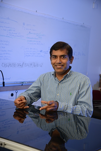
“What we’re interested in is the tools that allow us to understand the world at a very small scale,” said UCF professor Laurene Tetard, formerly of the Oak Ridge National Laboratory. “Not just the shape of the object, but its mechanical properties, its composition and how it evolves in time.”

An Aug. 10, 2015 UCF news release (also on EurekAlert), which originated the news item, describes the limitations of atomic force microscopy and gives a few details about the hybrid microscope (Note: A link has been removed),

For more than two decades, scientists have used atomic force microscopy – a probe that acts like an ultra-sensitive needle on a record player – to determine the surface characteristics of samples at the microscopic scale. A “needle” that comes to an atoms-thin point traces a path over a sample, mapping the surface features at a sub-cellular level [nanoscale].

But that technology has its limits. It can determine the topographical characteristics of [a] sample, but it can’t identify its composition. And with the standard tools currently used for chemical mapping, anything smaller than roughly half a micron is going to look like a blurry blob, so researchers are out of luck if they want to study what’s happening at the molecular level.

A team led by Tetard has come up with a hybrid form of that technology that produces a much clearer chemical image. As described Aug. 10 in the journal Nature Nanotechnology, Hybrid Photonic-Nanomechanical Force Microscopy (HPFM) can discern a sample’s topographic characteristics together with the chemical properties at a much finer scale.

The HPFM method is able to identify materials based on differences in the vibration produced when they’re subjected to different wavelengths of light – essentially a material’s unique “fingerprint.”

“What we are developing is a completely new way of making that detection possible,” said Tetard, who has joint appointments to UCF’s Physics Department, Material Science and Engineering Department and the NanoScience Technology Center.

The researchers proved the effectiveness of HPFM while examining samples from an eastern cottonwood tree, a potential source of biofuel. By examining the plant samples at the nanoscale, the researchers for the first time were able to determine the molecular traits of both untreated and chemically processed cottonwood inside the plant cell walls.

The research team included Tetard; Ali Passian, R.H. Farahi and Brian Davison, all of Oak Ridge National Laboratory; and Thomas Thundat of the University of Alberta.

Long term, the results will help reveal better methods for producing the most biofuel from the cottonwood, a potential boon for industry. Likewise, the new method could be used to examine samples of myriad plants to determine whether they’re good candidates for biofuel production.

Potential uses of the technology go beyond the world of biofuel. Continued research may allow HPFM to be used as a probe so, for instance, it would be possible to study the effect of new treatments being developed to save plants such as citrus trees from bacterial diseases rapidly decimating the citrus industry, or study fundamental photonically-induced processes in complex systems such as in solar cell materials or opto-electronic devices.

Here’s a link to and a citation for the paper,

Opto-nanomechanical spectroscopic material characterization by L. Tetard, A. Passian, R. H. Farahi, T. Thundat, & B. H. Davison. Nature Nanotechnology (2015) doi:10.1038/nnano.2015.168 Published online 10 August 2015

This paper is behind a paywall.

*ETA August 27, 2015:

August 10, 2015 ORNL news release (Note: Funding information and a link to the paper [previously given] have been removed):

A microscope being developed at the Department of Energy’s Oak Ridge National Laboratory will allow scientists studying biological and synthetic materials to simultaneously observe chemical and physical properties on and beneath the surface.

The Hybrid Photonic Mode-Synthesizing Atomic Force Microscope is unique, according to principal investigator Ali Passian of ORNL’s Quantum Information System group. As a hybrid, the instrument, described in a paper published in Nature Nanotechnology, combines the disciplines of nanospectroscopy and nanomechanical microscopy.

“Our microscope offers a noninvasive rapid method to explore materials simultaneously for their chemical and physical properties,” Passian said. “It allows researchers to study the surface and subsurface of synthetic and biological samples, which is a capability that until now didn’t exist.”

ORNL’s instrument retains all of the advantages of an atomic force microscope while simultaneously offering the potential for discoveries through its high resolution and subsurface spectroscopic capabilities.

“The originality of the instrument and technique lies in its ability to provide information about a material’s chemical composition in the broad infrared spectrum of the chemical composition while showing the morphology of a material’s interior and exterior with nanoscale – a billionth of a meter – resolution,” Passian said.

Researchers will be able to study samples ranging from engineered nanoparticles and nanostructures to naturally occurring biological polymers, tissues and plant cells.

The first application as part of DOE’s BioEnergy Science Center was in the examination of plant cell walls under several treatments to provide submicron characterization. The plant cell wall is a layered nanostructure of biopolymers such as cellulose. Scientists want to convert such biopolymers to free the useful sugars and release energy.

An earlier instrument, also invented at ORNL, provided imaging of poplar cell wall structures that yielded unprecedented topological information, advancing fundamental research in sustainable biofuels.

Because of this new instrument’s impressive capabilities, the researcher team envisions broad applications.
“An urgent need exists for new platforms that can tackle the challenges of subsurface and chemical characterization at the nanometer scale,” said co-author Rubye Farahi. “Hybrid approaches such as ours bring together multiple capabilities, in this case, spectroscopy and high-resolution microscopy.”

Looking inside, the hybrid microscope consists of a photonic module that is incorporated into a mode-synthesizing atomic force microscope. The modular aspect of the system makes it possible to accommodate various radiation sources such as tunable lasers and non-coherent monochromatic or polychromatic sources.

ETA2 August 27, 2015: I’ve received an email from one of the paper’s authors (RH Farahi of the US Oak Ridge National Laboratory [ORNL]) who claims some inaccuracies in this piece.  The news release supplied by the University of Central Florida states that Dr. Tetard led the team and that is not so. According to Dr. Farahi, she had a postdoctoral position on the team which she left two years ago. You might also get the impression that some of the work was performed at the University of Central Florida. That is not so according to Dr. Farahi.  As a courtesy Dr. Tetard was retained as first author of the paper.

*Nov. 4, 2015: I suspect some of the misunderstanding was due to overeagerness and/or time pressures. Whoever wrote the news release may have made some assumptions. It’s very easy to make a mistake when talking to an ebullient scientist who can unintentionally lead you to believe something that’s not so. I worked in a high tech company and believed that there was some new software being developed which turned out to be a case of high hopes. Luckily, I said something that triggered a rapid rebuttal to the fantasies. Getting back to this situation, other contributing factors could include the writer not having time to get the news release reviewed the scientist or the scientist skimming the release and missing a few bits due to time pressure.*

New $1 test for early stage prostate cancer more sensitive and exact than standard tests

An April 5, 2015 news item on Nanotechnology Now describes an exciting development in testing for cancer,

The simple test developed by University of Central Florida scientist Qun “Treen” Huo holds the promise of earlier detection of one of the deadliest cancers among men. It would also reduce the number of unnecessary and invasive biopsies stemming from the less precise PSA test that’s now used.

“It’s fantastic,” said Dr. Inoel Rivera, a urologic oncologist at Florida Hospital Cancer Institute, which collaborated with Huo on the recent pilot studies. “It’s a simple test. It’s much better than the test we have right now, which is the PSA, and it’s cost-effective.”

An April 3, 2015 University of Central Florida (UCF) news release by Mark Schlueb (also on EurekAlert), which originated the news item, describes the test in more detail,

When a cancerous tumor begins to develop, the body mobilizes to produce antibodies. Huo’s test detects that immune response using gold nanoparticles about 10,000 times smaller than a freckle.

When a few drops of blood serum from a finger prick are mixed with the gold nanoparticles, certain cancer biomarkers cling to the surface of the tiny particles, increasing their size and causing them to clump together.

Among researchers, gold nanoparticles are known for their extraordinary efficiency at absorbing and scattering light. Huo and her team at UCF’s NanoScience Technology Center developed a technique known as nanoparticle-enabled dynamic light scattering assay (NanoDLSay) to measure the size of the particles by analyzing the light they throw off. That size reveals whether a patient has prostate cancer and how advanced it may be.

And although it uses gold, the test is cheap. A small bottle of nanoparticles suspended in water costs about $250, and contains enough for about 2,500 tests.

“What’s different and unique about our technique is it’s a very simple process, and the material required for the test is less than $1,” Huo said. “And because it’s low-cost, we’re hoping most people can have this test in their doctor’s office. If we can catch this cancer in its early stages, the impact is going to be big.”

After lung cancer, prostate cancer is the second-leading killer cancer among men, with more than 240,000 new diagnoses and 28,000 deaths every year. The most commonly used screening tool is the PSA, but it produces so many false-positive results – leading to painful biopsies and extreme treatments – that one of its discoverers recently called it “hardly more effective than a coin toss.”

Pilot studies found Huo’s technique is significantly more exact. The test determines with 90 to 95 percent confidence that the result is not false-positive. When it comes to false-negatives, there is 50 percent confidence – not ideal, but still significantly higher than the PSA’s 20 percent – and Huo is working to improve that number.

The results of the pilot studies were published recently in ACS Applied Materials & Interfaces. Huo is also scheduled to present her findings in June at the TechConnect World Innovation Summit & Expo in suburban Washington, D.C.

Huo’s team is pursuing more extensive clinical validation studies with Florida Hospital and others, including the VA Medical Center Orlando. She hopes to complete major clinical trials and see the test being used by physicians in two to three years.

Huo also is researching her technique’s effectiveness as a screening tool for other tumors.

“Potentially, we could have a universal screening test for cancer,” she said. “Our vision is to develop an array of blood tests for early detection and diagnosis of all major cancer types, and these blood tests are all based on the same technique and same procedure.”

Huo co-founded Nano Discovery Inc., a startup company headquartered in a UCF Business Incubator, to commercialize the new diagnostic test. The company manufacturers a test device specifically for medical research and diagnostic purposes.

Here’s a link to and a citation for the study,

Gold Nanoparticle-Enabled Blood Test for Early Stage Cancer Detection and Risk Assessment by Tianyu Zheng, Nickisha Pierre-Pierre, Xin Yan, Qun Huo, Alvin J.O. Almodovar, Felipe Valerio, Inoel Rivera-Ramirez, Elizabeth Griffith, David D. Decker, Sixue Chen, and Ning Zhu. ACS Appl. Mater. Interfaces, 2015, 7 (12), pp 6819–6827 DOI: 10.1021/acsami.5b00371

Publication Date (Web): March 10, 2015

This paper is behind a paywall.

You can find out more about Huo’s company, Nano Discovery Inc. here.

Citrus canker, Florida, and Zinkicide

Found in Florida orchards in 2005, a citrus canker, citrus greening, poses a serious threat to the US state’s fruit industry. An April 2, 2105 news item on phys.org describes a possible solution to the problem,

Since it was discovered in South Florida in 2005, the plague of citrus greening has spread to nearly every grove in the state, stoking fears among growers that the $10.7 billion-a-year industry may someday disappear.

Now the U.S. Department of Agriculture has awarded the University of Florida a $4.6 million grant aimed at testing a potential new weapon in the fight against citrus greening: Zinkicide, a bactericide invented by a nanoparticle researcher at the University of Central Florida.

An April 2, 2015 University of Central Florida news release by Mark Schlueb (also on EurekAlert), which originated the news item, describes the problem and the solution (Zinkicide),

Citrus greening – also known by its Chinese name, Huanglongbing, or HLB – causes orange, grapefruit and other citrus trees to produce small, bitter fruit that drop prematurely and is unsuitable for sale or juice. Eventually, infected trees die. Florida has lost tens of thousands of acres to the disease.

“It’s a hundred-year-old disease, but to date there is no cure. It’s a killer, a true killer for the citrus industry,” said Swadeshmukul Santra, associate professor in the NanoScience Technology Center at UCF.

The bacteria that causes HLB is carried by the Asian citrus psyllid, a tiny insect that  feeds on leaves and stems of infected citrus trees, then carries the bacteria to healthy trees.

Zinkicide, developed by Santra, is designed to kill the bacteria.

The $4.6 million grant is the largest of five totaling $23 million that were recently announced by the USDA’s National Institute of Food and Agriculture.

The evaluation of Zinkicide is a multi-institute project involving 13 investigators from six institutions. Evan Johnson of UF’s [University of Florida] Citrus Research and Education Center at Lake Alfred is the project director, and there are a dozen co-principal investigators from UF, UCF, Oak Ridge National Laboratory (ORNL), Auburn University, New Mexico State University and The Ohio State University.

”Managing systemic diseases like HLB is a difficult challenge that has faced plant pathologists for many years,” said Johnson “It is a privilege to work with an excellent team of researchers from many different disciplines with the goal of developing new tools that are both effective and safe.”

A portion of the grant money, $1.4 million, flows to UCF, where Santra leads a team that also includes Andre Gesquiere, Laurene Tetard and the Oak Ridge National Laboratory collaborator, Loukas Petridis.

HLB control is difficult because current bactericidal sprays, such as copper, simply leave a protective film on the outside of a plant. The insect-transmitted bacteria bypasses that barrier and lives inside a tree’s fruit, stems and roots, in the vascular tissue known as the phloem. There, it deprives the tree of carbohydrate and nutrients, causing root loss and ultimately death. For a bactericide to be effective against HLB, it must be able to move within the plant, too.

Zinkicide is a nanoparticle smaller than a single microscopic cell, and researchers are cautiously optimistic it will be able to move systemically from cell to cell to kill the bacteria that cause HLB.

“The bacteria hide inside the plant in the phloem region,” Santra said. “If you spray and your compound doesn’t travel to the phloem region, then you cannot treat HLB.”

Zinkicide is derived from ingredients which are found in plants, and is designed to break down and be metabolized after its job is done. [emphasis mine]

It’s the first step in a years-long process to bring a treatment to market. UF will lead five years of greenhouse and field trials on grapefruit and sweet orange to determine the effectiveness of Zinkicide and the best method and timing of application.

The project also includes research to study where the nanoparticles travel within the plant, understand how they interact with plant tissue and how long they remain before breaking down. [emphasis mine]

If effective, the bactericide could have a substantial role in combatting HLB in Florida, and in other citrus-producing states and countries. It would also likely be useful for control of other bacterial pathogens infecting other crops.

The Zinkicide project builds as a spinoff from previous collaborations between Santra and UF’s Jim Graham, at the Citrus Research and Education Center to develop alternatives to copper for citrus canker control.

The previous Citrus Research and Education Foundation (CRDF)-funded Zinkicide project has issued three reports, for June 30, 2014, Sept. 30, 2014, and Dec. 31, 2014. This project’s completion date is May 2015. The reports which are remarkably succinct, consisting of two paragraphs, can be found here.

Oddly, the UCF news release doesn’t mention that Zinkicide (although it can be inferred) is a zinc particulate (I’m guessing they mean zinc nanoparticle) as noted on the CRDF project webpage. Happily, they are researching what happens after the bactericide has done its work on the infection. It’s good to see a life cycle approach to this research.

3D television is resurrected by way of a nanocomposite

A Feb. 10, 2014 University of Central Florida news release by Barbara Abney (also on EurekAlert) tells the tale of a researcher working on the development of 3D images on television,

Gone are the goofy glasses required of existing sets. Instead, assistant professor Jayan Thomas is working on creating the materials necessary to create a 3-D image that could be seen from 360 degrees with no extra equipment.

“The TV screen should be like a table top,” Thomas said. “People would sit around and watch the TV from all angles like sitting around a table. Therefore, the images should be like real-world objects. If you watch a football game on this 3-D TV, you would feel like it is happening right in front of you. A holographic 3-D TV is a feasible direction to accomplish this without the need of glasses.”

His work is so far along that the National Science Foundation has given him a $400,000 grant over five years to develop the materials needed to produce display screens.

Here’s an image of Thomas sitting mimicking the experience of his 3D television at a tabletop,

UCF Researcher Jaden Thomas uses nantechnology to bring 3-D television back to life.

UCF [University of Central Florida] Researcher Jaden Thomas uses nantechnology to bring 3-D television back to life.

Thomas’ work comes at a very interesting juncture for the industry (from the news release),

When 3-D TVs first came on the market in 2010, there was a lot of hype and the market expected the new sets would take off. Several broadcasters even pledged to create special channels for 3-D programming, such as ESPN and the BBC.

But in the past year, those broadcasters have canceled plans because sales have lagged and the general public hasn’t adopted the sets as hoped. Some say that’s because the television sets are expensive and require bulky equipment and glasses.

Here’s how Thomas’ approach differs, in very general terms (from the news release),

Thomas’ approach would use new plastic composites made with nanotechnology to make the 3-D image recording process multitudes faster than currently possible. This would eliminate the need for glasses.

Thomas and his colleagues have developed the specific plastic composite needed to create the display screens necessary for effectively showing the 3-D images. That work has been published in the journals Nature and Advanced Materials.

There’s more about Dr. Thomas along with listings of his publications on his NanoScience Technology Center faculty page.

ETA Feb. 14, 2014: You may want to read Dexter Johnson’s Feb. 14, 2014 posting on his Nanoclast blog (on the IEEE [Institute for Electrical and Electronics Engineers]) concerning 3D televisions and Thomas’ work (Note: A link has been removed),

 At this year’s Consumer Electronics Show (CES), it became clear that the much-ballyhooed age of 3-D TV was coming to a quiet and uncelebrated end. One of the suggested causes of its demise was the cost of the 3D glasses. If you wanted to invite a group over to watch the big sporting event, you had better have a lot of extra pairs on hand, which might cost you a small fortune.

Eliminating the glasses from the experience has been proposed from the first moment 3-D TVs were introduced to the marketplace.

Dexter goes on to provide technical context for Thomas’ work as he expands on his theme.