Tag Archives: Ulsan National Institute of Science and Technology (UNIST)

Memristor artificial neural network learning based on phase-change memory (PCM)

Caption: Professor Hongsik Jeong and his research team in the Department of Materials Science and Engineering at UNIST. Credit: UNIST

I’m pretty sure that Professor Hongsik Jeong is the one on the right. He seems more relaxed, like he’s accustomed to posing for pictures highlighting his work.

Now on to the latest memristor news, which features the number 8.

For anyone unfamiliar with the term memristor, it’s a device (of sorts) which scientists, involved in neuromorphic computing (computers that operate like human brains), are researching as they attempt to replicate brainlike processes for computers.

From a January 22, 2021 Ulsan National Institute of Science and Technology (UNIST) press release (also on EurekAlert but published March 15, 2021),

An international team of researchers, affiliated with UNIST has unveiled a novel technology that could improve the learning ability of artificial neural networks (ANNs).

Professor Hongsik Jeong and his research team in the Department of Materials Science and Engineering at UNIST, in collaboration with researchers from Tsinghua University in China, proposed a new learning method to improve the learning ability of ANN chips by challenging its instability.

Artificial neural network chips are capable of mimicking the structural, functional and biological features of human neural networks, and thus have been considered the technology of the future. In this study, the research team demonstrated the effectiveness of the proposed learning method by building phase change memory (PCM) memristor arrays that operate like ANNs. This learning method is also advantageous in that its learning ability can be improved without additional power consumption, since PCM undergoes a spontaneous resistance increase due to the structural relaxation after amorphization.

ANNs, like human brains, use less energy even when performing computation and memory tasks, simultaneously. However, the artificial neural network chip in which a large number of physical devices are integrated has a disadvantage that there is an error. The existing artificial neural network learning method assumes a perfect artificial neural network chip with no errors, so the learning ability of the artificial neural network is poor.

The research team developed a memristor artificial neural network learning method based on a phase-change memory, conceiving that the real human brain does not require near-perfect motion. This learning method reflects the “resistance drift” (increased electrical resistance) of the phase change material in the memory semiconductor in learning. During the learning process, since the information update pattern is recorded in the form of increasing electrical resistance in the memristor, which serves as a synapse, the synapse additionally learns the association between the pattern it changes and the data it is learning.

The research team showed that the learning method developed through an experiment to classify handwriting composed of numbers 0-9 has an effect of improving learning ability by about 3%. In particular, the accuracy of the number 8, which is difficult to classify handwriting, has improved significantly. [emphasis mine] The learning ability improved thanks to the synaptic update pattern that changes differently according to the difficulty of handwriting classification.

Researchers expect that their findings are expected to promote the learning algorithms with the intrinsic properties of memristor devices, opening a new direction for development of neuromorphic computing chips.

Here’s a link to and a citation for the paper,

Spontaneous sparse learning for PCM-based memristor neural networks by Dong-Hyeok Lim, Shuang Wu, Rong Zhao, Jung-Hoon Lee, Hongsik Jeong & Luping Shi. Nature Communications volume 12, Article number: 319 (2021) DOI: https://doi.org/10.1038/s41467-020-20519-z Published 12 January 2021

This paper is open access.

Eradicating bacteria biofilm with nanocrystals

A January 8, 2021 news item on ScienceDaily announces new work from South Korea’s Pohang University of Science & Technology (POSTECH),

The COVID-19 pandemic is raising fears of new pathogens such as new viruses or drug-resistant bacteria. To this, a Korean research team has recently drawn attention for developing the technology for removing antibiotic-resistant bacteria by controlling the surface texture of nanomaterials.

A joint research team from POSTECH and UNIST [Ulsan National Institute of Science and Technology] has introduced mixed-FeCo-oxide-based surface-textured nanostructures (MTex) as highly efficient magneto-catalytic platform in the international journal Nano Letters. The team consisted of professors In Su Lee and Amit Kumar with Dr. Nitee Kumari of POSTECH’s Department of Chemistry and Professor Yoon-Kyung Cho and Dr. Sumit Kumar of UNIST’s Department of Biomedical Engineering.

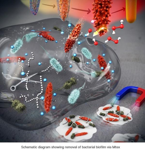
Caption: Schematic diagram showing removal of bacterial biofilm via Mtex Credit: POSTECH

A January 8, 2021 POSTECH press release (also on EurkeAlert), which originated the news item, delves further,

First, the researchers synthesized smooth surface nanocrystals in which various metal ions were wrapped in an organic polymer shell and heated them at a very high temperature. While annealing the polymer shell, a high-temperature solid-state chemical reaction induced mixing of other metal ions on the nanocrystal surface, creating a number of few-nm-sized branches and holes on it. This unique surface texture was found to catalyze a chemical reaction that produced reactive oxygen species (ROS) that kills the bacteria. It was also confirmed to be highly magnetic and easily attracted toward the external magnetic field. The team had discovered a synthetic strategy for converting normal nanocrystals without surface features into highly functional mixed-metal-oxide nanocrystals.

The research team named this surface topography – with branches and holes that resembles that of a ploughed field – “MTex.” This unique surface texture has been verified to increase the mobility of nanoparticles to allow efficient penetration into biofilm matrix while showing high activity in generating reactive oxygen species (ROS) that are lethal to bacteria.

This system produces ROS over a broad pH range and can effectively diffuse into the biofilm and kill the embedded bacteria resistant to antibiotics. And since the nanostructures are magnetic, biofilm debris can be scraped out even from the hard-to-reach microchannels.

“This newly developed MTex shows high catalytic activity, distinct from the stable smooth-surface of the conventional spinel forms,” explained Dr. Amit Kumar, one of the corresponding authors of the paper. “This characteristic is very useful in infiltrating biofilms even in small spaces and is effective in killing the bacteria and removing biofilms.”

“This research allows to regulate the surface nanotexturization, which opens up possibilities to augment and control the exposure of active sites,” remarked Professor In Su Lee who led the research. “We anticipate the nanoscale-textured surfaces to contribute significantly in developing a broad array of new enzyme-like properties at the nano-bio interface.”

Here’s a link to and a citation for the paper,

Surface-Textured Mixed-Metal-Oxide Nanocrystals as Efficient Catalysts for ROS Production and Biofilm Eradication by Nitee Kumari, Sumit Kumar, Mamata Karmacharya, Sateesh Dubbu, Taewan Kwon, Varsha Singh, Keun Hwa Chae, Amit Kumar, Yoon-Kyoung Cho, and In Su Lee. Nano Lett. 2021, 21, 1, 279–287 DOI: https://doi.org/10.1021/acs.nanolett.0c03639 Publication Date: December 11, 2020 Copyright © 2020 American Chemical Society

This paper is behind a paywall.

Cosmetics breakthrough for Ulsan National Institute of Science and Technology (UNIST)?

Cosmetics would not have been my first thought on reading the title for the paper (“Rates of cavity filling by liquids”) produced  by scientists from Ulsan National Institute of Science and Technology (UNIST).

A September 17, 2018 news item on Nanowerk announces the research,

A research team, affiliated with Ulsan National Institute of Science and Technology (UNIST) has examined the rates of liquid penetration on rough or patterned surfaces, especially those with pores or cavities. Their findings provide important insights into the development of everyday products, including cosmetics, paints, as well as industrial applications, like enhanced oil recovery.

This study has been jointly led by Professor Dong Woog Lee and his research team in the School of Energy and Chemical Engineering at UNIST and a research team in the University of California, Santa Barbara. Published online in the July 19th issue of the Proceedings of the National Academy of Sciences (“Rates of cavity filling by liquids”), the study identifies five variables that control the cavity-filling (wetting transition) rates, required for liquids to penetrate into the cavities.

A July 26, 2018 UNIST press release (also on EurekAlert but published on September 17, 2018), which originated the news item, delves further into the work,

In the study, Professor Lee fabricated silicon wafers with cylindrical cavities of different geometries. After immersing them in bulk water, they observed the details of, and the rates associated with, water penetration into the cavities from the bulk, using bright-field and confocal fluorescence microscopy. Cylindrical cavities are like skin pores with narrow entrance and specious interior. The cavity filling generally progresses when bulk water is spread above a hydrophilic, reentrant cavity. As described in “Wetting Transition from the Cassie–Baxter State to Wenzel State”, the liquid droplet that sits on top of the textured surface with trapped air underneath will be completely absorbed by the rough surface cavities.

Their findings revealed that the cavity-filling rates are affected by the following variables: (i) the intrinsic contact angle, (ii) the concentration of dissolved air in the bulk water phase, (iii) the liquid volatility that determines the rate of capillary condensation inside the cavities, (iv) the types of surfactants, and (v) the cavity geometry.

“Our results can used in the manufacture of special-purpose cosmetic products,” says Professor Lee. “For instance, pore minimizing face primers and facial cleansers that remove sebum need to reduce the amount of dissolved air, so that they can penetrate into the pores quickly.”

On the other hand, beauty products, like sunscreens should be designed to protect the skin from harmful sun, while preventing pores clogging. Because, clogged pores hinder the skin’s function of breathing or exchange of carbon dioxide and then cause further irritation, pimples, and blemished areas on your skin. In this case, it is better to reduce volatility and increase the amount of dissolved air in the cosmetic products, as opposed to facial cleansers.

“This knowledge of how cavities under bulk water are filled and what variables control the rate of filling can provide insights into the engineering of temporarily or permanently superhydrophobic surfaces, and the designing and manufacturing of various products that are applied to rough, textured, or patterned surfaces,” says Professor Lee. “Many of the fundamental insights gained can also be applied to other liquids (e.g., oils), contact angles, and cavities or pores of different dimensions or geometries.”

This study has been supported by the National Research Foundation of Korea (NRF) grant, funded by the Ministry of Science and ICT.

Here’s a link to and a citation for the paper,

Rates of cavity filling by liquids by Dongjin Seo, Alex M. Schrader, Szu-Ying Chen, Yair Kaufman, Thomas R. Cristiani, Steven H. Page, Peter H. Koenig, Yonas Gizaw, Dong Woog Lee, and Jacob N. Israelachvili. PNAS August 7, 2018 115 (32) 8070-8075 https://doi.org/10.1073/pnas.1804437115 Published ahead of print July 19, 2018

This paper is behind a paywall.

A new type of diode from South Korea’s Ulsan National Institute of Science and Technology

A Feb. 8, 2017 news item on phys.org features a ‘dream’ diode from Ulsan National Institute of Science and Technology,

A team of researchers, affiliated with UNIST [Ulsan National Institute of Science and Technology] has created a new technique that greatly enhances the performance of Schottky Diodes (metal-semiconductor junction) used in electronic devices. Their research findings have attracted considerable attention within the scientific community by solving the contact resistance problem of metal-semiconductor, which had remained unsolved for almost 50 years.

As described in the January [2017] issue of Nano Letters, the researchers have created a new type of diode with a graphene insertion layer sandwiched between metal and semiconductor. This new technique blows all previous attemps out the water, as it is expected to significantly contribute to the semiconductor industry’s growth.

A Jan. 27, 2017 UNIST press release, (also on EurekAlert), which originated the news item, describes the research in greater detail,

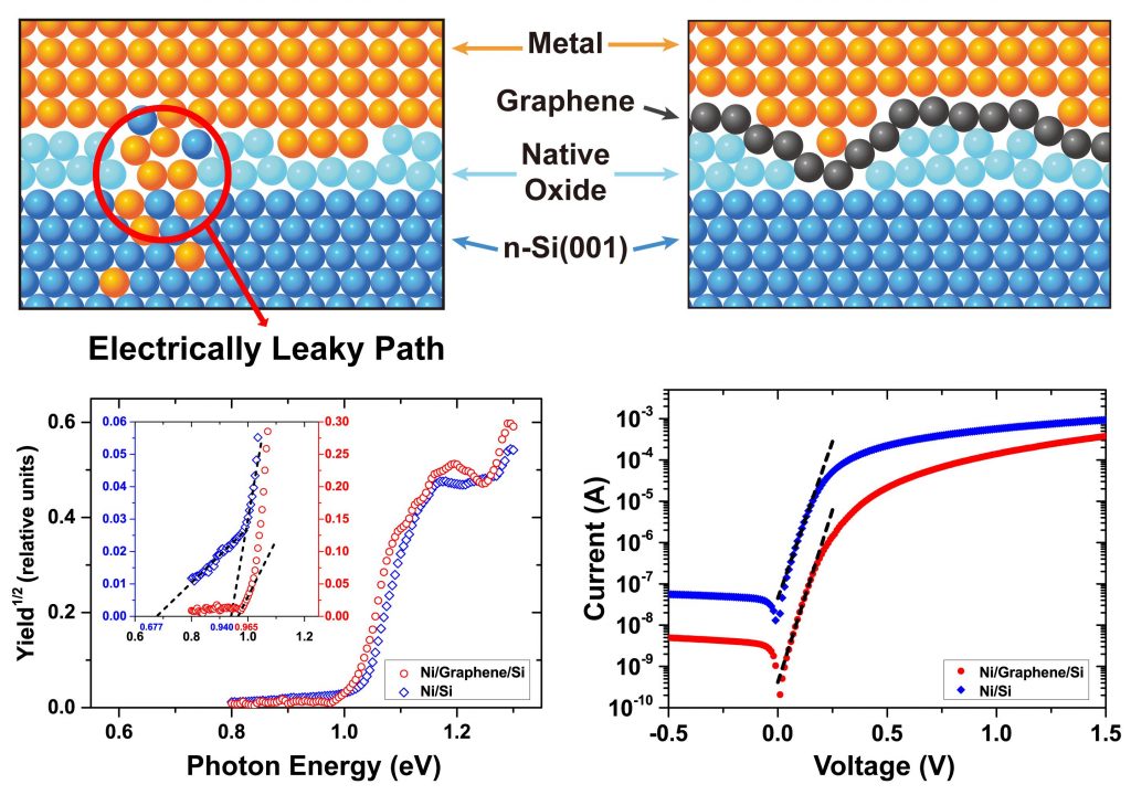
The Schottky diode is one of the oldest and most representative semiconductor devices, formed by the junction of a semiconductor with a metal.  However, due to the atomic intermixing along the interface between two materials, it has been impossible to produce an ideal diode. (An ideal diode acts like a perfect conductor when voltage is applied forward biased and like a perfect insulator when voltage is applied reverse biased.)

graphene interlayer 2

The schematic view of internal photoemission (IPE) measurements on metal/n-Si(001) junctions with Ni, Pt, and Ti electrodes for with and without a graphene insertion layer.

Professor Kibog Park of Natural Science solved this problem by inserting a graphene layer at the metal-semiconductor interface. In the study, the research team demonstrated that this graphene layer, consisting of a single layer of carbon atoms can not only suppress the material intermixing substantially, but also matches well with the theoretical prediction.

“The sheets of graphene in graphite have a space between each sheet that shows a high electron density of quantum mechanics in that no atoms can pass through,” says Professor Park. “Therefore, with this single-layer graphene sandwiched between metal and semiconductor, it is possible to overcome the inevitable atomic diffusion problem.”

The study also has the physiological meaning of confirming the theoretical prediction that “In the case of silicon semiconductors, the electrical properties of the junction surfaces hardly change regardless of the type of metal they use,” according to Hoon Hahn Yoon (Combined M.S./Ph.D. student of Natural Science), the first author of the study.

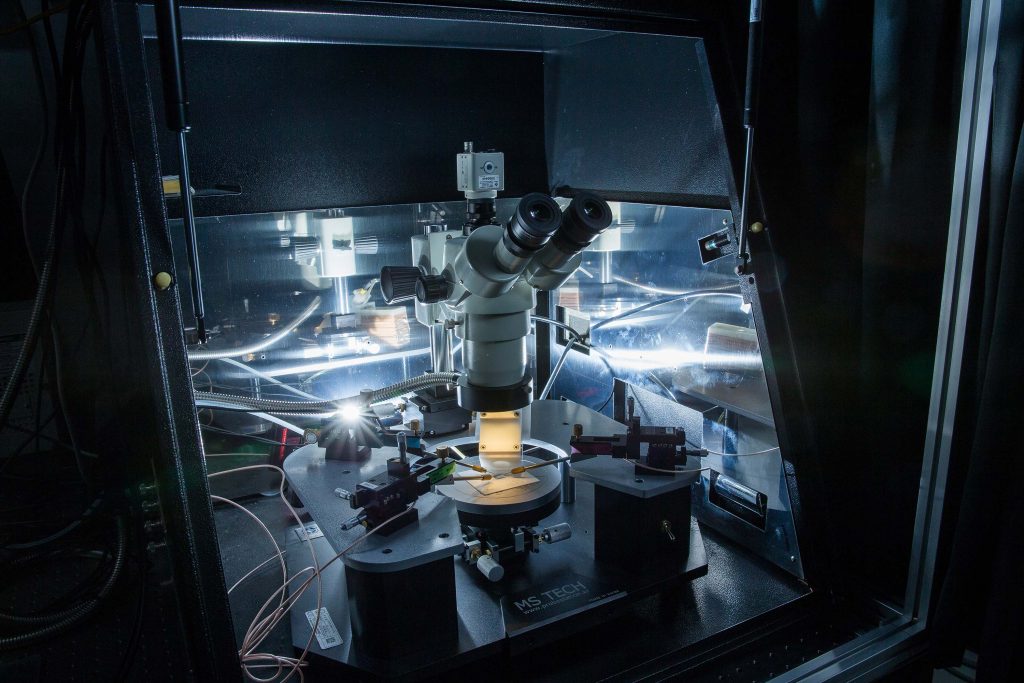
The internal photoemission method was used to measure the electronic energy barrier of the newly-fabricated metal/graphene/n-Si(001) junction diodes. The Internal Photoemission (IPE) Measurement System in the image shown above has contributed greatly to these experiments. This system has been developed by four UNIST graduate students (Hoon Han Yoon, Sungchul Jung, Gahyun Choi, and Junhyung Kim), which was carried out as part of an undergraduate research project in 2012 and was supported by the Korea Foundation for the Advancement of Science and Creativity (KOFAC).

This is the internal photoemission (IPE) measurement system, developed by Hoon Hahn Yoon of Physics at UNIST.

Shown above is the Internal Photoemission (IPE) Measurement System, developed by Hoon Hahn Yoon, combined M.S./Ph.D. student of Natural Science at UNIST.

“Students have teamed up and carried out all the necessary steps for the research since they were undergraduates,” Professor Park says. “Therefore, this research is a perfect example of time, persistence, and patience paying off.”

This study has been jointly conducted by Professor Hu Young Jeong of the UNIST Central Research Facilities (UCRF), Professor Kwanpyo Kim of Natural Science, Professor Soon-Yong Kwon of Materials Science and Engineering, and Professor Yong Soo Kim of Ulsan University. It has been also supported by the National Research Foundation of Korea, Nuclear Research Basis Expansion Project, as well as the Global Ph.D Fellowship (GPF).

Here’s a link to and a citation for the paper,

Strong Fermi-Level Pinning at Metal/n-Si(001) Interface Ensured by Forming an Intact Schottky Contact with a Graphene Insertion Layer by Hoon Hahn Yoon, Sungchul Jung, Gahyun Choi, Junhyung Kim, Youngeun Jeon, Yong Soo Kim, Hu Young Jeong, Kwanpyo Kim, Soon-Yong Kwon, and Kibog Park. Nano Lett., 2017, 17 (1), pp 44–49 DOI: 10.1021/acs.nanolett.6b03137 Publication Date (Web): December 14, 2016

Copyright © 2016 American Chemical Society

This paper is behind a paywall.

New design strategy for synthesizing metal-organic frameworks (MOFs)

A Jan. 24, 2017 news item on Nanowerk announces new research from South Korea,

The accurate interpretation of particle sizes and shapes in nanoporus materials is essential to understanding and optimizing the performance of porous materials used in many important existing and potentially new applications. However, only a few experimental techniques have been developed for this purpose.

A team of researchers, led by Professor Wonyoung Choe of Natural Science and Professor Ja Hun Kwak of Energy and Chemical Engineering [ at Ulsan National Institute of Science and Technology {UNIST}] has recently developed a novel design strategy for synthesizing various forms of functional materials, especially for metal-organic materials (MOMs).

The research team expects that this synthetic approach might open up a new direction for the development of diverse forms in MOMs, with highly advanced areas such as sequential drug delivery/release and heterogeneous cascade catalysis targeted in the foreseeable future.

A Jan. 6, 2017 UNIST press release, which originated the news item, provides more detail,

In the last decades, much research has been developed to the synthesis and design of functional materials, but only a few of them could control the walls of the interior of the particles within the nanoporous materials.

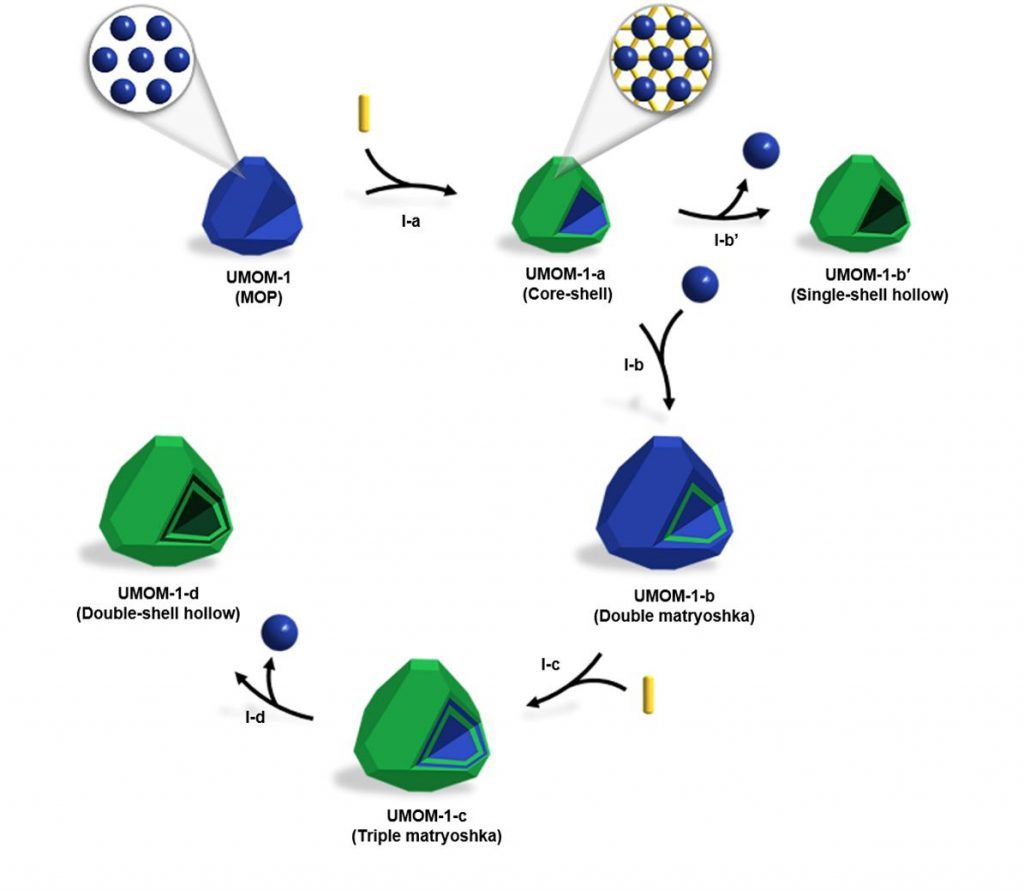
In the study, Professor Choe and his team denomstrated sequential self-assembly strategy for synthesizing various forms of MOM crystals, including double-shell hollow MOMs, based on single-crystal to single-crystal transformation from MOP to MOF.

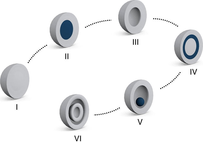
Schematic representation of various forms of micro-/nanostructures. From left are Solid, core-shell, hollow, matryoshka, yolk-shell and multi-shell hollow structures.

Porous materials are highly utilized as catalysts or gas capture materials because they supply abundant surface active sites for chemical reaction. Although materials, like Zeolites, which can be obtained from nature, have the ability to act as catalysts for chemical reactions, they suffer from the difficulty of controlling pore sizes and shapes.

As one solution, scientists have developed self-assembled porous materials using organic molecules and metals. Metal-Organic Frameworks (MOFs) and Metal-Organic Polyhedral (MOPs) are notable examples and they both have holes all over their surfaces. MOPs dissolve easily in chemical solvent, while MOFs are practically insoluble.

“MOFs take the form of three-dimensional (3D) structure, linking metals with organic molecules, while MOPs agglomerate together to form larger clusters,” says Jiyoung Lee, the first contributor of the study and a graduate student in the combined master-doctoral program from Chemistry department.

Schematic illustration of form evolution.

Schematic illustration of form evolution.

According to the research team, this synthetic strategy also yields other forms, such as solid, core-shell, double and triple matryoshka, and single-shell hollow MOMs, thereby exhibiting form evolution in MOMs.

“The best feature of this technique is that it allows two very different substances to coexist within a single crystal,” says Professor Choe. “This technique also permits greater control over size and shape of the pore, which can be then used to regulate the entrance and exit of molecules.”

This particular synthetic approach also has the potential to generate new type of porous materials containing micropores with diameters less than 2nm, macropores with diameters between 20 to 50nm, as well as pores of larger than 50 nm. Such hierarchical pore structure plays a critical role during catalysis, adsorption, and separation processes.

Here’s a link to and a citation for the paper,

Evolution of form in metal–organic frameworks by Jiyoung Lee, Ja Hun Kwak & Wonyoung Choe. Nature Communications 8, Article number: 14070 (2017) doi:10.1038/ncomms14070 Published online: 04 January 2017

This is an open access paper.

Not exactly ‘Prey’: self-organizing materials that can mimic swarm behaviour

Prey, a 2002 novel by Michael Crichton, focused on nanotechnology and other emerging technologies and how their development could lead to unleashing swarms of nanobots with agendas of their own. Crichton’s swarms had collective artificial intelligence, and could massive themselves together to take on different macroscale shapes to achieve their own ends. This latest development has nowhere near that potential—not yet and probably never. From a July 21, 2016 news item on ScienceDaily,

A new study by an international team of researchers, affiliated with Ulsan National Institute of Science and Technology (UNIST) [Korea] has announced that they have succeeded in demonstarting control over the interactions occurring among microscopic spheres, which cause them to self-propel into swarms, chains, and clusters.

The research published in the current online edition of Nature Materials takes lessons from cooperation in nature, including that observed in honey bee swarms and bacterial clusters. In the study, the team has successfully demonstrated the self-organizing pattern formation in active materials at microscale by modifying only one parameter.

A July 21, 2016 UNIST press release, which originated the news item, expands on the theme,

This breakthrough comes from a research, conducted by Dr. Steve Granick (School of Natural Science, UNIST) of IBS Center for Soft and Living Matter in collaboration with Dr. Erik Luijten from Northwestern University. Ming Han, a PhD student in Luijten’s laboratory, and Jing Yan, a former graduate student at the University of Illinois, served as co-first authors of the paper.

Researchers expect that such active particles could open a new class of technologies with applications in medicine, chemistry, and engineering as well as advance scientists’ fundamental understanding of collective, dynamic behavior in systems.

According to the research team, the significance of team work was stressed by both Dr. Luijten and Dr. Granick as this current breakthrough is part of a longtime partnership using a new class of soft-matter particles known as Janus colloids, which Dr. Granick had earlier created in his laboratory. The theoretical computer simulations were completed by the team, led by Dr. Luijten and Dr. Granick used these colloids to experimentally test the collective, dynamic behavior in the laboratory.

The micron-sized spheres, typically suspended in solution, were named after the Roman god with two faces as they have attractive interactions on one side and negative charges on the other side.

The electrostatic interactions between the two sides of the self-propelled spheres could be manipulated by subjecting the colloids to an electric field. Some experienced stronger repulsions between their forward-facing sides, while others went through the opposite. Along with them, another set remained completely neutral. This imbalance caused the self-propelled particles to swim and self-organize into one of the following patterns, which are swarms, chains, clusters and isotropic gases.

To avoid head-to-head collisions, head-repulsive particles swam side-by-side, forming into swarms. Depending on the electric-field frequency, tail-repulsive particles positioned their tails apart, thus encouraging them to face each other to form jammed clusters of high local density. Also, swimmers with equal-and-opposite charges attracted one another into connected chains.

Dr. Granick states, “This truly is a joint work of the technological know-how by the Korean IBS and the University of Illinois, as well as the computer simulations technology by Northwestern University.” He expects that this breakthrough has probable application in sensing, drug delivery, or even microrobotics.

With this discovery, a drug could be placed within particles, for instance, that cluster into the delivery spot. Moreover, alterations in the environment could be perceived if the system unexpectedly switches from swarming to forming chains.

Here’s a link to and a citation for the paper,

Reconfiguring active particles by electrostatic imbalance by Jing Yan, Ming Han, Jie Zhang, Cong Xu, Erik Luijten, & Steve Granick. Nature Materials (2016)  doi:10.1038/nmat4696 Published online 11 July 2016

This paper is behind a paywall.

Inspiration from the sea for titanium implants (mussels) and adhesive panels for flexible sensors (octopuses/octopi/octopodes)

I have two sea-inspired news bits both of which concern adhesion.

Mussels and titanium implants

A July 8, 2016 news item on ScienceDaily features some mussel-inspired research from Japan into how to make better titanium implants,

Titanium is used medically in applications such as artificial joints and dental implants. While it is strong and is not harmful to tissues, the metal lacks some of the beneficial biological properties of natural tissues such as bones and natural teeth. Now, based on insights from mussels–which are able to attach themselves very tightly to even metallic surfaces due to special proteins found in their byssal threads–scientists from RIKEN have successfully attached a biologically active molecule to a titanium surface, paving the way for implants that can be more biologically beneficial.

A July 11, 2016 RIKEN press release (also on EurekAlert but dated July 8, 2016), which originated the news item, provides more information,

The work began from earlier discoveries that mussels can attach to smooth surfaces so effectively thanks to a protein, L-DOPA, which is known to be able to bind very strongly to smooth surfaces such as rocks, ceramics, or metals (…). Interestingly, the same protein functions in humans as a precursor to dopamine, and is used as a treatment for Parkinson’s disease.

According to Chen Zhang of the RIKEN Nano Medical Engineering Laboratory, the first author of the paper published in Angewandte Chemie, “We thought it would be interesting to try to use various techniques to attach a biologically active protein—in our case we chose insulin-like growth factor-1, a promoter of cell proliferation—to a titanium surface like those used in implants” (…).

Using a combination of recombinant DNA technology and treatment with tyrosinase, they were able to create a hybrid protein that contained active parts of both the growth factor and L-DOPA. Tests showed that the proteins were able to fold normally, and further experiments in cell cultures demonstrated that the IGF-1 was still functioning normally. Thanks to the incorporation of the L-DOPA, the team was able to confirm that the proteins bound strongly to the titanium surface, and remained attached even when the metal was washed with phosphate-buffered saline, a water-based solution. Zhang says, “This is similar to the powerful properties of mussel adhesive, which can remain fixed to metallic materials even underwater.”

According to Yoshihiro Ito, Team Leader of the Emergent Bioengineering Research Team of the RIKEN Center for Emergent Matter Science, “We are very excited by this finding, because the modification process is a universal one that could be used with other proteins. It could allow us to prepare new cell-growth enhancing materials, with potential applications in cell culture systems and regenerative medicine. And it is particularly interesting that this is an example of biomimetics, where nature can teach us new ways to do things. The mussel has given us insights that could be used to allow us to live healthier lives.”

The work was done by RIKEN researchers in collaboration with Professor Peibiao Zhang of the Changchun Institute of Applied Chemistry, Chinese Academy of Sciences, and Professor Yi Wang of the School of Pharmaceutical Sciences, Jilin University. The work was partially supported by the Japan Society for the Promotion of Science KAKENHI (Grant Number 15H01810 and 22220009), CAS-JSPS joint fund (GJHZ1519), and RIKEN MOST joint project.

Here’s a link to and a citation for the paper,

A Bioorthogonal Approach for the Preparation of a Titanium-Binding Insulin-like Growth-Factor-1 Derivative by using Tyrosinase by Chen Zhang, Hideyuki Miyatake, Yu Wang, Takehiko Inaba, Yi Wang, Peibiao Zhang, and Prof. Yoshihiro Ito. Angewandte Chemie International Edition DOI: 10.1002/anie.201603155 Version of Record online: 6 JUL 2016

© 2016 WILEY-VCH Verlag GmbH & Co. KGaA, Weinheim

This paper is behind a paywall.

Octopuses/octopi/octopodes and adhesive panels

Before launching into the science part of this news bit, here’s some grammar (from the Octopus Wikipedia entry; Note: Links have been removed),

The standard pluralized form of “octopus” in the English language is “octopuses” /ˈɒktəpʊsɪz/,[10] although the Ancient Greek plural “octopodes” /ɒkˈtɒpədiːz/, has also been used historically.[9] The alternative plural “octopi” — which misguidedly assumes it is a Latin “-us”-word — is considered grammatically incorrect.[11][12][13][14] It is nevertheless used enough to make it notable, and was formally acknowledged by the descriptivist Merriam-Webster 11th Collegiate Dictionary and Webster’s New World College Dictionary. The Oxford English Dictionary (2008 Draft Revision)[15] lists “octopuses”, “octopi”, and “octopodes”, in that order, labelling “octopodes” as rare and noting that “octopi” derives from the apprehension that octōpus comes from Latin.[16] In contrast, New Oxford American Dictionary (3rd Edition 2010) lists “octopuses” as the only acceptable pluralization, with a usage note indicating “octopodes” as being still occasionally used but “octopi” as being incorrect.[17]

Now the news. A July 12, 2016 news item on Nanowerk highlights some research into adhesives and octopuses,

With increased study of bio-adhesives, a significant effort has been made in search for novel adhesives that will combine reversibility, repeated usage, stronger bonds and faster bonding time, non-toxic, and more importantly be effective in wet and other extreme conditions.

A team of Korean scientists-made up of scientists from Korea Institute of Science and Technology (KIST) and UNIST has recently found a way to make building flexible pressure sensors easier–by mimicking the suction cups on octopus’s tentacles.

A July 5, 2016 UNIST (Ulsan National Institute of Science and Technology) press release, which originated the news item, provides more information,

According to the research team, “Although flexible pressure sensors might give future prosthetics and robots a better sense of touch, building them requires a lot of laborious transferring of nano- and microribbons of inorganic semiconductor materials onto polymer sheets.”

In search of an easier way to process this transfer printing, Prof. Hyunhyub Ko (School of Energy and Chemical Engineering, UNIST) and his colleagues turned to the octopus suction cups for inspiration.

An octopus uses its tentacles to move to a new location and uses suction cups underneath each tentacle to grab onto something. Each suction cup contains a cavity whose pressure is controlled by surrounding muscles. These can be made thinner or thicker on demand, increasing or decreasing air pressure inside the cup, allowing for sucking and releasing as desired.

By mimicking muscle actuation to control cavity-pressure-induced adhesion of octopus suckers, Prof. Ko and his team engineered octopus-inspired smart adhesive pads. They used the rubbery material polydimethylsiloxane (PDMS) to create an array of microscale suckers, which included pores that are coated with a thermally responsive polymer to create sucker-like walls.

The team discovered that the best way to replicate organic nature of muscle contractions would be through applied heat. Indeed, at room temperature, the walls of each pit sit in an ‘open’ state, but when the mat is heated to 32°C, the walls contract, creating suction, therby allowing the entire mate to adhere to a material (mimicking the suction function of an octopus). The adhesive strength also spiked from .32 kilopascals to 94 kilopascals at high temperature.

The team reports that the mat worked as envisioned—they made some indium gallium arsenide transistors that sat on a flexible substrate and also used it to move some nanomaterials to a different type of flexible material.

Prof. Ko and his team expect that their smart adhesive pads can be used as the substrate for wearable health sensors, such as Band-Aids or sensors that stick to the skin at normal body temperatures but fall off when rinsed under cold water.

Here’s a link to and a citation for the paper,

Octopus-Inspired Smart Adhesive Pads for Transfer Printing of Semiconducting Nanomembranes by Hochan Lee, Doo-Seung Um, Youngsu Lee, Seongdong Lim, Hyung-jun Kim,  and Hyunhyub Ko. Advanced Materials DOI: 10.1002/adma.201601407 Version of Record online: 20 JUN 2016

© 2016 WILEY-VCH Verlag GmbH & Co. KGaA, Weinheim

This paper is behind a paywall.

Black gold: ultralight, high density nanoporous gold

South Korean researchers have found a way to fabricate a new kind of gold nanoparticle according to a March 28, 2016 news item on ScienceDaily,

A new material is more solid and 30 percent lighter than standard gold, scientists report. In their study, the team investigated grain boundaries in nanocrystalline np-Au and found a way to overcome the weakening mechanisms of this material, thereby suggesting its usefulness.

A March 28, 2016 Ulsan National Institute of Science and Technology (UNIST) press release (also on EurekAlert) by Chorok Oh, which originated the news item, provides more information,

A team of Korean research team, led by Professor Ju-Young Kim (School of Materials Science and Engineering) of Ulsan National Institute of Science and Technology (UNIST), South Korea has recently announced that they have successfully developed a way to fabricate an ultralight, high-dense nanoporous gold (np-Au).

In a new paper, published in Nano Letters on March 22, the team reported that this newly developed material, which they have dubbed “Black Gold” is twice more solid and 30% lighter than standard gold.

According to Prof. Kim, “This particular nanoporous gold has a 100,000 times wider surface when compared to standard gold. Moreover, due to its chemically stablity, it is also harmless to humans.”

The surfaces of np-Au are rough and the metal loses its shine and eventually turns black when they are at sizes less than 100 nanometres (nm). This is the reason that they are called “Black Gold”.

In their study, the team investigated grain boundaries in nanocrystalline np-Au and found a way to overcome the weakening mechanisms of this material, thereby suggesting its usefulness.

The team used a ball milling technique to increase the flexural strength of the three gold-silver precursor alloys. Then, using free corrosion dealloying of silver from gold-silver alloys, they were able to achieve the nanoporous surface. According to the team, “The size of the pores can be controlled by the temperature and concentration of nitrate.” Moreover, they also note that this crack-free nanoporous gold samples are reported to exhibit excellent durability in three-point bending tests.

Prof. Kim’s team notes, “Ball-milled np-Au has a much greater density of two-dimensional defects than annealed and prestrained np-Au, where intergranular fracture is preferred.” They continue, “Therefore, the probable existence of grain boundary opening in the highest tensile region is attributed to the flexural strength of np-Au.”

They suggest that this newly developed technique can be also applied to many other metal, as the np-Au produced by this technique have shown increased strength and durability while still maintaining the good qualities of standard gold.

This means that this technique can be also used in other technologies, like catalytic-converting as observed by platinum, the automobile catalyst and palladium, the hydrogen sensor catalyst.

Here’s a link to and a citation for the paper,

Weakened Flexural Strength of Nanocrystalline Nanoporous Gold by Grain Refinement by Eun-Ji Gwak and Ju-Young Kim. Nano Lett., Article ASAP DOI: 10.1021/acs.nanolett.6b00062 Publication Date (Web): March 16, 2016

Copyright © 2016 American Chemical Society

This paper is behind a paywall.

Turning sunlight into hydrogen (a Korean project)

A Feb. 17, 2016 news item on Nanowerk describes a new technique for solar water-splitting (turning sunlight into hydrogen),

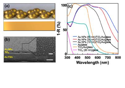
A team of Korean researchers, affiliated with UNIST [Ulsan National Institute of Science and Technology] has recently pioneered in developing a new type of multilayered (Au NPs/TiO2/Au) photoelectrode that boosts the ability of solar water-splitting to produce hydrogen. According to the research team, this special photoelectrode, inspired by the way plants convert sunlight into energy is capable of absorbing visible light from the sun, and then using it to split water molecules (H2O) into hydrogen and oxygen.

A Feb. 1, 2016 UNIST news release, which originated the news item, expands on the theme,

This multilayered photoelectrode takes the form of two-dimensional hybrid metal-dielectric structure, which mainly consists of three layers of gold (Au) film, ultrathin TiO2 layer (20 nm), and gold nanoparticles (Au NPs). In a study, reported in the January 21, 2016 issue of Nano Energy, the team reported that this promising photoelectrode shows high light absorption of about 90% in the visible range 380–700 nm, as well as significant enhancement in photo-catalytic applications.

The researchers have made an image illustrating their work available,

Two-dimensional metastructured film with Titanium Oxide is fabricated as a photo-catalytic photoanode with exceptional visible light absorption. Courtesy: UNIST

Two-dimensional metastructured film with Titanium Oxide is fabricated as a photo-catalytic photoanode with exceptional visible light absorption. Courtesy: UNIST

Back to the news release,

Many structural designs, such as hierarchical and branched assemblies of nanoscale materials have been suggested to increase the UV-visible absorption and to enhance water-splitting efficiency. However, through incorporation of plasmonic metal nanoparticles (i.e. Au) to TiO2 structures, their photoelectrodes have shown to enhance the photoactivity in the entire UV-visible region of solar spectrum when compared with the existing ones, the team reports.

Prof. Jeong Min Baik of UNIST (School of Materials Science and Engineering) states, “Several attemps have been made to use UV-based photoelectrodes for hydrogen production, but this is the first time to use the metal-dielectric hybrid-structured film with TiO2 for oxygen production.” Moreover, according to Prof. Baik, this special type of photoelectrode uses approximately 95% of the visible spectrum of sunlight, which makes up a substantial portion (40%) of full sunlight. He adds, “The developed technology is expected to improve hydrogen production efficiency.”

Prof. Heon Lee (Korean University) states, “This metal-dielectric hybrid-structured film is expected to further reduce the overall cost of producing hydrogen, as it doesn’t require complex operation processes.” He continues by saying, “Using nanoimprint lithography, mass production of hydrogen will be soon possible.”

Prof. Baik adds, “This simple system may serve as an efficient platform for solar energy conversion, utilizing the whole UV-visible range of solar spectrum based on two-dimensional plasmonic photoelectrodes.”

Here’s a link to and a citation for the paper,

Two-dimensional metal-dielectric hybrid-structured film with titanium oxide for enhanced visible light absorption and photo-catalytic application by Joonmo Park, Hee Jun Kim, SangHyeon Nam, Hyowook Kim, Hak-Jong Choi, Youn Jeong Jang, Jae Sung Lee, Jonghwa Shin, Heon Lee, Jeong Min Baik. Nano Energy Volume 21, March 2016, Pages 115–122 doi:10.1016/j.nanoen.2016.01.004

This paper is behind a paywall.

Save those coffee grounds, they can be used for fuel storage

A September 1, 2015 news item on Nanowerk features research from Korea that could point the way to using coffee grounds for methane storage (Note: A link has been removed),

Scientists have developed a simple process to treat waste coffee grounds to allow them to store methane. The simple soak and heating process develops a carbon capture nanomaterial with the additional environmental benefits of recycling a waste product.

The results are published today, 03 September 2015, in the journal Nanotechnology (“Activated carbon derived from waste coffee grounds for stable methane storage”). [emphasis mine]

Methane capture and storage provides a double environmental return – it removes a harmful greenhouse gas from the atmosphere that can then be used as a fuel that is cleaner than other fossil fuels.

The process developed by the researchers, based at the Ulsan National Institute of Science and Technology (UNIST), South Korea, involves soaking the waste coffee grounds in sodium hydroxide and heating to 700-900 °C in a furnace. This produced a stable carbon capture material in less than a day – a fraction of the time it takes to produce carbon capture materials.

I wonder if someone meant to embargo this news release as the paper isn’t due to be published until Thurs., Sept. 3, 2015.

In any event, the Institute of Physics (IOP) Sept. 1, 2015 news release on Alpha Galileo and elsewhere is making the rounds. Here’s more from the news release,

“The big thing is we are decreasing the fabrication time and we are using cheap materials,” explains Christian Kemp, an author of the paper now based at Pohang University of Science and Technology, Korea. “The waste material is free compared compared to all the metals and expensive organic chemicals needed in other processes – in my opinion this is a far easier way to go.”

Kemp found inspiration in his cup of coffee whilst discussing an entirely different project with colleagues at UNIST. “We were sitting around drinking coffee and looked at the coffee grounds and thought ‘I wonder if we can use this for methane storage?’” he continues.

The absorbency of coffee grounds may be the key to successful activation of the material for carbon capture. “It seems when we add the sodium hydroxide to form the activated carbon it absorbs everything,” says Kemp. “We were able to take away one step in the normal activation process – the filtering and washing – because the coffee is such a brilliant absorbant.”

The work also demonstrates hydrogen storage at cryogenic temperatures, and the researchers are now keen to develop hydrogen storage in the activated coffee grounds at less extreme temperatures.

Once the paper has been published I will return to add a link to and a citation for it.

ETA Sept. 3, 2015 (It seems I was wrong about the publication date):

Activated carbon derived from waste coffee grounds for stable methane storage by K Christian Kemp, Seung Bin Baek, Wang-Geun Lee, M Meyyappan, and Kwang S Kim. IOP Publishing Ltd • Nanotechnology, Volume 26, Number 38 doi:10.1088/0957-4484/26/38/385602) Published 2 September 2015 • © 2015

This is an open access paper.

Plus, there is a copy of the press release on EurekAlert.