Tag Archives: ultrasound

Using touch (bionic fingers) instead of x-rays

This is not the most exciting video but it is weirdly fascinating (thank you to ScientifiCult),

A February 15, 2023 news item on Nanowerk provides a textual description for what you’re seeing in the video, Note: A link has been removed,

What if, instead of using X-rays or ultrasound, we could use touch to image the insides of human bodies and electronic devices? In a study publishing in the journal Cell Reports Physical Science (“A smart bionic finger for subsurface tactile-tomography”), researchers present a bionic finger that can create 3D maps of the internal shapes and textures of complex objects by touching their exterior surface.

“We were inspired by human fingers, which have the most sensitive tactile perception that we know of,” says senior author Jianyi Luo, a professor at Wuyi University. “For example, when we touch our own bodies with our fingers, we can sense not only the texture of our skin, but also the outline of the bone beneath it.”

“Our bionic finger goes beyond previous artificial sensors that were only capable of recognizing and discriminating between external shapes, surface textures, and hardness,” says co-author Zhiming Chen, a lecturer at Wuyi University.

The bionic finger “scans” an object by moving across it and applying pressure—think of a constant stream of pokes or prods. With each poke, the carbon fibers compress, and the degree to which they compress provides information about the relative stiffness or softness of the object. Depending on the object’s material, it might also compress when poked by the bionic finger: rigid objects hold their shape, while soft objects will deform when enough pressure is applied. This information, along with the location at which it was recorded, is relayed to a personal computer and displayed onscreen as a 3D map.

A February 13, 2023 Cell Press news release on EurekAlert, which originated the news item, provides more details about the research and some hints at what the researchers may do next,

The researchers tested the bionic finger’s ability to map out the internal and external features of complex objects made of multiple types of material, such as a rigid “letter A” buried under a layer of soft silicon, as well as more abstractly shaped objects. When they used it to scan a small compound object made of three different materials—a rigid internal material, a soft internal material, and a soft outer coating—the bionic finger was able to discriminate between not only the soft outer coating and the internal hard ridges, but it could also tell the difference between the soft outer coating and the soft material that filled the internal grooves.

Next, the researchers tested the finger’s ability to sense and image simulated human tissue. They created this tissue— consisting of a skeletal component, made of three layers of hard polymer, and a soft silicone “muscle” layer—using 3D printing. The bionic finger was able to reproduce a 3D profile of the tissue’s structure and locate a simulated blood vessel beneath the muscle layer.

The team also explored the bionic finger’s ability to diagnose issues in electronic devices without opening them up. By scanning the surface of a defective electronic device with the bionic finger, the researchers were able to create a map of its internal electrical components and pinpoint the location at which the circuit was disconnected, as well as a mis-drilled hole, without breaking through the encapsulating layer.

“This tactile technology opens up a non-optical way for the nondestructive testing of the human body and flexible electronics,” says Luo. “Next, we want to develop the bionic finger’s capacity for omnidirectional detection with different surface materials.”

Here’s a link to and a citation for the paper,

A smart bionic finger for subsurface tactile tomography by Yizhou Li, Zhiming Chen, Youbin Chen, Hao Yang, Junyong Lu, Zhennan Li, Yongyao Chen, Dongyi Ding, Cuiying Zeng, Bingpu Zhou, Hongpeng Liang, Xingpeng Huang, Jiajia Hu, Jingcheng Huang, Jinxiu Wen, Jianyi Luo. Volume 4, Issue 2, 15 February 2023, 101257 DOI: https://doi.org/10.1016/j.xcrp.2023.101257 Published online: February 15, 2023 Version of Record 15 February 2023.

This paper is open access.
Eco-friendly nanocomposite catalyst and ultrasound to remove pollutants from water

The best part of this story is that they’re using biochar from rice hulls to create the nanocomposite catalyst. A July 19, 2019 news item on ScienceDaily reveals a few details about the research without discussing the rice hulls,

The research team of Dr. Jae-woo Choi and Dr. Kyung-won Jung of the Korea Institute of Science and Technology’s (KIST, president: Byung-gwon Lee) Water Cycle Research Center announced that it has developed a wastewater treatment process that uses a common agricultural byproduct to effectively remove pollutants and environmental hormones, which are known to be endocrine disruptors.

A July 19, 2019 Korea National Research Council of Science & Technology news release on EurekAlert, which originated the news item, provides more detail,

The sewage and wastewater that are inevitably produced at any industrial worksite often contain large quantities of pollutants and environmental hormones (endocrine disruptors). Because environmental hormones do not break down easily, they can have a significant negative effect on not only the environment but also the human body. To prevent this, a means of removing environmental hormones is required.

The performance of the catalyst that is currently being used to process sewage and wastewater drops significantly with time. Because high efficiency is difficult to achieve given the conditions, the biggest disadvantage of the existing process is the high cost involved. Furthermore, the research done thus far has mostly focused on the development of single-substance catalysts and the enhancement of their performance. Little research has been done on the development of eco-friendly nanocomposite catalysts that are capable of removing environmental hormones from sewage and wastewater.

The KIST research team, led by Dr. Jae-woo Choi and Dr. Kyung-won Jung, utilized biochar,** which is eco-friendly and made from agricultural byproducts, to develop a wastewater treatment process that effectively removes pollutants and environmental hormones. The team used rice hulls [emphasis mine] which are discarded during rice harvesting, to create a biochar that is both eco-friendly and economical. The surface of the biochar was coated with nano-sized manganese dioxide to create a nanocomposite. The high efficiency and low cost of the biochar-nanocomposite catalyst is based on the combination of the advantages of the biochar and manganese dioxide.

**Biochar: a term that collectively refers to substances that can be created through the thermal decomposition of diverse types of biomass or wood under oxygen-limited condition

The KIST team used the hydrothermal method, which is a type of mineral synthesis that uses high heat and pressure, when synthesizing the nanocomposite in order to create a catalyst that is highly active, easily replicable, and stable. It was confirmed that giving the catalyst a three-dimensional stratified structure resulted in the high effectiveness of the advanced oxidation process (AOP), due to the large surface area created.

When used under the same conditions in which the existing catalyst can remove only 80 percent of Bisphenol A (BPA), an environmental hormone, the catalyst developed by the KIST team removed over 95 percent in less than one hour. In particular, when combined with ultrasound (20kHz), it was confirmed that all traces of BPA were completely removed in less than 20 minutes. Even after many repeated tests, the BPA removal rate remained consistently at around 93 percent.

Dr. Kyung-won Jung of KIST’s Water Cycle Research Center said, “The catalyst developed through this study makes use of a common agricultural byproduct. Therefore, we expect that additional research on alternative substances will lead to the development of catalysts derived from various types of organic waste biomass.” Dr. Jae-woo Choi, also of KIST’s Water Cycle Research Center, said, “We have high hopes that future studies aimed at achieving process optimization and increasing removal rates will allow for the development an environmental hormone removal system that is both eco-friendly and low-cost.”

Here’s a link to and a citation for the paper,

Ultrasound-assisted heterogeneous Fenton-like process for bisphenol A removal at neutral pH using hierarchically structured manganese dioxide/biochar nanocomposites as catalysts by Kyung-Won Jung, Seon Yong Lee, Young Jae Lee, Jae-Woo Choi. Ultrasonics Sonochemistry
Volume 57, October 2019, Pages 22-28 DOI: https://doi.org/10.1016/j.ultsonch.2019.04.039 Available online 29 April 2019

This paper is behind a paywall.

Acoustic nanomotors deliver Cas9-sgRNA complex to the cell

The gene editing tool .CRISPR (clustered regularly interspaced short palindromic repeats) does feature in this story but only as a minor character; the real focus is on the delivery system. From a February 9, 2018 news item on Nanowerk ()Note: A link has been removed),

In cancer research, the “Cas-9–sgRNA” complex is an effective genomic editing tool, but its delivery across the cell membrane to the target (tumor) genome has not yet been satisfactorily solved.

American and Danish scientists have now developed an active nanomotor for the efficient transport, delivery, and release of this gene scissoring system. As detailed in their paper in the journal Angewandte Chemie (“Active Intracellular Delivery of a Cas9/sgRNA Complex Using Ultrasound-Propelled Nanomotors”), their nanovehicle is propelled towards its target by ultrasound.

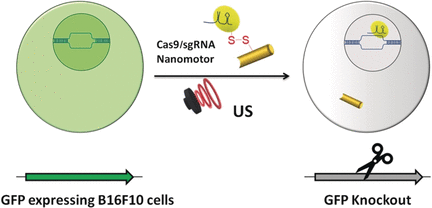
The publisher (Wiley) has made this image illustrating the work available,

Courtesy: Wiley

A February 9, 2018 Wiley Publications news release (also on EurekAlert), which originated the news item, provides more information,

Genomic engineering as a promising cancer therapeutic approach has experienced a tremendous surge since the discovery of the adaptive bacterial immune defense system “CRISPR” and its potential as a gene editing tool over a decade ago. Engineered CRISPR systems for gene editing now contain two main components, a single guide RNA or sgRNA and Cas-9 nuclease. While the sgRNA guides the nuclease to the specified gene sequence, Cas-9 nuclease performs its editing with surgical efficiency. However, the delivery of the large machinery to the target genome is still problematic. The authors of the Angewandte Chemie study, Liangfang Zhang and Joseph Wang from the University of California San Diego, and their colleagues now propose ultrasound-propelled gold nanowires as an active transport/release vehicle for the Cas9-sgRNA complex over the membrane.

Gold nanowires may cross a membrane passively, but thanks to their rod- or wirelike asymmetric shape, active motion can be triggered by ultrasound. “The asymmetric shape of the gold nanowire motor, given by the fabrication process, is essential for the acoustic propulsion,” the authors remarked. They assembled the vehicle by attaching the Cas-9 protein/RNA complex to the gold nanowire through sulfide bridges. These reduceable linkages have the advantage that inside the tumor cell, the bonds would be broken by glutathione, a natural reducing compound enriched in tumor cells. The Cas9-sgRNA would be released and sent to the nucleus to do its editing work, for, example, the knockout of a gene.

As a test system, the scientists monitored the suppression of fluorescence emitted by green fluorescence protein expressing melanoma B16F10 cells. Ultrasound was applied for five minutes, which accelerated the nanomotor carrying the Cas9-sgRNA complex across the membrane, accelerating it even inside the cell, as the authors noted. Moreover, they observed their Cas9-sgRNA complex effectively suppressing fluorescence with only tiny concentrations of the complex needed.

Thus, both the effective use of an acoustic nanomotor as an active transporter and the small payload needed for efficient gene knockout are intriguing results of the study. The simplicity of the system, which uses only few and readily available components, is another remarkable achievement.

Here’s a link to and a citation for the paper,

Active Intracellular Delivery of a Cas9/sgRNA Complex Using Ultrasound-Propelled Nanomotors by Malthe Hansen-Bruhn, Dr. Berta Esteban-Fernández de Ávila, Dr. Mara Beltrán-Gastélum, Prof. Jing Zhao, Dr. Doris E. Ramírez-Herrera, Pavimol Angsantikul, Prof. Kurt Vesterager Gothelf, Prof. Liangfang Zhang, and Prof. Joseph Wang. Angewandte Chemie International Edition Vol. 57 Issue 7 DOI: 10.1002/anie.201713082 Version of Record online: 6 FEB 2018

© 2018 Wiley-VCH Verlag GmbH & Co. KGaA, Weinheim

This paper is behind a paywall.

‘Neural dust’ could lead to introduction of electroceuticals

In case anyone is wondering, the woman who’s manipulating a prosthetic arm so she can eat or a drink of coffee probably has a bulky implant/docking station in her head. Right now that bulky implant is the latest and greatest innovation for tetraplegics (aka, quadriplegics) as it frees, to some extent, people who’ve had no independent movement of any kind. By virtue of the juxtaposition of the footage of the woman with the ‘neural dust’ footage, they seem to be suggesting that neural dust might some day accomplish the same type of connection. At this point, hopes for the ‘neural dust’ are more modest.

An Aug. 3, 2016 news item on ScienceDaily announces the ‘neural dust’,

University of California, Berkeley engineers have built the first dust-sized, wireless sensors that can be implanted in the body, bringing closer the day when a Fitbit-like device could monitor internal nerves, muscles or organs in real time.

Because these batteryless sensors could also be used to stimulate nerves and muscles, the technology also opens the door to “electroceuticals” to treat disorders such as epilepsy or to stimulate the immune system or tamp down inflammation.

An Aug. 3, 2016 University of California at Berkeley news release (also on EurekAlert) by Robert Sanders, which originated the news item, explains further and describes the researchers’ hope that one day the neural dust could be used to control implants and prosthetics,

The so-called neural dust, which the team implanted in the muscles and peripheral nerves of rats, is unique in that ultrasound is used both to power and read out the measurements. Ultrasound technology is already well-developed for hospital use, and ultrasound vibrations can penetrate nearly anywhere in the body, unlike radio waves, the researchers say.

“I think the long-term prospects for neural dust are not only within nerves and the brain, but much broader,“ said Michel Maharbiz, an associate professor of electrical engineering and computer sciences and one of the study’s two main authors. “Having access to in-body telemetry has never been possible because there has been no way to put something supertiny superdeep. But now I can take a speck of nothing and park it next to a nerve or organ, your GI tract or a muscle, and read out the data.“

Maharbiz, neuroscientist Jose Carmena, a professor of electrical engineering and computer sciences and a member of the Helen Wills Neuroscience Institute, and their colleagues will report their findings in the August 3 [2016] issue of the journal Neuron.

The sensors, which the researchers have already shrunk to a 1 millimeter cube – about the size of a large grain of sand – contain a piezoelectric crystal that converts ultrasound vibrations from outside the body into electricity to power a tiny, on-board transistor that is in contact with a nerve or muscle fiber. A voltage spike in the fiber alters the circuit and the vibration of the crystal, which changes the echo detected by the ultrasound receiver, typically the same device that generates the vibrations. The slight change, called backscatter, allows them to determine the voltage.

Motes sprinkled thoughout the body

In their experiment, the UC Berkeley team powered up the passive sensors every 100 microseconds with six 540-nanosecond ultrasound pulses, which gave them a continual, real-time readout. They coated the first-generation motes – 3 millimeters long, 1 millimeter high and 4/5 millimeter thick – with surgical-grade epoxy, but they are currently building motes from biocompatible thin films which would potentially last in the body without degradation for a decade or more.

While the experiments so far have involved the peripheral nervous system and muscles, the neural dust motes could work equally well in the central nervous system and brain to control prosthetics, the researchers say. Today’s implantable electrodes degrade within 1 to 2 years, and all connect to wires that pass through holes in the skull. Wireless sensors – dozens to a hundred – could be sealed in, avoiding infection and unwanted movement of the electrodes.

“The original goal of the neural dust project was to imagine the next generation of brain-machine interfaces, and to make it a viable clinical technology,” said neuroscience graduate student Ryan Neely. “If a paraplegic wants to control a computer or a robotic arm, you would just implant this electrode in the brain and it would last essentially a lifetime.”

In a paper published online in 2013, the researchers estimated that they could shrink the sensors down to a cube 50 microns on a side – about 2 thousandths of an inch, or half the width of a human hair. At that size, the motes could nestle up to just a few nerve axons and continually record their electrical activity.

“The beauty is that now, the sensors are small enough to have a good application in the peripheral nervous system, for bladder control or appetite suppression, for example,“ Carmena said. “The technology is not really there yet to get to the 50-micron target size, which we would need for the brain and central nervous system. Once it’s clinically proven, however, neural dust will just replace wire electrodes. This time, once you close up the brain, you’re done.“

The team is working now to miniaturize the device further, find more biocompatible materials and improve the surface transceiver that sends and receives the ultrasounds, ideally using beam-steering technology to focus the sounds waves on individual motes. They are now building little backpacks for rats to hold the ultrasound transceiver that will record data from implanted motes.

They’re also working to expand the motes’ ability to detect non-electrical signals, such as oxygen or hormone levels.

“The vision is to implant these neural dust motes anywhere in the body, and have a patch over the implanted site send ultrasonic waves to wake up and receive necessary information from the motes for the desired therapy you want,” said Dongjin Seo, a graduate student in electrical engineering and computer sciences. “Eventually you would use multiple implants and one patch that would ping each implant individually, or all simultaneously.”

Ultrasound vs radio

Maharbiz and Carmena conceived of the idea of neural dust about five years ago, but attempts to power an implantable device and read out the data using radio waves were disappointing. Radio attenuates very quickly with distance in tissue, so communicating with devices deep in the body would be difficult without using potentially damaging high-intensity radiation.

Marharbiz hit on the idea of ultrasound, and in 2013 published a paper with Carmena, Seo and their colleagues describing how such a system might work. “Our first study demonstrated that the fundamental physics of ultrasound allowed for very, very small implants that could record and communicate neural data,” said Maharbiz. He and his students have now created that system.

“Ultrasound is much more efficient when you are targeting devices that are on the millimeter scale or smaller and that are embedded deep in the body,” Seo said. “You can get a lot of power into it and a lot more efficient transfer of energy and communication when using ultrasound as opposed to electromagnetic waves, which has been the go-to method for wirelessly transmitting power to miniature implants”

“Now that you have a reliable, minimally invasive neural pickup in your body, the technology could become the driver for a whole gamut of applications, things that today don’t even exist,“ Carmena said.

Here’s a link to and a citation for the team’s latest paper,

Wireless Recording in the Peripheral Nervous System with Ultrasonic Neural Dust by Dongjin Seo, Ryan M. Neely, Konlin Shen, Utkarsh Singhal, Elad Alon, Jan M. Rabaey, Jose M. Carmena. and Michel M. Maharbiz. Neuron Volume 91, Issue 3, p529–539, 3 August 2016 DOI: http://dx.doi.org/10.1016/j.neuron.2016.06.034

This paper appears to be open access.