Tag Archives: ultrasound waves

Sound waves for wearable patches that deliver drugs painlessly

While watching this video I started wondering if they were testing their research on students but that’s not the case; these wearable patches were tested on porcine (pig) skin, which is quite similar to human skin, Note: They tested a B vitamin called niacinamide so, it’s highly unlikely the pigs suffered from it,

An April 20, 2023 news item on ScienceDaily announces the research into using ultrasonic waves for drug delivery,

The skin is an appealing route for drug delivery because it allows drugs to go directly to the site where they’re needed, which could be useful for wound healing, pain relief, or other medical and cosmetic applications. However, delivering drugs through the skin is difficult because the tough outer layer of the skin prevents most small molecules from passing through it.

In hopes of making it easier to deliver drugs through the skin, MIT [Massachusetts Institute of Technology] researchers have developed a wearable patch that applies painless ultrasonic waves to the skin, creating tiny channels that drugs can pass through. This approach could lend itself to delivery of treatments for a variety of skin conditions, and could also be adapted to deliver hormones, muscle relaxants, and other drugs, the researchers say.

An April 20, 2023 Massachusetts Institute of Technology (MIT) news release (also on EurekAlert), which originated the news item, provides technical details about the research, Note: A link has been removed,

“The ease-of-use and high-repeatability offered by this system provides a game-changing alternative to patients and consumers suffering from skin conditions and premature skin aging,” says Canan Dagdeviren, an associate professor in MIT’s Media Lab and the senior author of the study. “Delivering drugs this way could offer less systemic toxicity and is more local, comfortable, and controllable.”

MIT research assistants Chia-Chen Yu and Aastha Shah are the lead authors of the paper, which appears in Advanced Materials, as part of the journal’s “Rising Stars” series, which showcases the outstanding work of researchers in the early stages of their independent careers. Other MIT authors include Research Assistant Colin Marcus and postdoc Md Osman Goni Nayeem. Nikta Amiri, Amit Kumar Bhayadia, and Amin Karami of the University of Buffalo are also authors of the paper.

A boost from sound waves

The researchers began this project as an exploration of alternative ways to deliver drugs. Most drugs are delivered orally or intravenously, but the skin is a route that could offer much more targeted drug delivery for certain applications.

“The main benefit with skin is that you bypass the whole gastrointestinal tract. With oral delivery, you have to deliver a much larger dose in order to account for the loss that you would have in the gastric system,” Shah says. “This is a much more targeted, focused modality of drug delivery.”

Ultrasound exposure has been shown to enhance the skin’s permeability to small-molecule drugs, but most of the existing techniques for performing this kind of drug delivery require bulky equipment. The MIT team wanted to come up with a way to perform this kind of transdermal drug delivery with a lightweight, wearable patch, which could make it easier to use for a variety of applications.

The device that they designed consists of a patch embedded with several disc-shaped piezoelectric transducers, which can convert electric currents into mechanical energy. Each disc is embedded in a polymeric cavity that contains the drug molecules dissolved in a liquid solution. When an electric current is applied to the piezoelectric elements, they generate pressure waves in the fluid, creating bubbles that burst against the skin. These bursting bubbles produce microjets of fluid that can penetrate through the skin’s tough outer layer, the stratum corneum.

“This works open the door to using vibrations to enhance drug delivery. There are several parameters that result in generation of different kinds of waveform patterns. Both mechanical and biological aspects of drug delivery can be improved by this new toolset,” Karami says.

The patch is made of PDMS, a silicone-based polymer that can adhere to the skin without tape. In this study, the researchers tested the device by delivering a B vitamin called niacinamide, an ingredient in many sunscreens and moisturizers.

In tests using pig skin, the researchers showed that when they delivered niacinamide using the ultrasound patch, the amount of drug that penetrated the skin was 26 times greater than the amount that could pass through the skin without ultrasonic assistance.

The researchers also compared the results from their new device to microneedling, a technique sometimes used for transdermal drug delivery, which involves puncturing the skin with miniature needles. The researchers found that their patch was able to deliver the same amount of niacinamide in 30 minutes that could be delivered with microneedles over a six-hour period.

Local delivery

With the current version of the device, drugs can penetrate a few millimeters into the skin, making this approach potentially useful for drugs that act locally within the skin. These could include niacinamide or vitamin C, which is used to treat age spots or other dark spots on the skin, or topical drugs used to heal burns.

With further modifications to increase the penetration depth, this technique could also be used for drugs that need to reach the bloodstream, such as caffeine, fentanyl, or lidocaine. Dagdeviren also envisions that this kind of patch could be useful for delivering hormones such as progesterone. In addition, the researchers are now exploring the possibility of implanting similar devices inside the body to deliver drugs to treat cancer or other diseases.

The researchers are also working on further optimizing the wearable patch, in hopes of testing it soon on human volunteers. They also plan to repeat the lab experiments they did in this study, with larger drug molecules.

“After we characterize the drug penetration profiles for much larger drugs, we would then see which candidates, like hormones or insulin, can be delivered using this technology, to provide a painless alternative for those who are currently bound to self-administer injections on a daily basis,” Shah says.

Here’s a link to and a citation for the paper,

A Conformable Ultrasound Patch for Cavitation-Enhanced Transdermal Cosmeceutical Delivery by Chia-Chen Yu, Aastha Shah, Nikta Amiri, Colin Marcus, Md Osman Goni Nayeem, Amit Kumar Bhayadia, Amin Karami, Canan Dagdeviren. Advanced Materials Volume35, Issue 23 June 8, 2023 2300066 DOI: https://doi.org/10.1002/adma.202300066 First published online: 19 March 2023

This paper is open access.
Making longer lasting bandages with sound and bubbles

This research into longer lasting bandages described in an August 12, 2022 news item on phys.org comes from McGill University (Montréal, Canada)

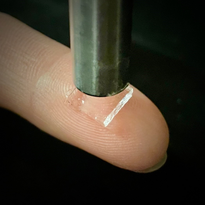
Researchers have discovered that they can control the stickiness of adhesive bandages using ultrasound waves and bubbles. This breakthrough could lead to new advances in medical adhesives, especially in cases where adhesives are difficult to apply such as on wet skin.

“Bandages, glues, and stickers are common bioadhesives that are used at home or in clinics. However, they don’t usually adhere well on wet skin. It’s also challenging to control where they are applied and the strength and duration of the formed adhesion,” says McGill University Professor Jianyu Li, who led the research team of engineers, physicists, chemists, and clinicians.

Caption: Adhesive hydrogel applied on skin under ultrasound probe. Credit: Ran Huo and Jianyu Li

An August 12, 2022 McGill University news release (also on EurekAlert), which originated the news item, delves further into the work,

“We were surprised to find that by simply playing around with ultrasonic intensity, we can control very precisely the stickiness of adhesive bandages on many tissues,” says lead author Zhenwei Ma, a former student of Professor Li and now a Killam Postdoctoral Fellow at the University of British Columbia.

Ultrasound induced bubbles control stickiness

In collaboration with physicists Professor Outi Supponen and Claire Bourquard from the Institute of Fluid Dynamics at ETH Zurich, the team experimented with ultrasound induced microbubbles to make adhesives stickier. “The ultrasound induces many microbubbles, which transiently push the adhesives into the skin for stronger bioadhesion,” says Professor Supponen. “We can even use theoretical modeling to estimate exactly where the adhesion will happen.”

Their study, published in the journal Science, shows that the adhesives are compatible with living tissue in rats. The adhesives can also potentially be used to deliver drugs through the skin. “This paradigm-shifting technology will have great implications in many branches of medicine,” says University of British Columbia Professor Zu-hua Gao. “We’re very excited to translate this technology for applications in clinics for tissue repair, cancer therapy, and precision medicine.”

“By merging mechanics, materials and biomedical engineering, we envision the broad impact of our bioadhesive technology in wearable devices, wound management, and regenerative medicine,” says Professor Li, who is also a Canada Research Chair in Biomaterials and Musculoskeletal Health.

Here’s a link to and a citation for the paper,

Controlled tough bioadhesion mediated by ultrasound by Zhenwei Ma, Claire Bourquard, Qiman Gao, Shuaibing Jiang, Tristan De Iure-Grimmel, Ran Huo, Xuan Li, Zixin He, Zhen Yang, Galen Yang, Yixiang Wang, Edmond Lam, Zu-hua Gao, Outi Supponen and Jianyu Li. Science 11 Aug 2022 Vol 377, Issue 6607 pp. 751-755 DOI: 10.1126/science.abn8699

This paper is behind a paywall.

I haven’t seen this before but it seems that one of the journal’s editors decided to add a standalone paragraph to hype some of the other papers about adhesives in the issue,

A sound way to make it stick

Tissue adhesives play a role in temporary or permanent tissue repair, wound management, and the attachment of wearable electronics. However, it can be challenging to tailor the adhesive strength to ensure reversibility when desired and to maintain permeability. Ma et al. designed hydrogels made of polyacrylamide or poly(N-isopropylacrylamide) combined with alginate that are primed using a solution containing nanoparticles of chitosan, gelatin, or cellulose nanocrystals (see the Perspective by Es Sayed and Kamperman). The application of ultrasound causes cavitation that pushes the primer molecules into the tissue. The mechanical interlocking of the anchors eventually results in strong adhesion between hydrogel and tissue without the need for chemical bonding. Tests on porcine or rat skin showed enhanced adhesion energy and interfacial fatigue resistance with on-demand detachment. —MSL

I like the wordplay and am guessing that MSL is:

Marc S. Lavine
Senior Editor
Education: BASc, University of Toronto; PhD, University of Cambridge
Areas of responsibility: Reviews; materials science, biomaterials, engineering

Brain surgery with no scalpel or incisions

A December 3, 2021 news item on ScienceDaily announces some very exciting work from the University of Virginia UVA) and Stanford University,

University of Virginia School of Medicine researchers have developed a noninvasive way to remove faulty brain circuits that could allow doctors to treat debilitating neurological diseases without the need for conventional brain surgery.

The UVA team, together with colleagues at Stanford University, indicate that the approach, if successfully translated to the operating room, could revolutionize the treatment of some of the most challenging and complex neurological diseases, including epilepsy, movement disorders and more. The approach uses low-intensity focused ultrasound waves combined with microbubbles to briefly penetrate the brain’s natural defenses and allow the targeted delivery of a neurotoxin. This neurotoxin kills the culprit brain cells while sparing other healthy cells and preserving the surrounding brain architecture.

A November 22, 2021 University of Virginia news release (also on EurekAlert but published on December 3, 2021), which originated the news item, offers technical details (Note: Links have been removed),

“This novel surgical strategy has the potential to supplant existing neurosurgical procedures used for the treatment of neurological disorders that don’t respond to medication,” said researcher Kevin S. Lee of UVA’s Departments of Neuroscience and Neurosurgery and the Center for Brain Immunology and Glia, or BIG. “This unique approach eliminates the diseased brain cells, spares adjacent healthy cells and achieves these outcomes without even having to cut into the scalp.”

The Power of PING

The new approach, called “PING,” has already demonstrated exciting potential in laboratory studies. For instance, one of the promising applications for PING could be for the surgical treatment of epilepsies that do not respond to medication. Approximately a third of patients with epilepsy do not respond to anti-seizure drugs, and surgery can reduce or eliminate seizures for some of them. Lee and his team, along with their collaborators at Stanford, have shown that PING can reduce or eliminate seizures in two research models of epilepsy. The findings raise the possibility of treating epilepsy in a carefully targeted and noninvasive manner without the need for traditional brain surgery. 

Another important potential advantage of PING is that it could encourage the surgical treatment of appropriate patients with epilepsy who are reluctant to undergo conventional invasive or ablative surgery.

In a scientific paper newly published in the Journal of Neurosurgery, Lee and his collaborators detail the ability of PING to focally eliminate neurons in a brain region, while sparing non-target cells in the same area. In contrast, currently available surgical approaches damage all cells in a treated brain region. 

A key advantage of the approach is its incredible precision. PING harnesses the power of magnetic-resonance imaging to let scientists peer inside the skull so that they can precisely guide sound waves to open the body’s natural blood-brain barrier exactly where needed. This barrier is designed to keep harmful cells and molecules out of the brain, but it also prevents the delivery of potentially beneficial treatments.

The UVA group’s new paper concludes that PING allows the delivery of a highly targeted neurotoxin, cleanly wiping out problematic neurons, a type of brain cell, without causing collateral damage. 

Another key advantage of the precision of this approach is that it can be used on irregularly shaped targets in areas that would be extremely difficult or impossible to reach through regular brain surgery. “If this strategy translates to the clinic,” the researchers write in their new paper, “the noninvasive nature and specificity of the procedure could positively influence both physician referrals for, and patient confidence in, surgery for medically intractable neurological disorders.”

“Our hope is that the PING strategy will become a key element in the next generation of very precise, noninvasive, neurosurgical approaches to treat major neurological disorders,” said Lee, who is part of the UVA Brain Institute.

About the Research

Lee’s groundbreaking research has been supported by the National Institutes of Health, the Chester Fund and the Charlottesville-based Focused Ultrasound Foundation. The work is part of an expansive effort at UVA Health to explore the potential of scalpel-free focused ultrasound to treat complex diseases throughout the body.

UVA’s pioneering research has already paved the way for the federal Food and Drug Administration to approve focused ultrasound for the treatment of essential tremor, a common movement disorder, and Parkinson’s disease symptoms. Research is underway on its potential applications for many more conditions, including breast cancer and glioblastoma, a deadly form of brain tumor. Learn more about UVA’s focused ultrasound research.

The research team included Yi Wang, Matthew J. Anzivino, Yanrong Zhang, Edward H. Bertram, James Woznak, Alexander L. Klibanov, Erik Dumont and Max Wintermark. 

An application to patent the PING procedure has been submitted by members of the research group. 

The research was funded by the National Institutes of Health, grants R01 NS102194 and R01 CA217953-01; the Chester Fund; and the Focused Ultrasound Foundation.

To keep up with the latest medical research news from UVA, subscribe to the Making of Medicine blog at http://makingofmedicine.virginia.edu.

Here’s a link to and a citation for the paper,

Noninvasive disconnection of targeted neuronal circuitry sparing axons of passage and nonneuronal cells by Yi Wang, Matthew J. Anzivino, Yanrong Zhang, Edward H. Bertram, James Woznak, Alexander L. Klibanov, Erik Dumont, Max Wintermark, and Kevin S. Lee. Journal of Neurosurgery DOI: https://doi.org/10.3171/2021.7.JNS21123 Online Publication Date: 19 Nov 2021

This paper is behind a paywall.

Skin as a touchscreen (“smart” hands)

An April 11, 2016 news item on phys.org highlights some research presented at the IEEE (Institute of Electrical and Electronics Engineers) Haptics (touch) Symposium 2016,

Using your skin as a touchscreen has been brought a step closer after UK scientists successfully created tactile sensations on the palm using ultrasound sent through the hand.

The University of Sussex-led study – funded by the Nokia Research Centre and the European Research Council – is the first to find a way for users to feel what they are doing when interacting with displays projected on their hand.

This solves one of the biggest challenges for technology companies who see the human body, particularly the hand, as the ideal display extension for the next generation of smartwatches and other smart devices.

Current ideas rely on vibrations or pins, which both need contact with the palm to work, interrupting the display.

However, this new innovation, called SkinHaptics, sends sensations to the palm from the other side of the hand, leaving the palm free to display the screen.

An April 11, 2016 University of Sussex press release (also on EurekAlert) by James Hakmer, which originated the news item, provides more detail,

The device uses ‘time-reversal’ processing to send ultrasound waves through the hand. This technique is effectively like ripples in water but in reverse – the waves become more targeted as they travel through the hand, ending at a precise point on the palm.

It draws on a rapidly growing field of technology called haptics, which is the science of applying touch sensation and control to interaction with computers and technology.

Professor Sriram Subramanian, who leads the research team at the University of Sussex, says that technologies will inevitably need to engage other senses, such as touch, as we enter what designers are calling an ‘eye-free’ age of technology.

He says: “Wearables are already big business and will only get bigger. But as we wear technology more, it gets smaller and we look at it less, and therefore multisensory capabilities become much more important.

“If you imagine you are on your bike and want to change the volume control on your smartwatch, the interaction space on the watch is very small. So companies are looking at how to extend this space to the hand of the user.

“What we offer people is the ability to feel their actions when they are interacting with the hand.”

The findings were presented at the IEEE Haptics Symposium [April 8 – 11] 2016 in Philadelphia, USA, by the study’s co-author Dr Daniel Spelmezan, a research assistant in the Interact Lab.

There is a video of the work (I was not able to activate sound, if there is any accompanying this video),

The consequence of watching this silent video was that I found the whole thing somewhat mysterious.