Tag Archives: University of California at Irvine

Nanobody could lead to treatment for retinitis pigmentosa (a condition that leads to blindness)

This is an image illustrating the work but you’ll probably need to read the news release to understand the explanation offered,

Caption: This image depicts the crystal structure of two nanobodies binding to a rhodopsin dimer. The rhodopsin molecules are shown in green and blue, with 11-cis-retinal displayed in red. The figure emphasizes the significant interactions between the nanobodies (represented in a semi-transparent surface cartoon) and the extracellular surface of rhodopsin, including its N-terminal glycans highlighted in orange.. Credit: UCI [University of California at Irvine] School of Medicine

The research from the University of California at Irvine (UCI) has been featured twice, in an August 31, 2023 news item on phys.org and again in a September 7, 2023 news item on ScienceDaily.

An August 29, 2023 UCI news release (also on EurekAlert but published Sept. 6, 2023), which originated the news items, provides information about RP and the nanobodies,

A team of scientists from the University of California, Irvine, believe they have discovered a special antibody which may lead to a treatment for Retinitis Pigmentosa, a condition that causes loss of central vision, as well as night and color vision. 

The study, Structural basis for the allosteric modulation of rhodopsin by nanobody binding to its extracellular domain, was published in Nature Communications. Authors of the study were Arum Wu, PhD, David Salom, PhD, John D. Hong, Aleksander Tworak, PhD, Philip D. Kiser, PharmD, PhD, and Krzysztof Palczewski, PhD, in the Department of Ophthalmology, Gavin Herbert Eye Institute, at the University of California, Irvine. Research was conducted  in collaboration with Jan Steyaert, PhD, at the Vrije Universiteit Brussel (VUB).

Retinitis Pigmentosa (RP) is a group of inherited eye diseases that affect the retina in the back of the eye. It is caused by the death of cells that detect light signals, known as photoreceptor cells. There is no known cure for RP, and the development of new treatments for this condition relies on cell and gene therapies. 

UCI researchers have targeted their study on a specific molecule which they believe will provide a treatment for Rhodopsin-associated autosomal dominant RP (adRP). The molecule, Rhodopsin, is a key light-sensing molecule in the human retina. It is found in rod photoreceptor cells, and mutations in the Rhodopsin gene are a primary cause of adRP. 

“More than 150 mutations in rhodopsin can cause Retinitis Pigmentosa, making it challenging to develop targeted gene therapies,” said Krzysztof Palczewski, PhD, Donald Bren Professor, UCI School of Medicine. “However due to the high prevalence of RP, there has been significant investment in research and development efforts to find novel treatments.”

Although Rhodopsin has been studied for over a century, key details of its mechanism for converting light into a cellular signal have been difficult to experimentally address.

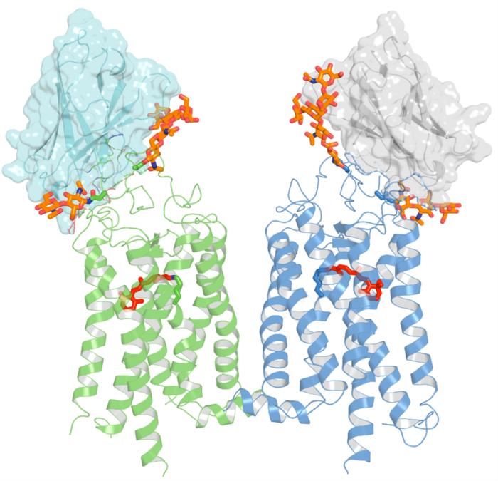
For this study, researchers used a special type of llama-derived antibody, known as a nanobody, that can halt the process of Rhodopsin photoactivation, allowing it to be investigated at high resolution. 

“Our team has developed nanobodies that work through a novel mechanism of action. These nanobodies have high specificity and can recognize the target rhodopsin extracellularly,” said David Salom , PhD, researcher and project scientist, UCI School of Medicine. “This enables us to lock this GPCR in a non-signaling state.” 

Scientists discovered that these nanobodies target an unexpected site on the Rhodopsin molecule, near the location where retinaldehyde binds. They also found that the stabilizing effect of these nanobodies can also be applied to Rhodopsin mutants that are associated with retinal disease, suggesting their use as therapeutics. 

“In the future, we hope to involve the in vitro evolution of these initial set of nanobodies,” said Arum Wu, PhD, researcher and project scientist, UCI School of Medicine. “We will also evaluate the safety and effectiveness of a future nanobody gene therapy for RP.”

Researchers hope to improve nanobodies’ ability to recognize Rhodopsin from other species including mice, for which several pre-clinical models of adRP are available. They also have plans to use these nanobodies to address a long-term goal in the field of structurally resolving the key intermediate states of Rhodopsin from the inactive state to the fully ligand-activated state.

Here’s a link to and a citation for the paper,

Structural basis for the allosteric modulation of rhodopsin by nanobody binding to its extracellular domain by Arum Wu, David Salom, John D. Hong, Aleksander Tworak, Kohei Watanabe, Els Pardon, Jan Steyaert, Hideki Kandori, Kota Katayama, Philip D. Kiser & Krzysztof Palczewski. Nature Communications volume 14, Article number: 5209 (2023) DOI: https://doi.org/10.1038/s41467-023-40911-9 Published: 25 August 2023

This paper is open access.

Keeping your hands cool and your coffee hot with a cup cozy inspired by squid skin

Researchers in the Department of Chemical and Biomolecular Engineering at the University of California, Irvine have invented a squid-skin inspired material that can wrap around a coffee cup to shield sensitive fingers from heat. They have also created a method for economically mass producing the adaptive fabric, making possible a wide range of uses. Credit: Melissa Sung Courtesy: University of California Irvine

I love that image. Melissa Sung, thank you. Sadly, squid-inspired cup cozies aren’t available yet according to a March 28, 2022 news item on phys.org but researchers are working on it, Note: Links have been removed,

In the future, you may have a squid to thank for your coffee staying hot on a cold day. Drawing inspiration from cephalopod skin, engineers at the University of California, Irvine invented an adaptive composite material that can insulate beverage cups, restaurant to-go bags, parcel boxes and even shipping containers.

The innovation is an infrared-reflecting metallized polymer film developed in the laboratory of Alon Gorodetsky, UCI associate professor of chemical and biomolecular engineering. In a paper published today [March 28, 2022] in Nature Sustainability, Gorodetsky and his team members describe a large-area composite material that regulates heat by means of reconfigurable metal structures that can reversibly separate from one another and come back together under different strain levels.

“The metal islands in our composite material are next to one another when the material is relaxed and become separated when the material is stretched, allowing for control of the reflection and transmission of infrared light or heat dissipation,” said Gorodetsky. “The mechanism is analogous to chromatophore expansion and contraction in a squid’s skin, which alters the reflection and transmission of visible light.”

Chromatophore size changes help squids communicate and camouflage their bodies to evade predators and hide from prey. Gorodetsky said by mimicking this approach, his team has enabled “tunable thermoregulation” in their material, which can lead to improved energy efficiency and protect sensitive fingers from hot surfaces.

A March 28, 2022 University of California at Irvine (UCI) news release (also on EurekAlert), which originated the news item, delves further into this squid-inspired research and its commercialization,

A key breakthrough of this project was the UCI researchers’ development of a cost-effective production method of their composite material at application-relevant quantities. The copper and rubber raw materials start at about a dime per square meter with the costs reduced further by economies of scale, according to the paper. The team’s fabrication technique involves depositing a copper film onto a reusable substrate such as aluminum foil and then spraying multiple polymer layers onto the copper film, all of which can be done in nearly any batch size imaginable.

“The combined manufacturing strategy that we have now perfected in our lab is a real game changer,” said Gorodetsky. “We have been working with cephalopod-inspired adaptive materials and systems for years but previously have only been able to fabricate them over relatively small areas. Now there is finally a path to making this stuff roll-by-roll in a factory.”

The developed strategy and economies of scale should make it possible for the composite material to be used in a wide range of applications, from the coffee cup cozy up to tents, or in any container in which tunable temperature regulation is desired.

The invention will go easy on the environment due its environmental sustainability, said lead author Mohsin Badshah, a former UCI postdoctoral scholar in chemical and biomolecular engineering. “The composite material can be recycled in bulk by removing the copper with vinegar and using established commercial methods to repurpose the remaining stretchable polymer,” he said.

The team conducted universally relatable coffee cup testing in their laboratory on the UCI campus, where they proved they could control the cooling of the coffee. They were able to accurately and theoretically predict and then experimentally confirm the changes in temperature for the beverage-filled cups. The was also able to achieve a 20-fold modulation of infrared radiation transmittance and a 30-fold regulation of thermal fluxes under standardized testing conditions. The stable material even worked well for high levels of mechanical deformation and after repeated mechanical cycling.

“There is an enormous array of applications for this material,” said Gorodetsky. “Think of all the perishable goods that have been delivered to people’s homes during the pandemic. Any package that Amazon or another company sends that needs to be temperature-controlled can use a lining made from our squid-inspired adaptive composite material. Now that we can make large sheets of it at a time, we have something that can benefit many aspects of our lives.”

Joining Gorodetsky and Badshah on this project were Erica Leung, who recently graduated UCI with a Ph.D. in chemical and biomolecular engineering, and Aleksandra Strzelecka and Panyiming Liu, who are current UCI graduate students. The research was funded by the Defense Advanced Research Projects Agency, the Advanced Research Projects Agency – Energy and the Air Force Office of Scientific Research. A provisional patent for the technology and manufacturing process has been applied for.

Here’s a link to and a citation for the paper,

Scalable manufacturing of sustainable packaging materials with tunable thermoregulability by Mohsin Ali Badshah, Erica M. Leung, Panyiming Liu, Aleksandra Anna Strzelecka & Alon A. Gorodetsky. Nature Sustainability (2022) DOI: https://doi.org/10.1038/s41893-022-00847-2 Published: 28 March 2022

This paper is behind a paywall.

Documentary “NNI Retrospective Video: Creating a National Initiative” celebrates the US National Nanotechnology Initiative (NNI) and a lipid nanoparticle question

i stumbled across an August 4, 2022 tvworldwide.com news release about a video celbrating the US National Nanotechnology Initiative’s (NNI) over 20 years of operation, (Note: A link has been removed),

TV Worldwide, since 1999, a pioneering web-based global TV network, announced that it was releasing a video trailer highlighting a previously released documentary on NNI over the past 20 years, entitled, ‘NNI Retrospective Video: Creating a National Initiative’.

The video and its trailer were produced in cooperation with the National Nanotechnology Initiative (NNI), the National Science Foundation and the University of North Carolina Greensboro.

Video Documentary Synopsis

Nanotechnology is a megatrend in science and technology at the beginning of the 21 Century. The National Nanotechnology Initiative (NNI) has played a key role in advancing the field after it was announced by President Clinton in January 2000. Neil Lane was Presidential Science Advisor. Mike Roco proposed the initiative at the White House in March 1999 on behalf of the Interagency Working Group on Nanotechnology and was named the founding Chair of NSET to implement NNI beginning with Oct. 2000. NSF led the preparation of this initiative together with other agencies including NIH, DoD, DOE, NASA, and EPA. Jim Murday was named the first Director of NNCO to support NSET. The scientific and societal success of NNI has been recognized in the professional communities, National Academies, PCAST, and Congress. Nanoscale science, engineering and technology are strongly connected and collectively called Nanotechnology.

This video documentary was made after the 20th NNI grantees conference at NSF. It is focused on creating and implementing NNI, through video interviews. The interviews focused on three questions: (a) Motivation and how NNI started; (b) The process and reason for the success in creating NNI; (c) Outcomes of NNI after 20 years, and how the initial vision has been realized.

About the National Nanotechnology Initiative (NNI)

The National Nanotechnology Initiative (NNI) is a U.S. Government research and development (R&D) initiative. Over thirty Federal departments, independent agencies, and commissions work together toward the shared vision of a future in which the ability to understand and control matter at the nanoscale leads to ongoing revolutions in technology and industry that benefit society. The NNI enhances interagency coordination of nanotechnology R&D, supports a shared infrastructure, enables leveraging of resources while avoiding duplication, and establishes shared goals, priorities, and strategies that complement agency-specific missions and activities.

The NNI participating agencies work together to advance discovery and innovation across the nanotechnology R&D enterprise. The NNI portfolio encompasses efforts along the entire technology development pathway, from early-stage fundamental science through applications-driven activities. Nanoscience and nanotechnology are prevalent across the R&D landscape, with an ever-growing list of applications that includes nanomedicine, nanoelectronics, water treatment, precision agriculture, transportation, and energy generation and storage. The NNI brings together representatives from multiple agencies to leverage knowledge and resources and to collaborate with academia and the private sector, as appropriate, to promote technology transfer and facilitate commercialization. The breadth of NNI-supported infrastructure enables not only the nanotechnology community but also researchers from related disciplines.

In addition to R&D efforts, the NNI is helping to build the nanotechnology workforce of the future, with focused efforts from K–12 through postgraduate research training. The responsible development of nanotechnology has been an integral pillar of the NNI since its inception, and the initiative proactively considers potential implications and technology applications at the same time. Collectively, these activities ensure that the United States remains not only the place where nanoscience discoveries are made, but also where these discoveries are translated and manufactured into products to benefit society.

I’m embedding the trailer here and a lipid nanoparticle question follows (The origin story told in Vancouver [Canada] is that the work was started at the University of British Columbia by Pieter Quilty.),

I was curious about what involvement the US NNI had with the development of lipid nanoparticles (LNPs) and found a possible answer to that question on Wikipedia The LNP Wikipedia entry certainly gives the bulk of the credit to Quilty but there was work done prior to his involvement (Note: Links have been removed),

A significant obstacle to using LNPs as a delivery vehicle for nucleic acids is that in nature, lipids and nucleic acids both carry a negative electric charge—meaning they do not easily mix with each other.[19] While working at Syntex in the mid-1980s,[20] Philip Felgner [emphasis mine] pioneered the use of artificially-created cationic lipids (positively-charged lipids) to bind lipids to nucleic acids in order to transfect the latter into cells.[21] However, by the late 1990s, it was known from in vitro experiments that this use of cationic lipids had undesired side effects on cell membranes.[22]

During the late 1990s and 2000s, Pieter Cullis of the University of British Columbia [emphasis mine] developed ionizable cationic lipids which are “positively charged at an acidic pH but neutral in the blood.”[8] Cullis also led the development of a technique involving careful adjustments to pH during the process of mixing ingredients in order to create LNPs which could safely pass through the cell membranes of living organisms.[19][23] As of 2021, the current understanding of LNPs formulated with such ionizable cationic lipids is that they enter cells through receptor-mediated endocytosis and end up inside endosomes.[8] The acidity inside the endosomes causes LNPs’ ionizable cationic lipids to acquire a positive charge, and this is thought to allow LNPs to escape from endosomes and release their RNA payloads.[8]

From 2005 into the early 2010s, LNPs were investigated as a drug delivery system for small interfering RNA (siRNA) drugs.[8] In 2009, Cullis co-founded a company called Acuitas Therapeutics to commercialize his LNP research [emphasis mine]; Acuitas worked on developing LNPs for Alnylam Pharmaceuticals’s siRNA drugs.[24] In 2018, the FDA approved Alnylam’s siRNA drug Onpattro (patisiran), the first drug to use LNPs as the drug delivery system.[3][8]

By that point in time, siRNA drug developers like Alnylam were already looking at other options for future drugs like chemical conjugate systems, but during the 2010s, the earlier research into using LNPs for siRNA became a foundation for new research into using LNPs for mRNA.[8] Lipids intended for short siRNA strands did not work well for much longer mRNA strands, which led to extensive research during the mid-2010s into the creation of novel ionizable cationic lipids appropriate for mRNA.[8] As of late 2020, several mRNA vaccines for SARS-CoV-2 use LNPs as their drug delivery system, including both the Moderna COVID-19 vaccine and the Pfizer–BioNTech COVID-19 vaccines.[3] Moderna uses its own proprietary ionizable cationic lipid called SM-102, while Pfizer and BioNTech licensed an ionizable cationic lipid called ALC-0315 from Acuitas.[8] [emphases mine]

You can find out more about Philip Felgner here on his University of California at Irvine (UCI) profile page.

I wish they had been a little more careful about some of the claims that Thomas Kalil made about lipid nanoparticles in both the trailer and video but, getting back to the trailer (approx. 3 mins.) and the full video (approx. 25 mins.), either provides insight into a quite extraordinary effort.

Bravo to the US NNI!

Revival of dead pig brains raises moral questions about life and death

The line between life and death may not be what we thought it was according to some research that was reported in April 2019. Ed Wong’s April 17, 2019 article (behind a paywall) for The Atlantic was my first inkling about the life-death questions raised by some research performed at Yale University, (Note: Links have been removed)

The brain, supposedly, cannot long survive without blood. Within seconds, oxygen supplies deplete, electrical activity fades, and unconsciousness sets in. If blood flow is not restored, within minutes, neurons start to die in a rapid, irreversible, and ultimately fatal wave.

But maybe not? According to a team of scientists led by Nenad Sestan at Yale School of Medicine, this process might play out over a much longer time frame, and perhaps isn’t as inevitable or irreparable as commonly believed. Sestan and his colleagues showed this in dramatic fashion—by preserving and restoring signs of activity in the isolated brains of pigs that had been decapitated four hours earlier.

The team sourced 32 pig brains from a slaughterhouse, placed them in spherical chambers, and infused them with nutrients and protective chemicals, using pumps that mimicked the beats of a heart. This system, dubbed BrainEx, preserved the overall architecture of the brains, preventing them from degrading. It restored flow in their blood vessels, which once again became sensitive to dilating drugs. It stopped many neurons and other cells from dying, and reinstated their ability to consume sugar and oxygen. Some of these rescued neurons even started to fire. “Everything was surprising,” says Zvonimir Vrselja, who performed most of the experiments along with Stefano Daniele.

… “I don’t see anything in this report that should undermine confidence in brain death as a criterion of death,” says Winston Chiong, a neurologist at the University of California at San Francisco. The matter of when to declare someone dead has become more controversial since doctors began relying more heavily on neurological signs, starting around 1968, when the criteria for “brain death” were defined. But that diagnosis typically hinges on the loss of brainwide activity—a line that, at least for now, is still final and irreversible. After MIT Technology Review broke the news of Sestan’s work a year ago, he started receiving emails from people asking whether he could restore brain function to their loved ones. He very much cannot. BrainEx isn’t a resurrection chamber.

“It’s not going to result in human brain transplants,” adds Karen Rommelfanger, who directs Emory University’s neuroethics program. “And I don’t think this means that the singularity is coming, or that radical life extension is more possible than before.”

So why do the study? “There’s potential for using this method to develop innovative treatments for patients with strokes or other types of brain injuries, and there’s a real need for those kinds of treatments,” says L. Syd M Johnson, a neuroethicist at Michigan Technological University. The BrainEx method might not be able to fully revive hours-dead brains, but Yama Akbari, a critical-care neurologist at the University of California at Irvine, wonders whether it would be more successful if applied minutes after death. Alternatively, it could help to keep oxygen-starved brains alive and intact while patients wait to be treated. “It’s an important landmark study,” Akbari says.

Yong notes that the study still needs to be replicated in his article which also probes some of the ethical issues associated with the latest neuroscience research.

Nature published the Yale study,

Restoration of brain circulation and cellular functions hours post-mortem by Zvonimir Vrselja, Stefano G. Daniele, John Silbereis, Francesca Talpo, Yury M. Morozov, André M. M. Sousa, Brian S. Tanaka, Mario Skarica, Mihovil Pletikos, Navjot Kaur, Zhen W. Zhuang, Zhao Liu, Rafeed Alkawadri, Albert J. Sinusas, Stephen R. Latham, Stephen G. Waxman & Nenad Sestan. Nature 568, 336–343 (2019) DOI: https://doi.org/10.1038/s41586-019-1099-1 Published 17 April 2019 Issue Date 18 April 2019

This paper is behind a paywall.

Two neuroethicists had this to say (link to their commentary in Nature follows) as per an April 71, 2019 news release from Case Western Reserve University (also on EurekAlert), Note: Links have been removed,

The brain is more resilient than previously thought. In a groundbreaking experiment published in this week’s issue of Nature, neuroscientists created an artificial circulation system that successfully restored some functions and structures in donated pig brains–up to four hours after the pigs were butchered at a USDA food processing facility. Though there was no evidence of restored consciousness, brains from the pigs were without oxygen for hours, yet could still support key functions provided by the artificial system. The result challenges the notion that mammalian brains are fully and irreversibly damaged by a lack of oxygen.

“The assumptions have always been that after a couple minutes of anoxia, or no oxygen, the brain is ‘dead,'” says Stuart Youngner, MD, who co-authored a commentary accompanying the study with Insoo Hyun, PhD, both professors in the Department of Bioethics at Case Western Reserve University School of Medicine. “The system used by the researchers begs the question: How long should we try to save people?”

In the pig experiment, researchers used an artificial perfusate (a type of cell-free “artificial blood”), which helped brain cells maintain their structure and some functions. Resuscitative efforts in humans, like CPR, are also designed to get oxygen to the brain and stave off brain damage. After a period of time, if a person doesn’t respond to resuscitative efforts, emergency medical teams declare them dead.

The acceptable duration of resuscitative efforts is somewhat uncertain. “It varies by country, emergency medical team, and hospital,” Youngner said. Promising results from the pig experiment further muddy the waters about the when to stop life-saving efforts.

At some point, emergency teams must make a critical switch from trying to save a patient, to trying to save organs, said Youngner. “In Europe, when emergency teams stop resuscitation efforts, they declare a patient dead, and then restart the resuscitation effort to circulate blood to the organs so they can preserve them for transplantation.”

The switch can involve extreme means. In the commentary, Youngner and Hyun describe how some organ recovery teams use a balloon to physically cut off blood circulation to the brain after declaring a person dead, to prepare the organs for transplantation.

The pig experiment implies that sophisticated efforts to perfuse the brain might maintain brain cells. If technologies like those used in the pig experiment could be adapted for humans (a long way off, caution Youngner and Hyun), some people who, today, are typically declared legally dead after a catastrophic loss of oxygen could, tomorrow, become candidates for brain resuscitation, instead of organ donation.

Said Youngner, “As we get better at resuscitating the brain, we need to decide when are we going to save a patient, and when are we going to declare them dead–and save five or more who might benefit from an organ.”

Because brain resuscitation strategies are in their infancy and will surely trigger additional efforts, the scientific and ethics community needs to begin discussions now, says Hyun. “This study is likely to raise a lot of public concerns. We hoped to get ahead of the hype and offer an early, reasoned response to this scientific advance.”

Both Youngner and Hyun praise the experiment as a “major scientific advancement” that is overwhelmingly positive. It raises the tantalizing possibility that the grave risks of brain damage caused by a lack of oxygen could, in some cases, be reversible.
“Pig brains are similar in many ways to human brains, which makes this study so compelling,” Hyun said. “We urge policymakers to think proactively about what this line of research might mean for ongoing debates around organ donation and end of life care.”

Here’s a link to and a citation to the Nature commentary,

Pig experiment challenges assumptions around brain damage in people by Stuart Youngner and Insoo Hyun. Nature 568, 302-304 (2019) DOI: 10.1038/d41586-019-01169-8 April 17, 2019

This paper is open access.

I was hoping to find out more about BrainEx, but this April 17, 2019 US National Institute of Mental Health news release is all I’ve been able to find in my admittedly brief online search. The news release offers more celebration than technical detail.

Quick comment

Interestingly, there hasn’t been much of a furor over this work. Not yet.

Snakebite? Roll out the nanoparticles

An October 4, 2018 news item on Nanowerk highlights some recent research into treating snakebites (Note: A link has been removed),

Venomous snakebites affect 2.5 million people, and annually cause more than 100,000 deaths and leave 400,000 individuals with permanent physical and psychological trauma each year.

Researchers reporting in PLOS Neglected Tropical Diseases (“Engineered nanoparticles bind elapid snake venom toxins and inhibit venom-induced dermonecrosis”) have now described a new approach to treating snake bites [sic], using nanoparticles to bind to venom toxins and prevent the spread of venom through the body.

Caption: “Synthetic polymer nanoparticles bind elapid snake venom toxins and inhibit venom-induced dermonecrosis.” Credit: Shea, et al. CC BY 4.0: Redistribution allowed with credit

An October 4, 2018 PLOS news release on EurekAlert, which originated the news item, expands on the theme,

The standard treatment for snakebites is the intravenous administration of IgG immune molecules that recognize venoms. However, such antivenom therapies must be administered quickly–and by trained healthcare workers– to be effective and are highly specific to particular venoms. There is an ongoing need for a snakebite treatment which can be used in a rural setting and works against the bites of diverse venomous snakes.

In the new work, Kenneth Shea, of the University of California, Irvine, and colleagues engineered nanoparticles that bind to and sequester an array of phospholipases A2 (PLA2) and three-finger toxin (3FTX) molecules found in Elapidae snake venoms. The Elapidae family is a large family of venomous snakes that includes cobras, kraits, tiger snakes, sea snakes, coral snakes and mambas, among other species. The researchers tested the ability of the nanoparticles to block Naja nigricollis (black-necked spitting cobra) venom in mice that received varying doses of the nanoparticles, injected into the skin. Envenomings by this snake in sub-Saharan Africa inflict serious cutaneous necrosis that may leave permanent tissue damage in the victims.

In experiments on isolated cells, the nanoparticles were found to sequester a wide range of Elapidae PLA and 3FTX venoms. Moreover, with collaborator José María Gutiérrez from the Instituto Clodomiro Picado (Universidad de Costa Rica), experiments with mice demonstrated that injections of the nanoparticles at the site of venom injection significantly mitigated the typical necrotic effects–including blistering and ulcers– of the spitting cobra venom. The nanoparticles administered to mice that had not received venom did not have an effect on skin and did not induce systemic toxicity.

“The stable, low-cost nanoparticles have the potential to be administered subcutaneously immediately after the bite at the site of envenoming by this spitting cobra to halt or reduce the extent of local damage and mitigate the systemic distribution of toxins post-envenoming,” the researchers say.

Here’s a link to and a citation for the paper,

Engineered nanoparticles bind elapid snake venom toxins and inhibit venom-induced dermonecrosis by Jeffrey O’Brien, Shih-Hui Lee, José María Gutiérrez, Kenneth J. Shea. PLOS Neglected Tropical Diseases 12(10): e0006736 DOI: https://doi.org/10.1371/journal.pntd.0006736 Published: October 4, 2018

This paper is open access. By the way, PLOS stands for Public Library of Science.

Making lead look like gold (so to speak)

Apparently you can make lead ‘look’ like gold if you can get it to reflect light in the same way. From a Feb. 28, 2017 news item on Nanowerk (Note: A link has been removed),

Since the Middle Ages, alchemists have sought to transmute elements, the most famous example being the long quest to turn lead into gold. Transmutation has been realized in modern times, but on a minute scale using a massive particle accelerator.

Now, theorists at Princeton University have proposed a different approach to this ancient ambition — just make one material behave like another. A computational theory published Feb. 24 [2017] in the journal Physical Review Letters (“How to Make Distinct Dynamical Systems Appear Spectrally Identical”) demonstrates that any two systems can be made to look alike, even if just for the smallest fraction of a second.

In this context, for two objects to “look” like each other, they need to reflect light in the same way. The Princeton researchers’ method involves using light to make non-permanent changes to a substance’s molecules so that they mimic the reflective properties of another substance’s molecules. This ability could have implications for optical computing, a type of computing in which electrons are replaced by photons that could greatly enhance processing power but has proven extremely difficult to engineer. It also could be applied to molecular detection and experiments in which expensive samples could be replaced by cheaper alternatives.

A Feb. 28, 2017 Princeton University news release (also on EurekAlert) by Tien Nguyen, which originated the news item, expands on the theme (Note: Links have been removed),

“It was a big shock for us that such a general statement as ‘any two objects can be made to look alike’ could be made,” said co-author Denys Bondar, an associate research scholar in the laboratory of co-author Herschel Rabitz, Princeton’s Charles Phelps Smyth ’16 *17 Professor of Chemistry.

The Princeton researchers posited that they could control the light that bounces off a molecule or any substance by controlling the light shone on it, which would allow them to alter how it looks. This type of manipulation requires a powerful light source such as an ultrafast laser and would last for only a femtosecond, or one quadrillionth of a second. Unlike normal light sources, this ultrafast laser pulse is strong enough to interact with molecules and distort their electron cloud while not actually changing their identity.

“The light emitted by a molecule depends on the shape of its electron cloud, which can be sculptured by modern lasers,” Bondar said. Using advanced computational theory, the research team developed a method called “spectral dynamic mimicry” that allowed them to calculate the laser pulse shape, which includes timing and wavelength, to produce any desired spectral output. In other words, making any two systems look alike.

Conversely, this spectral control could also be used to make two systems look as different from one another as possible. This differentiation, the researchers suggested, could prove valuable for applications of molecular detections such as identifying toxic versus safe chemicals.

Shaul Mukamel, a chemistry professor at the University of California-Irvine, said that the Princeton research is a step forward in an important and active research field called coherent control, in which light can be manipulated to control behavior at the molecular level. Mukamel, who has collaborated with the Rabitz lab but was not involved in the current work, said that the Rabitz group has had a prominent role in this field for decades, advancing technology such as quantum computing and using light to drive artificial chemical reactivity.

“It’s a very general and nice application of coherent control,” Mukamel said. “It demonstrates that you can, by shaping the optical paths, bring the molecules to do things that you want beforehand — it could potentially be very significant.”

Since the Middle Ages, alchemists have sought to transmute elements, the most famous example being the long quest to turn lead into gold. Now, theorists at Princeton University have proposed a different approach to this ancient ambition — just make one material behave like another, even if just for the smallest fraction of a second. The researchers are, left to right, Renan Cabrera, an associate research scholar in chemistry; Herschel Rabitz, Princeton’s Charles Phelps Smyth ’16 *17 Professor of Chemistry; associate research scholar in chemistry Denys Bondar; and graduate student Andre Campos. (Photo by C. Todd Reichart, Department of Chemistry)

Here’s a link to and a citation for the paper,

How to Make Distinct Dynamical Systems Appear Spectrally Identical by
Andre G. Campos, Denys I. Bondar, Renan Cabrera, and Herschel A. Rabitz.
Phys. Rev. Lett. 118, 083201 (Vol. 118, Iss. 8) DOI:https://doi.org/10.1103/PhysRevLett.118.083201 Published 24 February 2017

© 2017 American Physical Society

This paper is behind a paywall.

Bedbugs: a bean-based solution from the Balkans or an artificial spider web solution from Fibertrap

Today (Apr. 10, 2013), I came across two news items about ridding oneself of bedbugs. Given the amount of coverage the pests and their growing ubiquity have been receiving the last few years, it seems that at some point everyone will experience an infestation. So, it’s good to see that scientists and entrepreneurs are working on solutions.

First up, there’s a team of scientists who are studying how people in the Balkans rid themselves of bedbugs, from the Apr. 9, 2013 news item on ScienceDaily,

Inspired by a traditional Balkan bedbug remedy, researchers have documented how microscopic hairs on kidney bean leaves effectively stab and trap the biting insects, according to findings published online today [Apr. 9, 2013] in the Journal of the Royal Society Interface. Scientists at UC [University of California] Irvine and the University of Kentucky are now developing materials that mimic the geometry of the leaves.

I knew they were a problem but I hadn’t realized how very hardy the bugs are, from the news item,

Bedbugs have made a dramatic comeback in the U.S. in recent years, infesting everything from homes and hotels to schools, movie theaters and hospitals. Although not known to transmit disease, their bites can cause burning, itching, swelling and psychological distress. It helps to catch infestations early, but the nocturnal parasites’ ability to hide almost anywhere, breed rapidly and “hitchhike” from place to place makes detection difficult. They can survive as long as a year without a blood meal.

Current commercial prevention methods, including freezing, extreme heating, vacuuming and pesticides, can be costly and unreliable. Many sufferers resort to ineffective, potentially dangerous measures, such as spraying nonapproved insecticides themselves rather than hiring a professional.

The University of California Irvine Apr. 9, 2013 news release, which originated the news item, describes the researchers’ [Doctoral student Megan Szyndler, entomologist Catherine Loudon and chemist Robert Corn of UC Irvine and entomologists Kenneth Haynes and Michael Potter of the University of Kentucky] inspiration, the bean leaves, at more length and the proposed bedbug solution,

Their work was motivated by a centuries-old remedy for bedbugs used in Bulgaria, Serbia and other southeast European countries. Kidney bean leaves were strewn on the floor next to beds and seemed to ensnare the blood-seeking parasites on their nightly forays. The bug-encrusted greenery was burned the next morning to exterminate the insects.

Through painstaking detective work, the scientists discovered that the creatures are trapped within seconds of stepping on a leaf, their legs impaled by microscopic hooked hairs known botanically as trichomes.

Using the bean leaves as templates, the researchers have microfabricated materials that closely resemble them geometrically. The synthetic surfaces snag the bedbugs temporarily but do not yet stop them as effectively as real leaves, Loudon said, suggesting that crucial mechanics of the trichomes still need to be determined.

Theoretically, bean leaves could be used for pest control, but they dry out and don’t last very long. They also can’t easily be applied to locations other than a floor. Synthetic materials could provide a nontoxic alternative.

“Plants exhibit extraordinary abilities to entrap insects,” said Loudon, lead author of the paper. “Modern scientific techniques let us fabricate materials at a microscopic level, with the potential to ‘not let the bedbugs bite’ without pesticides.”

“Nature is a hard act to follow, but the benefits could be enormous,” Potter said. “Imagine if every bedbug inadvertently brought into a dwelling was captured before it had a chance to bite and multiply.”

Here’s a citation and link to the article,

Entrapment of bed bugs by leaf trichomes inspires microfabrication of biomimetic surfaces by Megan W. Szyndler,  Kenneth F. Haynes, Michael F. Potter, Robert M. Corn,
and Catherine Loudon. J. R. Soc. Interface. 2013 10 83 20130174; doi:10.1098/rsif.2013.0174 (published 10 April 2013) 1742-5662

This article is open access.

Moving onto the second bedbug item, Azonano features an Apr. 10, 2013 news item about Fibertrap and its artificial spider web trap for bedbugs,

A breakthrough and innovative solution to the growing plague of bedbugs is about to impact the lives of people suffering from one of the world’s most tenacious pests. Fibertrap is a New York based firm that has developed a revolutionary new way to stop bedbugs, termites and other pests without the use of harmful and toxic chemicals and instead by using an artificial, micro-fiber spider web.

Here’s more about how this solution works,

As the war against bedbugs rages on these nasty insects have become increasingly resistant to pesticides and other common methods of pest control. Fibertrap’s ground-breaking new method addresses the fundamental weakness in all bedbugs and pests: mobility. Utilizing micro-fibers 50 times thinner than human hair, Fibertrap entangles the bugs as they crawl trapping them in the man-made web. Without the ability to move and seek food the creatures will die, ceasing re-production and preventing the establishment of infestation.

Most often, bedbugs move between walls via electrical outlets to unsuspecting home and business owners. To help prevent bedbug migration, Fibertrap intends to produce easy to use traps and insulation products using this innovative new web-like material that will allow the consumer to protect their homes, apartments, offices and dorm rooms with ease and peace of mind.

You can read more about it at Azonano or you can try the Fibertrap website. I cannot find any information about purchasing a Fibertrap product. I think this is publicity designed to excite interest and further investment so these materials ,which are currently at a prototype stage, can be brought to market.

I hope someone is able to get a pest control product for bedbugs to us soon.

MORPHONANO, an art/sci exhibit in California

This description of the event (MORPHONANO) which is being held at the Beall Center at the University of California (Irvine) comes from the Beall Center’s home page,

MORPHONANO explores a number of art works created by media artist Victoria Vesna and nanoscientist James Gimzewski. Their collaborative works create an intersection of space, time and embodiment by employing a very subtle and responsive energetic exchange. Participants interact with the works in mindful ways resulting in rich visual and sonic experiences within a meditative space. By reversing the scale of nanotechnology to the realm of human experience, the artist and scientist create a sublime reversal of space-time.

Here’s an image depicting one of the exhibits in the show,

ZERO@WAVEFUNCTION plays with the idea of scale and molecular manipulation from a distance with the participant changing the structures of the buckyballs with their shadows, a real time interactive metaphor of the scanning tunneling microscope (STM).

It looks to me that the idea is to ’embody’ the nanoscale as per the caption “the participants changing the structures of the buckyballs with their shadows, a real time interactive metaphor of the scanning tunneling microscope.” There’s a larger version of the image and information about this exhibit in the Feb. 14, 2012 news item on Nanowerk,

BLUE MORPH is an interactive installation that uses nanoscale images combined with sounds derived from the microscopic undulations of a chrysalis during the period of its metamorphosis into a butterfly recorded using nanotechnology. The work is designed to be responsive to minute, subtle, mindful movements of the participant creating a rich visual and sonic experience of morphing. Most is revealed in complete stillness.

NANOMANDALA is a video projected onto a disk of sand, 8 feet in diameter. Visitors can touch the sand as images are projected in evolving scale from the molecular structure of a single grain of sand – achieved my means of photography, optical and scanning electron microscopy (SEM) – to the recognizable image of the complete mandala, and then back again. The original Chakrasamvara mandala was created by monks of the Ghaden Lhopa Khangsten monastery. Patience will allow experiencing the whole.

ZERO@WAVEFUNCTION plays with the idea of scale and molecular manipulation from a distance with the participant changing the structures of the buckyballs with their shadows, a real time interactive metaphor of the scanning tunneling microscope (STM). Slow motion makes change happen.

BRAIN STORMING: SOUNDS OF THINKING a premier of a work of self organization in progress focusing on scale invariant and the brain using biometric data. A number of brain storming sessions with cutting neuroscientists, nanotechnologists, philosophers and monks will take place throughout the exhibition. In many ways the works in this exhibition reverse the scale of nanotechnology to a visible realm and time in nano scale creating a sublime reversal of space-time.

The show opened Feb. 2 and closes May 6, 2012. The address is

Beall Center for Art + Technology
University of California, Irvine
Claire Trevor School of the Arts
712 Arts Plaza
Irvine, CA 92697-2775
www.beallcenter.uci.edu

Here are some details about the art/sci collaborators, Victoria Vesna and James Gimzewski, from the undated Beall Center news release,

Victoria Vesna is a media artist and Professor at the Department of Design | Media Arts at the UCLA School of the Arts and director of the UCLA Art|Sci center. Currently she is Visiting Professor at Art, Media + Technology, Parsons the New School for Design in New York and a senior researcher at IMéRA – Institut Méditerranéen de Recherches Avancées in Marseille, France. Her work can be defined as experimental creative research that resides between disciplines and technologies. She explores how communication technologies affect collective behavior and how perceptions of identity shift in relation to scientific innovation. Her most recent experiential installations — Blue Morph, Water Bowls, Hox Zodiac, all aim to raise consciousness around environmental issues natural and human-animal relations. …

James Gimzewski FRS is a distinguished Professor in the Dept. of Chemistry and Biochemistry at UCLA. He is director of Pico and Nano core laboratory at the California NanoSynstems Institute (CNSI). He is also scientific director of the Art | Sci center and a senior fellow of IMéRA. He is a satellite co-director and PI of materials nanoarchitectonics at the National Institute of Material Science in Tsukuba, Japan. Until February 2001, he was a group leader at the IBM Zurich Labs, where he was involved in Nanoscale science since 1983. He pioneered research on electrical contact with single atoms and molecules, light emission and molecular imaging using STM. His accomplishments include the first STM-manipulation of molecules at room temperature, the realization of molecular abacus using buckyballs, the discovery of single molecule rotors and the development of nanomechanical sensors based on nanotechnology, which explore the ultimate limits of sensitivity and measurement. …

I have mentioned Gimzewski previously in a post (Oct. 17, 2011) about a three-part nanotechnology series on Canadian television.