Tag Archives: University of Illinois at Chicago

The roles mathematics and light play in cellular communication

These are two entirely different types of research but taken together they help build a picture about how the cells in our bodies function.

Cells and light

An April 30, 2018 news item on phys.org describes work on controlling biology with light,

Over the past five years, University of Chicago chemist Bozhi Tian has been figuring out how to control biology with light.

A longterm science goal is devices to serve as the interface between researcher and body—both as a way to understand how cells talk among each other and within themselves, and eventually, as a treatment for brain or nervous system disorders [emphasis mine] by stimulating nerves to fire or limbs to move. Silicon—a versatile, biocompatible material used in both solar panels and surgical implants—is a natural choice.

In a paper published April 30 in Nature Biomedical Engineering, Tian’s team laid out a system of design principles for working with silicon to control biology at three levels—from individual organelles inside cells to tissues to entire limbs. The group has demonstrated each in cells or mice models, including the first time anyone has used light to control behavior without genetic modification.

“We want this to serve as a map, where you can decide which problem you would like to study and immediately find the right material and method to address it,” said Tian, an assistant professor in the Department of Chemistry.

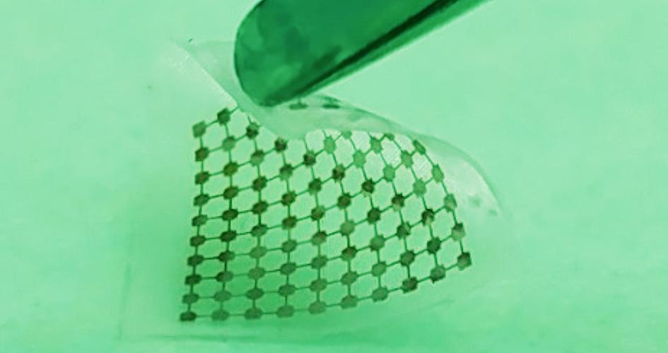
Researchers built this thin layer of silicon lace to modulate neural signals when activated by light. Courtesy of Yuanwen Jiang and Bozhi Tian

An April 30, 2018 University of Chicago news release by Louise Lerner, which originated the news item, describes the work in greater detail,

The scientists’ map lays out best methods to craft silicon devices depending on both the intended task and the scale—ranging from inside a cell to a whole animal.

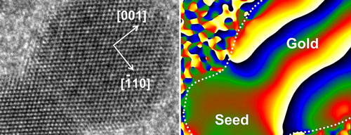
For example, to affect individual brain cells, silicon can be crafted to respond to light by emitting a tiny ionic current, which encourages neurons to fire. But in order to stimulate limbs, scientists need a system whose signals can travel farther and are stronger—such as a gold-coated silicon material in which light triggers a chemical reaction.

The mechanical properties of the implant are important, too. Say researchers would like to work with a larger piece of the brain, like the cortex, to control motor movement. The brain is a soft, squishy substance, so they’ll need a material that’s similarly soft and flexible, but can bind tightly against the surface. They’d want thin and lacy silicon, say the design principles.

The team favors this method because it doesn’t require genetic modification or a power supply wired in, since the silicon can be fashioned into what are essentially tiny solar panels. (Many other forms of monitoring or interacting with the brain need to have a power supply, and keeping a wire running into a patient is an infection risk.)

They tested the concept in mice and found they could stimulate limb movements by shining light on brain implants. Previous research tested the concept in neurons.

“We don’t have answers to a number of intrinsic questions about biology, such as whether individual mitochondria communicate remotely through bioelectric signals,” said Yuanwen Jiang, the first author on the paper, then a graduate student at UChicago and now a postdoctoral researcher at Stanford. “This set of tools could address such questions as well as pointing the way to potential solutions for nervous system disorders.”

Other UChicago authors were Assoc. Profs. Chin-Tu Chen and Chien-Min Kao, Asst. Prof Xiaoyang, postdoctoral researchers Jaeseok Yi, Yin Fang, Xiang Gao, Jiping Yue, Hsiu-Ming Tsai, Bing Liu and Yin Fang, graduate students Kelliann Koehler, Vishnu Nair, and Edward Sudzilovsky, and undergraduate student George Freyermuth.

Other researchers on the paper hailed from Northwestern University, the University of Illinois at Chicago and Hong Kong Polytechnic University.

The researchers have also made this video illustrating their work,

via Gfycat Tiny silicon nanowires (in blue), activated by light, trigger activity in neurons. (Courtesy Yuanwen Jiang and Bozhi Tian)

Here’s a link to and a citation for the paper,

Rational design of silicon structures for optically controlled multiscale biointerfaces by Yuanwen Jiang, Xiaojian Li, Bing Liu, Jaeseok Yi, Yin Fang, Fengyuan Shi, Xiang Gao, Edward Sudzilovsky, Ramya Parameswaran, Kelliann Koehler, Vishnu Nair, Jiping Yue, KuangHua Guo, Yin Fang, Hsiu-Ming Tsai, George Freyermuth, Raymond C. S. Wong, Chien-Min Kao, Chin-Tu Chen, Alan W. Nicholls, Xiaoyang Wu, Gordon M. G. Shepherd, & Bozhi Tian. Nature Biomedical Engineering (2018) doi:10.1038/s41551-018-0230-1 Published: 30 April 2018

This paper is behind a paywall.

Mathematics and how living cells ‘think’

This May 2, 2018 Queensland University of Technology (QUT; Australia) press release is also on EurekAlert,

How does the ‘brain’ of a living cell work, allowing an organism to function and thrive in changing and unfavourable environments?

Queensland University of Technology (QUT) researcher Dr Robyn Araujo has developed new mathematics to solve a longstanding mystery of how the incredibly complex biological networks within cells can adapt and reset themselves after exposure to a new stimulus.

Her findings, published in Nature Communications, provide a new level of understanding of cellular communication and cellular ‘cognition’, and have potential application in a variety of areas, including new targeted cancer therapies and drug resistance.

Dr Araujo, a lecturer in applied and computational mathematics in QUT’s Science and Engineering Faculty, said that while we know a great deal about gene sequences, we have had extremely limited insight into how the proteins encoded by these genes work together as an integrated network – until now.

“Proteins form unfathomably complex networks of chemical reactions that allow cells to communicate and to ‘think’ – essentially giving the cell a ‘cognitive’ ability, or a ‘brain’,” she said. “It has been a longstanding mystery in science how this cellular ‘brain’ works.

“We could never hope to measure the full complexity of cellular networks – the networks are simply too large and interconnected and their component proteins are too variable.

“But mathematics provides a tool that allows us to explore how these networks might be constructed in order to perform as they do.

“My research is giving us a new way to look at unravelling network complexity in nature.”

Dr Araujo’s work has focused on the widely observed function called perfect adaptation – the ability of a network to reset itself after it has been exposed to a new stimulus.

“An example of perfect adaptation is our sense of smell,” she said. “When exposed to an odour we will smell it initially but after a while it seems to us that the odour has disappeared, even though the chemical, the stimulus, is still present.

“Our sense of smell has exhibited perfect adaptation. This process allows it to remain sensitive to further changes in our environment so that we can detect both very feint and very strong odours.

“This kind of adaptation is essentially what takes place inside living cells all the time. Cells are exposed to signals – hormones, growth factors, and other chemicals – and their proteins will tend to react and respond initially, but then settle down to pre-stimulus levels of activity even though the stimulus is still there.

“I studied all the possible ways a network can be constructed and found that to be capable of this perfect adaptation in a robust way, a network has to satisfy an extremely rigid set of mathematical principles. There are a surprisingly limited number of ways a network could be constructed to perform perfect adaptation.

“Essentially we are now discovering the needles in the haystack in terms of the network constructions that can actually exist in nature.

“It is early days, but this opens the door to being able to modify cell networks with drugs and do it in a more robust and rigorous way. Cancer therapy is a potential area of application, and insights into how proteins work at a cellular level is key.”

Dr Araujo said the published study was the result of more than “five years of relentless effort to solve this incredibly deep mathematical problem”. She began research in this field while at George Mason University in Virginia in the US.

Her mentor at the university’s College of Science and co-author of the Nature Communications paper, Professor Lance Liotta, said the “amazing and surprising” outcome of Dr Araujo’s study is applicable to any living organism or biochemical network of any size.

“The study is a wonderful example of how mathematics can have a profound impact on society and Dr Araujo’s results will provide a set of completely fresh approaches for scientists in a variety of fields,” he said.

“For example, in strategies to overcome cancer drug resistance – why do tumours frequently adapt and grow back after treatment?

“It could also help understanding of how our hormone system, our immune defences, perfectly adapt to frequent challenges and keep us well, and it has future implications for creating new hypotheses about drug addiction and brain neuron signalling adaptation.”

Hre’s a link to and a citation for the paper,

The topological requirements for robust perfect adaptation in networks of any size by Robyn P. Araujo & Lance A. Liotta. Nature Communicationsvolume 9, Article number: 1757 (2018) doi:10.1038/s41467-018-04151-6 Published: 01 May 2018

This paper is open access.

Possible nanoparticle-based vaccine/microbiocide for herpes simplex virus-2

An April 27, 2016 news item on ScienceDaily describes a new therapeutic and preventative technology for herpes,

An effective vaccine against the virus that causes genital herpes has evaded researchers for decades. But now, researchers from the University of Illinois at Chicago [UIC] working with scientists from Germany have shown that zinc-oxide nanoparticles shaped like jacks can prevent the virus from entering cells, and help natural immunity to develop.

“We call the virus-trapping nanoparticle a microbivac, because it possesses both microbicidal and vaccine-like properties,” says corresponding author Deepak Shukla, professor of ophthalmology and microbiology & immunology in the UIC College of Medicine. “It is a totally novel approach to developing a vaccine against herpes, and it could potentially also work for HIV and other viruses,” he said.

The particles could serve as a powerful active ingredient in a topically-applied vaginal cream that provides immediate protection against herpes virus infection while simultaneously helping stimulate immunity to the virus for long-term protection, explained Shukla.

An April 27, 2016 UIC news release (also on EurekAlert), which originated the news item, provides more context for the work,

Herpes simplex virus-2, which causes serious eye infections in newborns and immunocompromised patients as well as genital herpes, is one of the most common human viruses. According to the Centers for Disease Control and Prevention, about 15 percent of people from ages 14-49 carry HSV-2, which can hide out for long periods of time in the nervous system. The genital lesions caused by the virus increase the risk for acquiring human immunodeficiency virus, or HIV.

“Your chances of getting HIV are three to four times higher if you already have genital herpes, which is a very strong motivation for developing new ways of preventing herpes infection,” Shukla said.

Treatments for HSV-2 include daily topical medications to suppress the virus and shorten the duration of outbreaks, when the virus is active and genital lesions are present. However, drug resistance is common, and little protection is provided against further infections. Efforts to develop a vaccine have been unsuccessful because the virus does not spend much time in the bloodstream, where most traditional vaccines do their work.

The news release goes on to provide technical details,

The tetrapod-shaped zinc-oxide nanoparticles, called ZOTEN, have negatively charged surfaces that attract the HSV-2 virus, which has positively charged proteins on its outer envelope. ZOTEN nanoparticles were synthesized using technology developed by material scientists at Germany’s Kiel University and protected under a joint patent with UIC.

When bound to the nanoparticles, HSV-2 cannot infect cells. But the bound virus remains susceptible to processing by immune cells called dendritic cells that patrol the vaginal lining. The dendritic cells “present” the virus to other immune cells that produce antibodies. The antibodies cripple the virus and trigger the production of customized killer cells that identify infected cells and destroy them before the virus can take over and spread.

The researchers showed that female mice swabbed with HSV-2 and an ointment containing ZOTEN had significantly fewer genital lesions than mice treated with a cream lacking ZOTEN. Mice treated with ZOTEN also had less inflammation in the central nervous system, where the virus can hide out.

The researchers were able to watch immune cells pry the virus off the nanoparticles for immune processing, using high-resolution fluorescence microscopy.

“It’s very clear that ZOTEN facilitates the development of immunity by holding the virus and letting the dendritic cells get to it,” Shukla said.

If found safe and effective in humans, a ZOTEN-containing cream ideally would be applied vaginally just prior to intercourse, Shukla said. But if a woman who had been using it regularly missed an application, he said, she may have already developed some immunity and still have some protection. Shukla hopes to further develop the nanoparticles to work against HIV, which like HSV-2 also has positively charged proteins embedded in its outer envelope.

ZOTEN particles are uniform in size and shape, making them attractive for use in other biomedical applications. The novel flame transport synthesis technology used to make them allows large-scale production, said Rainer Adelung, professor of nanomaterials at Kiel University. And, because no chemicals are used, the production process is green.

Adelung hopes to begin commercial production of ZOTEN through a startup company that will be run jointly with his colleagues at UIC.

Here’s an image of the particles, courtesy of UIC,

Zinc oxide tetrapod nanoparticles. Credit: Deepak Shukla

Zinc oxide tetrapod nanoparticles. Credit: Deepak Shukla

Here’s a link to and a citation for the paper,

Intravaginal Zinc Oxide Tetrapod Nanoparticles as Novel Immunoprotective Agents against Genital Herpes by Thessicar E. Antoine, Satvik R. Hadigal, Abraam M. Yakoub, Yogendra Kumar Mishra, Palash Bhattacharya, Christine Haddad, Tibor Valyi-Nagy, Rainer Adelung, Bellur S. Prabhakar, and Deepak Shukla. The Journal of Immunology April 27, 2016 1502373  doi: 10.4049/jimmunol.1502373 Published online before print April 27, 2016

This paper is behind a paywall.

One final comment, it’s a long from a mouse vagina in this study to a human one.

And the bacteria shall save us—nanobiobots

A March 24, 2015 University of Illinois at Chicago news release (also on EurekAlert) describes the NERD, a Nano-Electro-Robotic Device which employs bacteria and graphene quantum dots,

As nanotechnology makes possible a world of machines too tiny to see, researchers are finding ways to combine living organisms with nonliving machinery to solve a variety of problems.

Like other first-generation bio-robots, the new nanobot engineered at the University of Illinois at Chicago [UIC] is a far cry from Robocop. It’s a robotic germ.

UIC researchers created an electromechanical device–a humidity sensor–on a bacterial spore. They call it NERD, for Nano-Electro-Robotic Device. …

“We’ve taken a spore from a bacteria, and put graphene quantum dots on its surface–and then attached two electrodes on either side of the spore,” said Vikas Berry, UIC associate professor of chemical engineering and principal investigator on the study.

“Then we change the humidity around the spore,” he said.

When the humidity drops, the spore shrinks as water is pushed out. As it shrinks, the quantum dots come closer together, increasing their conductivity, as measured by the electrodes.

“We get a very clean response–a very sharp change the moment we change humidity,” Berry said. The response was 10 times faster, he said, than a sensor made with the most advanced man-made water-absorbing polymers.

There was also better sensitivity in extreme low-pressure, low-humidity situations.

“We can go all the way down to a vacuum and see a response,” said Berry, which is important in applications where humidity must be kept low, for example, to prevent corrosion or food spoilage. “It’s also important in space applications, where any change in humidity could signal a leak,” he said.

Currently available sensors increase in sensitivity as humidity rises, Berry said. NERD’s sensitivity is actually higher at low humidity.

“This is a fascinating device,” Berry said. “Here we have a biological entity. We’ve made the sensor on the surface of these spores, with the spore a very active complement to this device. The biological complement is actually working towards responding to stimuli and providing information.”

Interesting, yes? Here’s a link to and a citation for the research paper,

Graphene Quantum Dots Interfaced with Single Bacterial Spore for Bio-Electromechanical Devices: A Graphene Cytobot by T. S. Sreeprasad, Phong Nguyen, Ahmed Alshogeathri, Luke Hibbeler, Fabian Martinez, Nolan McNeil, & Vikas Berry. Scientific Reports 5, Article number: 9138 doi:10.1038/srep09138 Published 16 March 2015

This paper is open access.

Dexter Johnson provides more context for this research in a March 26, 2015 post on his Nanoclast blog (on the IEEE [institute of Electrical and Electronics Engineers]) where he notes,

Recently, James Tours’ group at Rice University, who were the first to develop GQCs [graphene quantum dots] in 2013, created an improved way to manufacture them that promised to open them up to a new range of applications in optics.

Dexter’s insights make for worthwhile reading.

Dumbbells at the nanoscale according to researchers at the (US) Argonne National Laboratory

Researchers at the US Dept. of Energy’s Argonne National Laboratory are providing new insight into how nanoparticles ‘grow’. From a Dec. 5, 2014 news item on Nanowerk,

Like snowflakes, nanoparticles come in a wide variety of shapes and sizes. The geometry of a nanoparticle is often as influential as its chemical makeup in determining how it behaves, from its catalytic properties to its potential as a semiconductor component.

Thanks to a new study from the U.S. Department of Energy’s (DOE) Argonne National Laboratory, researchers are closer to understanding the process by which nanoparticles made of more than one material – called heterostructured nanoparticles – form. This process, known as heterogeneous nucleation, is the same mechanism by which beads of condensation form on a windowpane.

The scientists have provided an image which illustrates their findings,

This picture combines a transmission electron microscope image of a nanodumbbell with a gold domain oriented in direction. The seed and gold domains in the dumbbell in the image on the right are identified by geometric phase analysis. Image credit: Soon Gu Kwon.

This picture combines a transmission electron microscope image of a nanodumbbell with a gold domain oriented in direction. The seed and gold domains in the dumbbell in the image on the right are identified by geometric phase analysis. Image credit: Soon Gu Kwon.

A Dec. 4, 2014 Argonne National Laboratory news release by Jared Sagoff, which originated the news item, describes the structures being examined and the reason for doing so,

Heterostructured nanoparticles can be used as catalysts and in advanced energy conversion and storage systems. Typically, these nanoparticles are created from tiny “seeds” of one material, on top of which another material is grown.  In this study, the Argonne researchers noticed that the differences in the atomic arrangements of the two materials have a big impact on the shape of the resulting nanoparticle.

“Before we started this experiment, it wasn’t entirely clear what’s happening at the interface when one material grows on another,” said nanoscientist Elena Shevchenko of Argonne Center for Nanoscale Materials, a DOE Office of Science user facility.

In this study, the researchers observed the formation of a nanoparticle consisting of platinum and gold.  The researchers started with a platinum seed and grew gold around it. Initially, the gold covered the platinum seed’s surface uniformly, creating a type of nanoparticle known as “core-shell.” However, as more gold was deposited, it started to grow unevenly, creating a dumbbell-like structure.

Thanks to state-of-the-art X-ray analysis provided by Argonne’s Advanced Photon Source (APS), a DOE Office of Science user facility, the researchers identified the cause of the dumbbell formation as “lattice mismatch,” in which the spacing between the atoms in the two materials doesn’t align.

“Essentially, you can think of lattice mismatch as having a row of smaller boxes on the bottom layer and larger boxes on the top layer.  When you try to fit the larger boxes into the space for a smaller box, it creates an immense strain,” said Argonne physicist Byeongdu Lee.

While the lattice mismatch is only fractions of a nanometer, the effect accumulates as layer after layer of gold forms on the platinum. The mismatch can be handled by the first two layers of gold atoms – creating the core-shell effect – but afterwards it proves too much to overcome. “The arrangement of atoms is the same in the two materials, but the distance between atoms is different,” said Argonne postdoctoral researcher Soon Gu Kwon. “Eventually, this becomes unstable, and the growth of the gold becomes unevenly distributed.”

As the gold continues to accumulate on one side of the seed nanoparticle, small quantities “slide” down the side of the nanoparticle like grains of sand rolling down the side of a sand hill, creating the dumbbell shape.

The advantage of the Argonne study comes from the researchers’ ability to perform in situ observations of the material in realistic conditions using the APS. “This is the first time anyone has been able to study the kinetics of this heterogeneous nucleation process of nanoparticles in real-time under realistic conditions,” said Argonne physicist Byeongdu Lee. “The combination of two X-ray techniques gave us the ability to observe the material at both the atomic level and the nanoscale, which gave us a good view of how the nanoparticles form and transform.” All conclusions made based on the X-ray studies were further confirmed using atomic-resolution microscopy in the group of Professor Robert Klie of the University of Illinois at Chicago.

This analysis of nanoparticle formation will help to lay the groundwork for the formation of new materials with different and controllable properties, according to Shevchenko. “In order to design materials, you have to understand how these processes happen at a very basic level,” she said.

Here’s a link to and a citation for the paper,

Heterogeneous nucleation and shape transformation of multicomponent metallic nanostructures by Soon Gu Kwon, Galyna Krylova, Patrick J. Phillips, Robert F. Klie, Soma Chattopadhyay, Tomohiro Shibata, Emilio E. Bunel, Yuzi Liu, Vitali B. Prakapenka, Byeongdu Lee, & Elena V. Shevchenko. Nature Materials (2014) doi:10.1038/nmat4115 Published online 02 November 2014

This paper is behind a paywall but there is a free preview via ReadCube Access.

Bringing the Nanoworld Together Workshop in Beijing, China, Sept. 24 – 25, 2014

The speakers currently confirmed for the ‘Bringing the Nanoworld Together Workshop organized by Oxford Instruments are from the UK, China, Canada, the US, and the Netherlands as per a July 2, 2014 news item on Nanowerk (Note: A link has been removed),

‘Bringing the Nanoworld Together’ is an event organised by Oxford Instruments to share the expertise of scientists in the field of Nanotechnology. It will be hosted at the IOS-CAS [Institute of Semiconductors-Chinese Academy of Sciences] Beijing.

Starting with half day plenary sessions on 2D materials with guest plenary speaker Dr Aravind Vijayaraghavan from the National Graphene Institute in Manchester, UK, and on Quantum Information Processing with guest plenary speaker Prof David Cory from the Institute for Quantum Computing, University of Waterloo, Canada, Oxford Instruments’ seminar at the IOP in Beijing from 24-25th September [2014] promises to discuss cutting edge nanotechnology solutions for multiple applications.

A July 1, 2014 Oxford Instruments press release, which originated the news item, describes the sessions and provides more details about the speakers,

Two parallel sessions will focus on thin film processing, & materials characterisation, surface science and cryogenic environments and a wide range of topics will be covered within each technical area. These sessions will include guest international and Chinese speakers from renowned research institutions, speakers from the host institute, and technical experts from Oxford Instruments. This will also present an excellent opportunity for networking between all participants.

Confirmed speakers include the following, but more will be announced soon:

Dr. Aravind Vijayaraghavan, National Graphene Institute, Manchester, UK
Prof David Cory, Institute for Quantum Computing, University of Waterloo, Canada
Prof Guoxing Miao, Institute for Quantum Computing, University of Waterloo, Canada
Prof. HE Ke, Tsinghua University, Institute of Physics, CAS, China
Dr. WANG Xiaodong, Institute of Semiconductors, CAS, China
Prof Erwin Kessels, Tue Eindhoven, Netherlands
Prof. ZENG Yi, Institute of Semiconductor, CAS, China
Prof Robert Klie, University of Illinois Chicago, USA
Prof. Xinran WANG, Nanjing University, China
Prof. Zhihai CHENG, National Centre for Nanoscience and Technology, China
Prof. Yeliang WANG, Institute of Physics, CAS, China

The thin film processing sessions will review latest etch and deposition technological advances, including: ALD, Magnetron Sputtering, ICP PECVD, Nanoscale Etch, MEMS, MBE and more.

Materials characterisation, Surface Science and Cryogenic Environment sessions will cover multiple topics and technologies including: Ultra high vacuum SPM, Cryo free low temperature solutions, XPS/ESCA, an introduction to atomic force microscopy (AFM) and applications such as nanomechanics, In-situ heating and tensile characterisation using EBSD, Measuring Layer thicknesses and compositions using EDS, Nanomanipulation and fabrication within the SEM / FIB.

The host of last year’s Nanotechnology Tools seminar in India, Prof. Rudra Pratap, Chairperson at the Centre for Nano Science and Engineering, Indian Institute of Science, IISC Bangalore commented, “This seminar has been extremely well organised with competent speakers covering a variety of processes and tools for nanofabrication. It is great to have practitioners of the art give talks and provide tips and solutions based on their experience, something that cannot be found in text books.”

“This workshop is a great opportunity for a wide range of scientists in research and manufacturing to discover practical aspects of many new and established processes, technologies and applications, directly from renowned scientists and a leading manufacturer with over 50 years in the industry”, comments Mark Sefton, Sector Head of Oxford Instruments NanoSolutions, “Delegates appreciate the informal workshop atmosphere of these events, encouraging delegates to participate through open discussion and sharing their questions and experiences.”

This seminar is free of charge but prior booking is essential.

You can register on the Oxford Instruments website’s Bringing the Nanoworld Together Workshop webpage,

SpiderSense and wearable computers

Nancy Owano in her Feb. 23, 2013 article for phys.org, Wearable display meets blindfold test for sensing danger, features a project (SpiderSense) from the University of Illinois at Chicago that will be presented at the Augmented Human ’13 conference to be held March 7 – 8, 2013 in Stuttgart, Germany,

The researchers behind SpiderSense define it as a wearable device that projects the wearer’s near environment on the skin. The suit gives the user a special directional awareness of surrounding objects. They have explored a scenario where multiple sites over the body, rather than just hands, are fitted with transducers. These transducers relay information about the wearer’s environment into tactile sensations.

Modules are distributed across the suit to give the wearer as near to 360-degree ultrasound coverage as possible. The system modules can scan the environment; they are controlled through a Controller Box. The box carries the power source, the electronics and the system logic. The modules and the Controller Box are connected by means of ten pin ribbon cables. The researchers said that, in the future, this could be replaced by a wireless Bluetooth connection.

You can find out more about SpiderSense from its presentation webpage on the University of Illinois at Chicago Electronic Visualization Laboratory (EVL) website,

Sensing the environment through SpiderSense     

authors: Mateevitsi,V., Haggadone, B., Leigh, J., Kunzer, B., Kenyon, R.V.

Augmented Human ’13, 4th International Conference in Cooperation with ACM SIGCHI, Stuttgart, Germany

Recent scientific advances allow the use of technology to expand the number of forms of energy that can be perceived by humans. Smart sensors can detect hazards that human sensors are unable to perceive, for example radiation. This fusing of technology to human’s forms of perception enables exciting new ways of perceiving the world around us. In this paper we describe the design of SpiderSense, a wearable device that projects the wearer’s near environment on the skin and allows for directional awareness of objects around him. The millions of sensory receptors that cover the skin presents opportunities for conveying alerts and messages. We discuss the challenges and considerations of designing similar wearable devices.

Victor Mateevisti wearing SpiderSense image provided by L. Long, EVL

Victor Mateevisti wearing SpiderSense
image provided by L. Long, EVL

A Feb. 22, 2013 article by Hal Hodson for New Scientist inspired Owano who acknowledges that to be the case in her end notes,

Mateevitsi [Victor Mateevitsi] tested the suit out on students, getting them to stand outside on campus, blindfolded, and “feel” for approaching attackers. Each wearer had ninja cardboard throwing stars to use whenever they sensed someone approaching them. “Ninety five per cent of the time they were able to sense someone approaching and throw the star at them,” says Mateevitsi.

The SpiderSense presentation is scheduled for March 7, 2013 at the Augmented Human ’13 conference or as it’s also known, the 4th International Conference in Cooperation with ACM SIGCHI (Association for Computing Machinery, Special Interest Group on Computer-Human Interaction). The team, as per Hal Hodson’s article,  hopes to start human trials of SpiderSense with visually impaired individuals.