Tag Archives: University of Jyväskylä

Joint Mexican/Finnish research team analyzes circulating currents inside gold nanoparticles

An April 30, 2021 news item on ScienceDaily announces the research,

Researchers in the Nanoscience Center of University of Jyvaskyla, in Finland and in the Guadalajara University in Mexico developed a method that allows for simulation and visualization of magnetic-field-induced electron currents inside gold nanoparticles. The method facilitates accurate analysis of magnetic field effects inside complex nanostructures in nuclear magnetic resonance measurements and establishes quantitative criteria for aromaticity of nanoparticles. The work was published 30.4.2021 as an Open Access article in Nature Communications.

An April 30, 2021 University of Jyväskylä – Jyväskylän yliopisto news release (also on EurekAlert), which originated the news item, describes the work in greater technical detail,

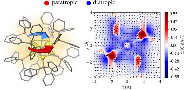
According to the classical electromagnetism, a charged particle moving in an external magnetic field experiences a force that makes the particle’s path circular. This basic law of physics is used, e.g., in designing cyclotrons that work as particle accelerators. When nanometer-size metal particles are placed in a magnetic field, the field induces a circulating electron current inside the particle. The circulating current in turn creates an internal magnetic field that opposes the external field. This physical effect is called magnetic shielding.

The strength of the shielding can be investigated by using nuclear magnetic resonance (NMR) spectroscopy. The internal magnetic shielding varies strongly in an atomic length scale even inside a nanometer-size particle. Understanding these atom-scale variations is possible only by employing quantum mechanical theory of the electronic properties of each atom making the nanoparticle.

Now, the research group of Professor Hannu Häkkinen in the University of Jyväskylä, in collaboration with University of Guadalajara in Mexico, developed a method to compute, visualize, and analyze the circulating electron currents inside complex 3D nanostructures. The method was applied to gold nanoparticles with a diameter of only about one nanometer. The calculations shed light onto unexplained experimental results from previous NMR measurements in the literature regarding how magnetic shielding inside the particle changes when one gold atom is replaced by one platinum atom.

A new quantitative measure to characterize aromaticity inside metal nanoparticles was also developed based on the total integrated strength of the shielding electron current.

“Aromaticity of molecules is one of the oldest concepts in chemistry, and it has been traditionally connected to ring-like organic molecules and to their delocalized valence electron density that can develop circulating currents in an external magnetic field. However, generally accepted quantitative criteria for the degree of aromaticity have been lacking. Our method yields now a new tool to study and analyze electron currents at the resolution of one atom inside any nanostructure, in principle. The peer reviewers of our work considered this as a significant advancement in the field”, says Professor Häkkinen who coordinated the research.

This image illustrates the work,

Caption: The atomic structure of a gold nanoparticle protected by phosphine molecules (left) and magnetic-field-induced electron currents in a plane intersecting the center of the particle (right). The total electron current consists of two (paratropic and diatropic) components circulating in opposite directions. Credit: University of Jyväskylä/Omar Lopez Estrada

Here’s a link to and a citation for the paper,

Magnetically induced currents and aromaticity in ligand-stabilized Au and AuPt superatoms by Omar López-Estrada, Bernardo Zuniga-Gutierrez, Elli Selenius, Sami Malola & Hannu Häkkinen . Nature Communications volume 12, Article number: 2477 (2021) DOI: https://doi.org/10.1038/s41467-021-22715 Published: 30 April 2021

This paper is open access.

Quantum entanglement in near-macroscopic objects

Researchers at Finland’s Aalto University seem excited in an April 25, 2018 news item on phys.org,

Perhaps the strangest prediction of quantum theory is entanglement, a phenomenon whereby two distant objects become intertwined in a manner that defies both classical physics and a common-sense understanding of reality. In 1935, Albert Einstein expressed his concern over this concept, referring to it as “spooky action at a distance.”

Today, entanglement is considered a cornerstone of quantum mechanics, and it is the key resource for a host of potentially transformative quantum technologies. Entanglement is, however, extremely fragile, and it has previously been observed only in microscopic systems such as light or atoms, and recently in superconducting electric circuits.

In work recently published in Nature, a team led by Prof. Mika Sillanpää at Aalto University in Finland has shown that entanglement of massive objects can be generated and detected.

The researchers managed to bring the motions of two individual vibrating drumheads—fabricated from metallic aluminium on a silicon chip—into an entangled quantum state. The macroscopic objects in the experiment are truly massive compared to the atomic scale—the circular drumheads have a diametre similar to the width of a thin human hair.

An April 20,2018 Aalto University press release (also on EurekAlert), which originated the news item, provides more detail,

‘The vibrating bodies are made to interact via a superconducting microwave circuit. The electromagnetic fields in the circuit carry away any thermal disturbances, leaving behind only the quantum mechanical vibrations’, says Professor Sillanpää, describing the experimental setup.

Eliminating all forms of external noise is crucial for the experiments, which is why they have to be conducted at extremely low temperatures near absolute zero, at –273 °C. Remarkably, the experimental approach allows the unusual state of entanglement to persist for long periods of time, in this case up to half an hour. In comparison, measurements on elementary particles have witnessed entanglement to last only tiny fractions of a second.

‘These measurements are challenging but extremely fascinating. In the future, we will attempt to teleport the mechanical vibrations. In quantum teleportation, properties of physical bodies can be transmitted across arbitrary distances using the channel of “spooky action at a distance”. We are still pretty far from Star Trek, though,’ says Dr. Caspar Ockeloen-Korppi, the lead author on the work, who also performed the measurements.

The results demonstrate that it is now possible to have control over the most delicate properties of objects whose size approaches the scale of our daily lives. The achievement opens doors for new kinds of quantum technologies, where the entangled drumheads could be used as routers or sensors. The finding also enables new studies of fundamental physics in, for example, the poorly understood interplay of gravity and quantum mechanics.

The team also included scientists from the University of New South Wales in Australia, the University of Chicago in the USA, and the University of Jyväskylä in Finland, whose theoretical innovations paved the way for the laboratory experiment.

An illustration has been made available,

An illustration of the 15-micrometre-wide drumheads prepared on silicon chips used in the experiment. The drumheads vibrate at a high ultrasound frequency, and the peculiar quantum state predicted by Einstein was created from the vibrations. Image: Aalto University / Petja Hyttinen & Olli Hanhirova, ARKH Architects.

Here’s a link to and a citation for the paper,

Stabilized entanglement of massive mechanical oscillators by C. F. Ockeloen-Korppi, E. Damskägg, J.-M. Pirkkalainen, M. Asjad, A. A. Clerk, F. Massel, M. J. Woolley & M. A. Sillanpää. Nature volume 556, pages478–482 (2018) doi:10.1038/s41586-018-0038-x Published online: 25 April 2018

This paper is behind a paywall.

Novel self-assembly at 102 atoms

A Jan. 13, 2017 news item on ScienceDaily announces a discovery about self-assembly of 102-atom gold nanoclusters,

Self-assembly of matter is one of the fundamental principles of nature, directing the growth of larger ordered and functional systems from smaller building blocks. Self-assembly can be observed in all length scales from molecules to galaxies. Now, researchers at the Nanoscience Centre of the University of Jyväskylä and the HYBER Centre of Excellence of Aalto University in Finland report a novel discovery of self-assembling two- and three-dimensional materials that are formed by tiny gold nanoclusters of just a couple of nanometres in size, each having 102 gold atoms and a surface layer of 44 thiol molecules. The study, conducted with funding from the Academy of Finland and the European Research Council, has been published in Angewandte Chemie.

A Jan. 13, 2017 Academy of Finland press release, which originated the news item, provides more technical information about the work,

The atomic structure of the 102-atom gold nanocluster was first resolved by the group of Roger D Kornberg at Stanford University in 2007 (2). Since then, several further studies of its properties have been conducted in the Jyväskylä Nanoscience Centre, where it has also been used for electron microscopy imaging of virus structures (3). The thiol surface of the nanocluster has a large number of acidic groups that can form directed hydrogen bonds to neighbouring nanoclusters and initiate directed self-assembly.

The self-assembly of gold nanoclusters took place in a water-methanol mixture and produced two distinctly different superstructures that were imaged in a high-resolution electron microscope at Aalto University. In one of the structures, two-dimensional hexagonally ordered layers of gold nanoclusters were stacked together, each layer being just one nanocluster thick. Modifying the synthesis conditions, also three-dimensional spherical, hollow capsid structures were observed, where the thickness of the capsid wall corresponds again to just one nanocluster size (see figure).

While the details of the formation mechanisms of these superstructures warrant further systemic investigations, the initial observations open several new views into synthetically made self-assembling nanomaterials.

“Today, we know of several tens of different types of atomistically precise gold nanoclusters, and I believe they can exhibit a wide variety of self-assembling growth patterns that could produce a range of new meta-materials,” said Academy Professor Hannu Häkkinen, who coordinated the research at the Nanoscience Centre. “In biology, typical examples of self-assembling functional systems are viruses and vesicles. Biological self-assembled structures can also be de-assembled by gentle changes in the surrounding biochemical conditions. It’ll be of great interest to see whether these gold-based materials can be de-assembled and then re-assembled to different structures by changing something in the chemistry of the surrounding solvent.”

“The free-standing two-dimensional nanosheets will bring opportunities towards new-generation functional materials, and the hollow capsids will pave the way for highly lightweight colloidal framework materials,” Postdoctoral Researcher Nonappa (Aalto University) said.

Professor Olli Ikkala of Aalto University said: “In a broader framework, it has remained as a grand challenge to master the self-assemblies through all length scales to tune the functional properties of materials in a rational way. So far, it has been commonly considered sufficient to achieve sufficiently narrow size distributions of the constituent nanoscale structural units to achieve well-defined structures. The present findings suggest a paradigm change to pursue strictly defined nanoscale units for self-assemblies.”

References:

(1)    Nonappa, T. Lahtinen, J.S. Haataja, T.-R. Tero, H. Häkkinen and O. Ikkala, “Template-Free Supracolloidal Self-Assembly of Atomically Precise Gold Nanoclusters: From 2D Colloidal Crystals to Spherical Capsids”, Angewandte Chemie International Edition, published online 23 November 2016, DOI: 10.1002/anie.201609036

(2)    P. Jadzinsky et al., “Structure of a thiol-monolayer protected gold nanoparticle at 1.1Å resolution”, Science 318, 430 (2007)

(3)    V. Marjomäki et al., “Site-specific targeting of enterovirus capsid by functionalized monodispersed gold nanoclusters”, PNAS 111, 1277 (2014)

Here’s the figure mentioned in the news release,

Figure: 2D hexagonal sheet-like and 3D capsid structures based on atomically precise gold nanoclusters as guided by hydrogen bonding between the ligands. The inset in the top left corner shows the atomic structure of one gold nanocluster.

Here’s a link to and a citation for the paper,

Template-Free Supracolloidal Self-Assembly of Atomically Precise Gold Nanoclusters: From 2D Colloidal Crystals to Spherical Capsids by Dr. Nonappa, Dr. Tanja Lahtinen, M. Sc. Johannes. S. Haataja, Dr. Tiia-Riikka Tero, Prof. Hannu Häkkinen, and Prof. Olli Ikkala. Angewandte Chemie International Edition Volume 55, Issue 52, pages 16035–16038, December 23, 2016 Version of Record online: 23 NOV 2016 DOI: 10.1002/anie.201609036

© 2016 Wiley-VCH Verlag GmbH & Co. KGaA, Weinheim

This paper is behind a paywall.

Ink toner on paper: research into topographies

An April 14, 2015 news item on Nanowerk about pen (in this case, ink toner) and paper,

A team of Finnish scientists has found a new way to examine the ancient art of putting ink to paper in unprecedented 3-D detail. The technique could improve scientists’ understanding of how ink sticks to paper and ultimately lead to higher quality, less expensive and more environmentally-friendly printed products.

Using modern X-ray and laser-based technologies, the researchers created a nano-scale map of the varying thickness of toner ink on paper. They discovered that wood fibers protruding from the paper received relatively thin coatings of ink. In general, they also found the toner thickness was dictated mainly by the local changes in roughness, rather than the chemical variations caused by the paper’s uneven glossy finish.

“We believe that this gives new insight, especially on how the topography of paper impacts the ink setting or consolidation,” said Markko Myllys, an applied physicist at the University of Jyvaskyla in Finland. “This in turn helps us understand how glossy and non-glossy printed surfaces should be made.”

An April 14, 2016 American Institute of Physics (AIP) news release (also on EurekAlert) by Catherine Myers, which originated the news item, describes the research in more detail,

To achieve their detailed picture of ink thickness, the researchers first examined the underlying paper with X-ray microtomography, a smaller cousin of the CT scanning technology used in hospitals to produce images of the inside of the body.

To analyze the cyan ink layers, the researchers used two additional technologies: optical profilometry, which bounced a light beam off the surface of the ink to obtain a surface profile, and laser ablation, which zapped away controlled amounts of ink with a laser to determine the ink depth.

Although none of the imaging techniques are themselves new, the researchers were the first to combine all three to achieve a complete, high-resolution 3-D image of the intricate ink and paper microstructures.

The final images resemble a rugged mountain landscape, with the higher peaks generally showing thinner coatings of ink, and the valleys showing thicker pools.

The researchers found the typical ink layer was approximately 2.5 micrometers deep, about 1/40 the thickness of an average sheet of paper, but with relatively large spatial variations between the thickest and thinnest areas.

Knowing how topographical variations affect ink thickness will help the printing industry create more environmentally-friendly and less energy-demanding ink and optimize the size distribution of ink particles, Myllys said. It could also help the papermaking industry design more sustainable paper and packaging, for example from recycled components, while still maintaining the quality needed to make ink stick well. Additionally, the papermaking industry could use the findings to help decide how best to incorporate smart and novel features into paper, Myllys said.

The team believes the imaging methods they used can also be adapted to effectively analyze the thickness variations in other types of thin films, including those found in microelectronics, wear-resistant coatings and solar panels.

“This result can certainly be generalized, and that makes it actually quite interesting,” Myllys said. “Thickness variations of thin films are crucial in many applications, but the 3-D analysis has been very difficult or impossible until now.”

Here’s a link to and a citation for the paper,

X-ray microtomography and laser ablation in the analysis of ink distribution in coated paper by  M. Myllys, H. Häkkänen, Korppi-Tommola, K. Backfolk, P. Sirviö, and J. Timonen1. J. Appl. Phys. 117, 144902 (2015); http://dx.doi.org/10.1063/1.4916588

This paper appears to be open access.

Gold atoms: sometimes they’re a metal and sometimes they’re a molecule

Fascinating work out of Finland shows that a minor change in the number of gold atoms in your gold nanoparticle can mean the difference between a metal and a molecule (coincidentally, this phenomenon is alluded to in my April 14, 2015 post (Nature’s patterns reflected in gold nanoparticles); more about that at the end of this piece. Getting back to Finland and when gold is metal and when it’s a molecule, here’s more from an April 10, 2015 news item on ScienceDaily,

Researchers at the Nanoscience Center at the University of Jyväskylä, Finland, have shown that dramatic changes in the electronic properties of nanometre-sized chunks of gold occur in well-defined size range. Small gold nanoclusters could be used, for instance, in short-term storage of energy or electric charge in the field of molecular electronics. Funded by the Academy of Finland, the researchers have been able to obtain new information which is important, among other things, in developing bioimaging and sensing based on metal-like clusters.

An April 10, 2015 news release (also on EurekAlert) on the Academy of Finland (Suomen Akatemia) website, which originated the news item, describes the work in more detail,

Two recent papers by the researchers at Jyväskylä (1, 2) demonstrate that the electronic properties of two different but still quite similar gold nanoclusters can be drastically different. The clusters were synthesised by chemical methods incorporating a stabilising ligand layer on their surface. The researchers found that the smaller cluster, with up to 102 gold atoms, behaves like a giant molecule while the larger one, with at least 144 gold atoms, already behaves, in principle, like a macroscopic chunk of metal, but in nanosize.

The fundamentally different behaviour of these two differently sized gold nanoclusters was demonstrated by shining a laser light onto solution samples containing the clusters and by monitoring how energy dissipates from the clusters into the surrounding solvent.

“Molecules behave drastically different from metals,” said Professor Mika Pettersson, the principal investigator of the team conducting the experiments. “The additional energy from light, absorbed by the metal-like clusters, transfers to the environment extremely rapidly, in about one hundred billionth of a second, while a molecule-like cluster is excited to a higher energy state and dissipates the energy into the environment with a rate that is at least 100 times slower. This is exactly what we saw: the 102-gold atom cluster is a giant molecule showing even a transient magnetic state while the 144-gold atom cluster is already a metal. We’ve thus managed to bracket an important size region where this fundamentally interesting change in the behaviour takes place.”

“These experimental results go together very well with what our team has seen from computational simulations on these systems,” said Professor Hannu Häkkinen, a co-author of the studies and the scientific director of the nanoscience centre. “My team predicted this kind of behaviour back in 2008-2009 when we saw big differences in the electronic structure of exactly these nanoclusters. It’s wonderful that robust spectroscopic experiments have now proved these phenomena. In fact, the metal-like 144-atom cluster is even more interesting, since we just published a theoretical paper where we saw a big enhancement of the metallic properties of just a few copper atoms mixed with gold.” (3)

Here are links to and citation for the papers,

Ultrafast Electronic Relaxation and Vibrational Cooling Dynamics of Au144(SC2H4Ph)60 Nanocluster Probed by Transient Mid-IR Spectroscopy by Satu Mustalahti, Pasi Myllyperkiö, Tanja Lahtinen, Kirsi Salorinne, Sami Malola, Jaakko Koivisto, Hannu Häkkinen, and Mika Pettersson. J. Phys. Chem. C, 2014, 118 (31), pp 18233–18239 DOI: 10.1021/jp505464z Publication Date (Web): July 3, 2014

Copyright © 2014 American Chemical Society

Copper Induces a Core Plasmon in Intermetallic Au(144,145)–xCux(SR)60 Nanoclusters by Sami Malola, Michael J. Hartmann, and Hannu Häkkinen. J. Phys. Chem. Lett., 2015, 6 (3), pp 515–520 DOI: 10.1021/jz502637b Publication Date (Web): January 22, 2015

Copyright © 2015 American Chemical Society

Molecule-like Photodynamics of Au102(pMBA)44 Nanocluster by Satu Mustalahti, Pasi Myllyperkiö, Sami Malola, Tanja Lahtinen, Kirsi Salorinne, Jaakko Koivisto, Hannu Häkkinen, and Mika Pettersson. ACS Nano, 2015, 9 (3), pp 2328–2335 DOI: 10.1021/nn506711a Publication Date (Web): February 22, 2015

Copyright © 2015 American Chemical Society

These papers are behind paywalls.

As for my April 14, 2015 post (Nature’s patterns reflected in gold nanoparticles), researchers at Carnegie Mellon University were researching patterns in different sized gold nanoparticles when this was noted in passing,

… Normally, gold is one of the best conductors of electrical current, but the size of Au133 is so small that the particle hasn’t yet become metallic. …

‘Scotch-tape’ technique for isolating graphene

The ‘scotch-tape’ technique is mythologized in the graphene origins story which has scientists, Andre Geim and Konstantin Novoselov, first isolating the material by using adhesive (aka ‘sticky’ tape or ‘scotch’ tape) as per my Oct. 7, 2010 posting,

The technique that Geim and Novoselov used to create the first graphene sheets both amuses and fascinates me (from the article by Kit Eaton on the Fast Company website),

The two scientists came up with the technique that first resulted in samples of graphene–peeling individual atoms-deep sheets of the material from a bigger block of pure graphite. The science here seems almost foolishly simple, but it took a lot of lateral thinking to dream up, and then some serious science to investigate: Geim and Novoselo literally “ripped” single sheets off the graphite by using regular adhesive tape. Once they’d confirmed they had grabbed micro-flakes of the material, Geim and Novoselo were responsible for some of the very early experiments into the material’s properties. Novel stuff indeed, but perhaps not so unexpected from a scientist (Geim) who the Nobel Committe notes once managed to make a frog levitate in a magnetic field.

A May 21, 2014 article about Geim who has won both a Nobel and an Ig Nobel (the only scientist to do so) and graphene by Sarah Lewis for Fast Company offers more details about the discovery,

The graphene FNE [Friday Night Experiments] began when Geim asked Da Jiang, a doctoral student from China, to polish a piece of graphite an inch across and a few millimeters thick down to 10 microns using a specialized machine. Partly due to a language barrier, Jiang polished the graphite down to dust, but not the ultimate thinness Geim wanted.

Helpfully, the Geim lab was also observing graphite using scanning tunneling microscopy (STM). The experimenters would clean the samples beforehand using Scotch tape, which they would then discard. “We took it out of the trash and just used it,” Novoselov said. The flakes of graphite on the tape from the waste bin were finer and thinner than what Jiang had found using the fancy machine. They weren’t one layer thick—that achievement came by ripping them some more with Scotch tape.

They swapped the adhesive for Japanese Nitto tape, “probably because the whole process is so simple and cheap we wanted to fancy it up a little and use this blue tape,” Geim said. Yet “the method is called the ‘Scotch tape technique.’ I fought against this name, but lost.”

Scientists elsewhere have been inspired to investigate the process in minute detail as per a June 27, 2014 news item on Nanowerk,

The simplest mechanical cleavage technique using a primitive “Scotch” tape has resulted in the Nobel-awarded discovery of graphenes and is currently under worldwide use for assembling graphenes and other two-dimensional (2D) graphene-like structures toward their utilization in novel high-performance nanoelectronic devices.

The simplicity of this method has initiated a booming research on 2D materials. However, the atomistic processes behind the micromechanical cleavage have still been poorly understood.

A June 27, 2014 MANA (International Center for Materials Nanoarchitectoinics) news release, which originated the news item, provides more information,

A joined team of experimentalists and theorists from the International Center for Young Scientists, International Center for Materials Nanoarchitectonics and Surface Physics and Structure Unit of the National Institute for Materials Science, National University of Science and Technology “MISiS” (Moscow, Russia), Rice University (USA) and University of Jyväskylä (Finland) led by Daiming Tang and Dmitri Golberg for the first time succeeded in complete understanding of physics, kinetics and energetics behind the regarded “Scotch-tape” technique using molybdenum disulphide (MoS2) atomic layers as a model material.

The researchers developed a direct in situ probing technique in a high-resolution transmission electron microscope (HRTEM) to investigate the mechanical cleavage processes and associated mechanical behaviors. By precisely manipulating an ultra-sharp metal probe to contact the pre-existing crystalline steps of the MoS2 single crystals, atomically thin flakes were delicately peeled off, selectively ranging from a single, double to more than 20 atomic layers. The team found that the mechanical behaviors are strongly dependent on the number of layers. Combination of in situ HRTEM and molecular dynamics simulations reveal a transformation of bending behavior from spontaneous rippling (< 5 atomic layers) to homogeneous curving (~ 10 layers), and finally to kinking (20 or more layers).

By considering the force balance near the contact point, the specific surface energy of a MoS2 monoatomic layer was calculated to be ~0.11 N/m. This is the first time that this fundamentally important property has directly been measured.

After initial isolation from the mother crystal, the MoS2 monolayer could be readily restacked onto the surface of the crystal, demonstrating the possibility of van der Waals epitaxy. MoS2 atomic layers could be bent to ultimate small radii (1.3 ~ 3.0 nm) reversibly without fracture. Such ultra-reversibility and extreme flexibility proves that they could be mechanically robust candidates for the advanced flexible electronic devices even under extreme folding conditions.

Here’s a link to and a citation for the research paper,

Nanomechanical cleavage of molybdenum disulphide atomic layers by Dai-Ming Tang, Dmitry G. Kvashnin, Sina Najmaei, Yoshio Bando, Koji Kimoto, Pekka Koskinen, Pulickel M. Ajayan, Boris I. Yakobson, Pavel B. Sorokin, Jun Lou, & Dmitri Golberg. Nature Communications 5, Article number: 3631 doi:10.1038/ncomms4631 Published 03 April 2014

This paper is behind a paywall but there is a free preview available through ReadCube Access.

Getting new information on trafficking viruses with gold nanoparticles

Finnish researchers have developed a new technique for studying viruses according to a Jan. 15, 2014 news release on EurekAlert,

Researchers at the Nanoscience Center (NSC) of University of Jyväskylä in Finland have developed a novel method to study enterovirus structures and their functions. The method will help to obtain new information on trafficking of viruses in cells and tissues as well as on the mechanisms of virus opening inside cells.

The news release explains enteroviruses and describes the technique in more detail,

Enteroviruses are pathogenic viruses infecting humans. This group consists of polioviruses, coxsackieviruses, echoviruses and rhinoviruses. Enteroviruses are the most common causes of flu, but they also cause serious symptoms such as heart muscle infections and paralysis. Recently, enteroviruses have been linked with chronic diseases such as diabetes (2).

The infection mechanisms and infectious pathways of enteroviruses are still rather poorly known. Previous studies in the group of Dr. Varpu Marjomäki at the NSC have focused on the cellular factors that are important for the infection caused by selected enteroviruses (3). The mechanistic understanding of virus opening and the release of the viral genome in cellular structures for starting new virus production is still largely lacking. Furthermore, the knowledge of infectious processes in tissues is hampered by the lack of reliable tools for detecting virus infection.

The newly developed method involves a chemical modification of a known thiol-stabilized gold nanoparticle, the so-called Au102 cluster that was first synthesized and structurally solved by the group of Roger D Kornberg in 2007 (4) and later characterized at NSC by the groups of prof. Hannu Häkkinen and prof. Mika Pettersson in collaboration with Kornberg. (5) The organic thiol surface of the Au102 particles is modified by attaching linker molecules that make a chemical bond to sulfur-containing cysteine residues that are part of the surface structure of the virus. Several tens of gold particles can bind to a single virus, and the binding pattern shows up as dark tags reflecting the overall shape and structure of the virus (see the figure). The gold particles allow for studies on the structural changes of the viruses during their lifespan.

The study showed also that the infectivity of the viruses is not compromised by the attached gold particles which indicates that the labeling method does not interfere with the normal biological functions of viruses inside cells. This facilitates new investigations on the virus structures from samples taken from inside cells during the various phases of the virus infection, and gives possibilities to obtain new information on the mechanisms of virus uncoating (opening and release of the genome). The new method allows also for tracking studies of virus pathways in tissues. This is important for further understanding of acute and chronic symptoms caused by viruses. Finally, the method is expected to be useful for developing of new antiviral vaccines that are based on virus-like particles.

The method was developed at the NSC as a wide cross-disciplinary collaboration between chemists, physicists and biologists.

Here’s an image provided by the researchers, which illustrates their work,

Left: transmission electron microscopy (TEM) image of a single CVB3 virus showing tens of gold nanoparticles attached to its surface. The particles form a distinct "tagging pattern" that reflects the shape and the structure of the virus. The TEM image can be correlated to the model of the virus (right), where the yellow spheres mark the possible binding sites of the gold particles. The diameter of the virus is about 35 nanometers (nanometer = one billionth of a millimeter). The figure is taken from the publication. Courtesy: University of Jyväskylä

Left: transmission electron microscopy (TEM) image of a single CVB3 virus showing tens of gold nanoparticles attached to its surface. The particles form a distinct “tagging pattern” that reflects the shape and the structure of the virus. The TEM image can be correlated to the model of the virus (right), where the yellow spheres mark the possible binding sites of the gold particles. The diameter of the virus is about 35 nanometers (nanometer = one billionth of a millimeter). The figure is taken from the publication. Courtesy: University of Jyväskylä

Unfortunately, the researchers have published in the Proceedings f the National Academy of Sciences (PNAS). I noted in a previous posting that this publisher has developed a time-consuming process for getting access to a paper and payment options for reading it. I can provide a link to and a citation to the abstract for this paper but I’m not willing to spend several minutes trying to bypass the block they’ve placed on accessing papers and their payment options,

Site-specific targeting of enterovirus capsid by functionalized monodisperse gold nanoclusters by Varpu Marjomäki, Tanja Lahtinen, Mari Martikainen, Jaakko Koivisto, Sami Malola, Kirsi Salorinne, Mika Pettersson, and Hannu Häkkinenb. Proc. Natl. Acad. Sci. USA (2014), www.pnas.org/cgi/doi/10.1073/pnas.1310973111.

The University of Jyväskylä Jan. ??, 2014 news release about this work provides references (scroll down) to previous papers published on this work.

Knotty molecules

I couldn’t resist the wordplay (knotty/naughty) when I saw the Nov. 7, 2011 news item on Nanowerk titled, Tying molecules in knots. From the news item,

A research team headed by Professor David Leigh of the University of Edinburgh (UK) and Academy Professor Kari Rissanen of the University of Jyväskylä (Finland) have made the most complex molecular knot to date, as reported in Nature Chemistry (“A synthetic molecular pentafoil knot”).

However, deliberately tying molecules into well-defined knots so that these effects can be studied is extremely difficult. Up to now, only the simplest type of knot – a trefoil knot – had been prepared by scientists. Now Professor David Leigh’s team (www.catenane.net) at the University of Edinburgh together with Academy Professor Kari Rissanen at the University of Jyväskylä have succeeded in preparing and characterizing a more complex type of knot – a pentafoil knot (also known as a cinquefoil knot or a Solomon’s seal knot) – a knot which looks like a five-pointed star.

Remarkably, the thread that is tied into the star-shaped knot is just 160 atoms in length – that is about 16 nanometers long (one nanometer is one millionth of a millimeter).

Will this repopularize macramé (making textile by knotting the fibres)?

Cavandoli Macramé_Keith Russell

I found the image in Macramé essay on Wikipedia and Cavandoli is a form of Italian macramé.