Tag Archives: University of Malaya

Constructing a liver

Chinese researchers have taken a step closer to constructing complex (lifelike) liver tissue according to a Jan. 27, 2016 American Chemical Society (ACS) news release (also on EurekAlert),

Engineered liver tissue could have a range of important uses, from transplants in patients suffering from the organ’s failure to pharmaceutical testing [this usage is sometimes known as liver-on-a-chip]. Now scientists report in ACS’ journal Analytical Chemistry the development of such a tissue, which closely mimics the liver’s complicated microstructure and function more effectively than existing models.

The liver serves a critical role in digesting food and detoxifying the body. But due to a variety of factors, including viral infections, alcoholism and drug reactions, the organ can develop chronic or acute problems. When it doesn’t work well, a person can suffer abdominal pain, swelling, nausea and other symptoms. Complete liver failure can be life-threatening and can require a transplant, a procedure that currently depends on human donors. To curtail this reliance and provide an improved model for predicting drugs’ side effects, scientists have been engineering liver tissue in the lab. But so far, they haven’t achieved the complex architecture of the real thing. Jinyi Wang and colleagues came up with a new approach.

Wang’s team built a microfluidics-based tissue that copies the liver’s complex lobules, the organ’s tiny structures that resemble wheels with spokes. They did this with human cells from a liver and an aorta, the body’s main artery. In the lab, the engineered tissue had a metabolic rate that was closer to real-life levels than other liver models, and it successfully simulated how a real liver would react to various drug combinations. The researchers conclude their approach could lead to the development of functional liver tissue for clinical applications and screening drugs for side effects and potentially harmful interactions.

Here’s a link to and a citation for the paper,

On-Chip Construction of Liver Lobule-like Microtissue and Its Application for Adverse Drug Reaction Assay by Chao Ma, Lei Zhao, En-Min Zhou, Juan Xu, Shaofei Shen, and Jinyi Wang. Northwest A&F University, China Anal. Chem., Article ASAP DOI: 10.1021/acs.analchem.5b03869 Publication Date (Web): January 7, 2016

Copyright © 2016 American Chemical Society

This paper is behind a paywall.

In a teleconference earlier this month (January 2016), I spoke to researchers at the University of Malaya, Universiti Teknologi Malaysia (UTM), and Harvard University about a joint lung and nanomedicine research project where I asked researcher Joseph Brain (Harvard) about using lung-on-a-chip testing in place of in vivo (animal) testing and he indicated more confidence in the ‘precision cut lung slices’ technique. (You can find out more about the Malaysian project in my Jan. 12, 2016 posting but there’s only a brief mention of Brain’s preferred alternative animal testing technique.)

University of Malaya (Malaysia) and Harvard University (US) partner on nanomedicine/prevention projects

Unusually for a ‘nanomedicine’ project, the talk turned to prevention during a Jan. 10, 2016 teleconference featuring Dr. Noor Hayaty Abu Kasim of the University of Malaya and Dr. Wong Tin Wui of the Universiti Teknologi Malaysia and Dr. Joseph Brain of  Harvard University in a discussion about Malaysia’s major investment in nanomedicine treatment for lung diseases.

A Jan. 11, 2016 Malaysian Industry-Government Group for High Technology (MIGHT) news release on EurekAlert announces both the lung project (University of Malaya/Harvard University) and others under Malaysia’s NanoMITe (Malaysia Institute for Innovative Nanotechnology) banner,

Malaysian scientists are joining forces with Harvard University experts to help revolutionize the treatment of lung diseases — the delivery of nanomedicine deep into places otherwise impossible to reach.

Under a five-year memorandum of understanding between Harvard and the University of Malaya, Malaysian scientists will join a distinguished team seeking a safe, more effective way of tackling lung problems including chronic obstructive pulmonary disease (COPD), the progressive, irreversible obstruction of airways causing almost 1 in 10 deaths today.

Treatment of COPD and lung cancer commonly involves chemotherapeutics and corticosteroids misted into a fine spray and inhaled, enabling direct delivery to the lungs and quick medicinal effect. However, because the particles produced by today’s inhalers are large, most of the medicine is deposited in the upper respiratory tract.

The Harvard team, within the university’s T.H. Chan School of Public Health, is working on “smart” nanoparticles that deliver appropriate levels of diagnostic and therapeutic agents to the deepest, tiniest sacs of the lung, a process potentially assisted by the use of magnetic fields.

Malaysia’s role within the international collaboration: help ensure the safety and improve the effectiveness of nanomedicine, assessing how nanomedicine particles behave in the body, what attaches to them to form a coating, where the drug accumulates and how it interacts with target and non-target cells.

Led by Joseph Brain, the Cecil K. and Philip Drinker Professor of Environmental Physiology, the research draws on extensive expertise at Harvard in biokinetics — determining how to administer medicine to achieve the proper dosage to impact target cells and assessing the extent to which drug-loaded nanoparticles pass through biological barriers to different organs.

The studies also build on decades of experience studying the biology of macrophages — large, specialized cells that recognize, engulf and destroy target cells as part of the human immune system.

Manipulating immune cells represents an important strategy for treating lung diseases like COPD and lung cancer, as well as infectious diseases including tuberculosis and listeriosis.

Dr. Brain notes that every day humans breathe 20,000 litres of air loaded with bacteria and viruses, and that the world’s deadliest epidemic — an outbreak of airborne influenza in the 1920s — killed tens of millions.

Inhaled nanomedicine holds the promise of helping doctors prevent and treat such problems in future, reaching the target area more swiftly than if administered orally or even intravenously.

This is particularly true for lung cancer, says Dr. Brain. “Experiments have demonstrated that a drug dose administered directly to the respiratory tract achieves much higher local drug concentrations at the target site.”

COPD meanwhile affects over 235 million people worldwide and is on the rise, with 80% of cases caused by cigarette smoking. Exacerbated by poor air quality, COPD is expected to rise from 5th to 3rd place among humanity’s most lethal health problems by 2030.

“Nanotechnology is making a significant impact on healthcare by delivering improvements in disease diagnosis and monitoring, as well as enabling new approaches to regenerative medicine and drug delivery,” says Prof. Zakri Abdul Hamid, Science Advisor to the Prime Minister of Malaysia.

“Malaysia, through NanoMITe, is proud and excited to join the Harvard team and contribute to the creation of these life-giving innovations.”

While neither Dr. Abu Kasim nor Dr. Wong are included in the news release both are key members of the Malaysian team tasked to work on nanomedicines for lung disease. Dr. Abu Kasim is a professor of restorative dentistry at the University of Malaya and familiar with nanotechnology-enabled materials and nanoparticles through her work in that field. She is also the project lead for NanoMITe’s Project 4: Consequences of Smoking among the Malaysian Population. From the project webpage,

Smoking is a prevalent problem worldwide but especially so in Asia where nearly more than half of the world population reside. Smoking kills half of its users and despite the many documented harm to health is still a major problem. Globally six million lives are lost each year because of this addiction. This number is estimated to increase to ten million within the next two decades. Apart from the mortality, smokers are at increased risk of health morbidities of smoking which is a major risk factor for many non-communicable diseases (NCD) such as heart diseases, respiratory conditions and even mental health. Together, smoking reduces life expectancy 10-15 years compared to a non-smoker. Those with mental health lose double the years, 20 -25 years of their life as a result of their smoking. The current Malaysia death toll is at 10,000 lives per year due to smoking related health complications.

Although the health impact of smoking has been reported at length, this information is limited nationally. Lung cancer for example is closely linked to smoking, however, the study of the link between the two is lacking in Malaysia. Lung cancer particularly in Malaysia is also often diagnosed late, usually at stages 3 and 4. These stages of cancer are linked with a poorer prognosis. As a result to the harms to health either directly or indirectly, the World Health Organization (WHO) has introduced a legal treaty, the first, called the Framework Convention for Tobacco Control (FCTC). This treaty currently ratified by 174 countries was introduced in 2005 and consists of 38 FCTC Articles which are evidence based policies, known to assist member countries to reduce their smoking prevalence. Malaysia is an early signatory and early adopter of the MPOWER strategy which are major articles of the FCTC. Among them are education and information dissemination informing the dangers of smoking which can be done through awareness campaigns of advocacy using civil society groups. Most campaigns have focused on health harms with little mention non-health or environmental harm as a result of smoking. Therefore there is an opportunity to further develop this idea as a strong advocacy point towards a smoke-free generation in the near future

It is difficult impossible to recall any other nanomedicine initiative that has so thoroughly embedded prevention as part of its mandate. As Dr. Brain puts it, “Malaysia’s commitment to better health for everyone—sometimes, I’m jealous.”

Getting back to nanomedicine, it’s Dr. Wong, an associate professor in the school of pharmaceutics at Universiti Teknologi Malaysia (UTM), who is developing polymeric nanoparticles designed to carry medications into the lungs and Brain who will work on the best method of transport. From Dr. Brain’s webpage,

Dr. Brain’s research emphasizes responses to inhaled gases, particulates, and microbes. His studies extend from the deposition of inhaled particles in the respiratory tract to their clearance by respiratory defense mechanisms. Of particular interest is the role of lung macrophages; this resident cell keeps lung surfaces clean and sterile. Moreover, the lung macrophage is also a critical regulator of inflammatory and immune responses. The context of these studies on macrophages is the prevention and pathogenesis of environmental lung disease as well as respiratory infection.

His research has utilized magnetic particles in macrophages throughout the body as a non-invasive tool for measuring cell motility and the response of macrophages to various mediators and toxins. …

It was difficult to get any specifics about the proposed lung nanomedicine effort as it seems to be at a very early stage.

  • Malaysia through the Ministry of Higher Education with matching funds from the University of Malaya is funding this effort with 1M Ringgits ($300,00 USD) per year over five years for a total of 5M Ringgits ($1.5M USD)
  • A Malaysian researcher will be going to Harvard to collaborate directly with Dr. Brain and others on his team. The first will be Dr. Wong who will come to Harvard in June 2016 where he will work with his polymeric nanoparticles (vehicles for medications) and where Brain will examine transport strategies (aerosol, intrathecal administration, etc.) for those nanoparticle-bearing medications.
  • There will be a series of comparative studies of smoking in Malaysia and the US and other information efforts designed to support prevention strategies.

One last tidbit about research, Dr. Brain will be testing the nanoparticle-bearing medication once it has entered the lung using the ‘precision cut lung slices’ technique, as an alternative to some, if not all, in vivo testing.

Final comments

Nanomedicine is highly competitive and the Malaysians are interested in commercializing their efforts which according to Dr. Abu Kasim is one of the reasons they approached Harvard and Dr. Brain.

Should you find any errors please do let me know.
Hopes for Malaysia’s electrical and electronics industry and the opening of the Nano Semiconductor Technology Centre

A July 31, 2015 article for The Sun Daily by Ee Ann Nee announces four memorandums of understanding (MOU) featuring nanotechnology and signed by Malaysia’s Science, Technology and Innovation Ministry deputy secretary-general Dr Zulkifli Mohamed Hashim,

The export for Malaysia’s electrical and electronics (E&E) products is expected to increase by 20-30% by 2020 with nanotechnology and the rise of Internet of Things (IoT).

Science, Technology and Innovation Ministry deputy secretary-general Dr Zulkifli Mohamed Hashim said in 2014, the total export for E&E products was RM256 billion [Malaysian Ringgit], driven by strong global demand for new semiconductor applications and the rapid emergence of IoT.

The first MoU signed yesterday was for a technology partnership between nanotechnology commercialisation agency NanoMalaysia Bhd and Mimos will see the two agencies jointly undertake R&D and commercialisation of technology products.

The second MoU was a tripartite collaboration between NanoMalaysia, Mimos and Penchem Technologies Sdn Bhd for R&D and commercialisation of smart sensors and advanced material applications for electronic products.

The third and fourth MoU were signed between Mimos and the University of Malaya and Multimedia University respectively for research, design and development of grapheme, a carbon-based nanomaterial with superlative properties.

Good luck to them!

The most recent posting here featuring Malaysia was a Jan. 26, 2015 piece about a Malaysian nanotechnology scientist’s award from an Islamic organization (Islamic Educational Scientific and Cultural Organization [ISESCO])  that parallels UNESCO (United Nations Educational, Scientific and Cultural Organization)

Bone implants and restorative dentistry at the University of Malaya

The research into biomedical implants at the University of Malaya is part of an international effort and is in response to a demographic reality, hugely increased populations of the aged. From a Sept. 18, 2014 news item on ScienceDaily,

A major success in developing new biomedical implants with the ability to accelerate bone healing has been reported by a group of scientists from the Department of Restorative Dentistry, University of Malaya. This stems from a project partly funded by HIR [High Impact Research] and also involves Mr. Alireza Yaghoubi, HIR Young Scientist.

According to WHO (World Health Organization), between 2000 and 2050, the world’s population over 60 years is expected to increase from 605 million to more than 2 billion. This trend is particularly more prominent in Asia and Europe where in some countries by 2050, the majority of people will be older than 50. That is why in recent years, regenerative medicine has been among the most active and well-funded research areas in many developing nations.

As part of this global effort to realize better treatments for age-related conditions, a group of scientists from the department of restorative dentistry, University of Malaya and four other universities in the US have recently reported a major success in developing new biomedical implants with the ability to accelerate bone healing.

Two studies were published according to the Sept.15, 2014 University of Malaya news release, which originated the news item,

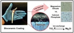
The two studies funded by the National Science Fund (NSF) in the US and the High Impact Research (HIR) program in Malaysia tackled the issue of bone-implant integration from different angles. In the first study appearing on the front cover of the July issue of Applied Surface Science, researchers demonstrated a mechanically superior bioactive coating based on magnesium silicates rather than the commercially available calcium phosphate which develops microcracks during preparation and delaminates under pressure. The new material owing to its lower thermal mismatch with titanium can prolong the durability of load-bearing orthopedic implants and reduce chances of post-surgery complications.

The other study published in the American Chemical Society’s Applied Materials & Interfaces reported a method for fabricating titanium implants with special surface topographies which double the chance of cell viability in early stages. The new technique is also much simpler as compared to the existing ones and therefore enables the preparation of personalized implants at the fraction of time and cost while offering a higher mechanical reliability.

Alireza Yaghoubi, the corresponding author of both studies believes that we are moving toward a future of personalized products. “It is very much like your taste in music and TV shows. People are different and the new trend in biotechnology is to make personalized medicine that matches the patient’s needs” Yaghoubi said. He continued “With regard to implants, we have the problem of variations in bone density in patients with osteoporosis and in some cases, even healthy individuals. Finding ways to integrate the implants with bone tissues can be challenging. There are also problems with the long-term performance of implants, such as release of debris from bioactive films which can potentially lead to osteolysis and chronic inflammation”.

The new technique employed by the scientists to create titanium implants with desirable surface properties uses microwave heating to create a porosity gradient on top of a dense core. The principles are very similar to a kitchen microwave and how it can make cooking easier, however apparently the fast heating capability is not only useful in cooking but it has numerous industrial applications. Prof. Bhaduri, the Director of Multi-functional materials laboratory at University of Toledo says that they have been using microwave for years to simplify fabrication of complex metallic components. “We needed a way to streamline the process and microwave sintering was a natural fit. With our new method, making the implant from titanium powder in custom sizes and with specific surface topographies is achieved through one easy step.” Bhaduri elaborated.

Researchers are hoping to carry out the clinical trial for this new generation of implants in order to make them available to the market soon. Dr. Kutty, one of the lead authors suggests that there is still room for improvement. Kutty concluded that “Roughened surfaces and bioceramics have desirable effects on osseointegration, but we are not stopping there. We are now developing new ways to use peptides for enhancing the performance of implants even further.”

This image provides an illustration of the proposed new material for implants,

The artwork appeared on the front cover of Applied Surface Science summarizes the benefits of a new bioceramic coating versus the commercially available Calcium Phosphate which develops microcracks during processing and may later cause osteolysis in load-bearing orthopedic implants. Courtesy: University of Malaya

The artwork appeared on the front cover of Applied Surface Science summarizes the benefits of a new bioceramic coating versus the commercially available Calcium Phosphate which develops microcracks during processing and may later cause osteolysis in load-bearing orthopedic implants. Courtesy: University of Malaya

Here are links to and citations for the papers,

Electrophoretic deposition of magnesium silicates on titanium implants: Ion migration and silicide interfaces by M. Afshar-Mohajer, A. Yaghoubi, S. Ramesh, A.R. Bushroa, K.M.C. Chin, C.C. Tin, and W.S. Chiu.  Applied Surface Science (2014) , Volume 307, 15 July 2014, Pages 1–6, DOI: 10.1016/j.apsusc.2014.04.033

Microwave-assisted Fabrication of Titanium Implants with Controlled Surface Topography for Rapid Bone Healing by Muralithran G. Kutty, Alok De, Sarit B. Bhaduri, and Alireza Yaghoubi. ACS Appl. Mater. Interfaces, 2014, 6 (16), pp 13587–13593 DOI: 10.1021/am502967n Publication Date (Web): August 6, 2014

Copyright © 2014 American Chemical Society

Both of these papers are behind paywalls.

New way to practice brain surgery skills before working on live patients

It’s a little disconcerting to learn that neurosurgeons don’t have many options to test drive their skills before they start practicing on patients as a Dec. 10, 2013 news release on EurekAlert about 3D printing (and a new way for neurosurgeons to practice) notes,,

Researchers from the University of Malaya in Malaysia, with collaboration from researchers from the University of Portsmouth and the University of Oxford in the United Kingdom, announce the creation of a cost-effective two-part model of the skull for use in practicing neurosurgical techniques. The model, produced using the latest generation of multimaterial 3D printers, is composed of a variety of materials that simulate the various consistencies and densities of human tissues encountered during neurosurgery. Details on the model are provided in “Utility of multimaterial 3D printers in creating models with pathological entities to enhance the training experience of neurosurgeons. Technical note.” By Vicknes Waran, F.R.C.S.(Neurosurgery), Vairavan Narayanan, F.R.C.S.(Neurosurgery), M.Surg., Ravindran Karuppiah, M.Surg., Sarah L. F. Owen, D.Phil., and Tipu Aziz, F.Med.Sci., published today [Dec. 10, 2013] online, ahead of print, in the Journal of Neurosurgery.

Here’s the disconcerting part (from the news release),

Neurosurgery is a difficult discipline to master. Trainees may spend as many as 10 years after graduation from medical school developing and honing their surgical skills before they can be designated as proficient in their specialty. The greater the number and variety of neurosurgical training sessions, the better the training experience. However, the authors point out that it is difficult to find suitable simulation models that offer accuracy and realism for neurosurgical training while keeping training costs down.

The news release provides a description of what makes the current generation of 3D printers particularly attractive for creating practice skulls, etc.,

Three-dimensional printers have been used to create models of normal and pathological human tissues and organs for physician training and patient instruction for some time. Until recently, however, only one material could be used in the creation of models. While useful for display purposes, one-material models have little value for hands-on training. With the advent of multimaterial 3D printers, the sophistication and versatility of the new models that could be created increased substantially, but so did their price.

Waran and colleagues tell us that this situation is now changing. They state that the newest generation of multimaterial 3D printers can aid neurosurgical training by creating models that simulate different diseases in a variety of body tissues, and they can do this in a cost-effective manner.

With the aid of an Objet500 Connex™ multimaterial 3D printer (Stratasys, Ltd.), researchers at the University of Malaya created a two-part model that can simulate pathological conditions in actual patients. The base piece of the model (the “head”) consists of one material. It has human features (a “face”) and the natural contours of a human skull. This piece is used to train the novice in neuronavigation techniques and can be reused again and again. The second part of the model defines the region in which simulated surgery is performed. This piece contains several different materials, which separately simulate skin, bone, dura mater, tumor, and normal brain tissue. The second piece fits into a slot in the base piece; this multi-textured piece can only be used once and is discarded after the practice session. Fortunately, it is easy to reproduce a steady stream of new pieces.

To make the training session valuable, the trainee must be able to see, feel, and even hear different “tissue” responses to surgical instruments and techniques during simulation surgery. The researchers tell us that the “skin” is designed to be pliable enough to be cut by a scalpel and repaired by sutures, yet sturdy enough to be held by a retractor; the “bone” has to be hard enough for the trainee to obtain experience using bone perforators and cutters; the “dura mater” must be thin and pliable—just like the real thing. The consistency and color of the “tumor” differ from those of the “brain” to simulate actual tissues. The researchers made the “tumor” softer than the “brain” and colored it orange, whereas they colored the brain light yellow.

To test the quality of the model produced by the printer and to make minor adjustments, the researchers from Malaysia were aided by other researchers from the UK. Three neurosurgeons and one expert in surgical simulations performed simulated surgery and assessed the model’s “tissue” components. All parts received ratings of “fair” or “good,” with most rated “good.”

The usefulness of the model in training neuronavigation techniques was also assessed. Since the two-part model was based on data from a real patient, it was no surprise that “neuroimaging” was rated “excellent” by the evaluating team. Two navigation systems were used, and in both cases “registration was accurate and planning possible.”

Waran and colleagues state that the reusable base piece of the model costs approximately US $2000 to fabricate and the disposable inset costs US $600. This makes these training models affordable. In addition, model designs are based on actual patient data, providing limitless variety.

Here’s a link to and a citation for the paper,

Waran V, Narayana V, Karuppiah R, Owen SLF, Aziz T: Utility of multimaterial 3D printers in creating models with pathological entities to enhance the training experience of neurosurgeons. Technical note. Journal of Neurosurgery, published online, ahead of print, December 10, 2013; DOI: 10.3171/2013.11.JNS131066.

This appears to be an open access paper.