Tag Archives: University of North Carolina at Chapel Hill

Robot that can maneuver through living lung tissue

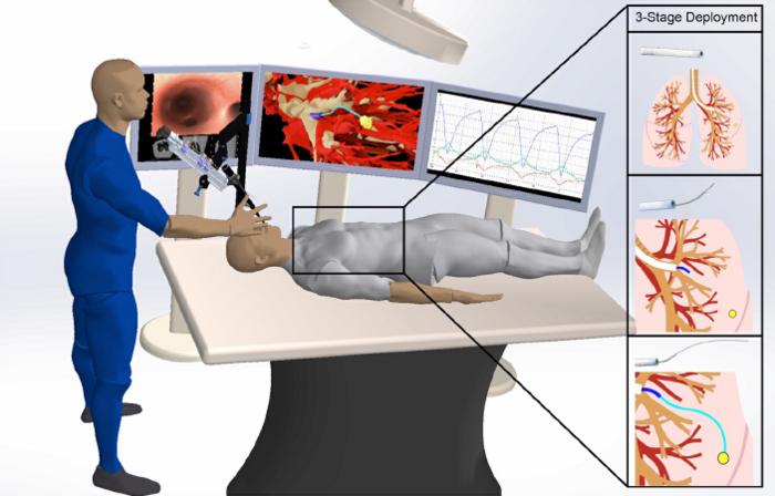
Caption: Overview of the semiautonomous medical robot’s three stages in the lungs. Credit: Kuntz et al.

This looks like one robot operating on another robot; I guess the researchers want to emphasize the fact that this autonomous surgical procedure isn’t currently being tested on human beings.

There’s more in a September 21, 2023 news item on ScienceDaily,

Scientists have shown that their steerable lung robot can autonomously maneuver the intricacies of the lung, while avoiding important lung structures.

Lung cancer is the leading cause of cancer-related deaths in the United States. Some tumors are extremely small and hide deep within lung tissue, making it difficult for surgeons to reach them. To address this challenge, UNC -Chapel Hill and Vanderbilt University researchers have been working on an extremely bendy but sturdy robot capable of traversing lung tissue.

Their research has reached a new milestone. In a new paper, published in Science Robotics, Ron Alterovitz, PhD, in the UNC Department of Computer Science, and Jason Akulian, MD MPH, in the UNC Department of Medicine, have proven that their robot can autonomously go from “Point A” to “Point B” while avoiding important structures, such as tiny airways and blood vessels, in a living laboratory model.

Thankfully there’s a September 21, 2023 University of North Carolina (UNC) news release (also on EurekAlert), which originated the news item, to provide more information, Note: Links have been removed,

“This technology allows us to reach targets we can’t otherwise reach with a standard or even robotic bronchoscope,” said Dr. Akulian, co-author on the paper and Section Chief of Interventional Pulmonology and Pulmonary Oncology in the UNC Division of Pulmonary Disease and Critical Care Medicine. “It gives you that extra few centimeters or few millimeters even, which would help immensely with pursuing small targets in the lungs.”

The development of the autonomous steerable needle robot leveraged UNC’s highly collaborative culture by blending medicine, computer science, and engineering expertise. In addition to Alterovitz and Akulian, the development effort included Yueh Z. Lee, MD, PhD, at the UNC Department of Radiology, as well as Robert J. Webster III at Vanderbilt University and Alan Kuntz at the University of Utah.

The robot is made of several separate components. A mechanical control provides controlled thrust of the needle to go forward and backward and the needle design allows for steering along curved paths. The needle is made from a nickel-titanium alloy and has been laser etched to increase its flexibility, allowing it to move effortlessly through tissue.

As it moves forward, the etching on the needle allows it to steer around obstacles with ease. Other attachments, such as catheters, could be used together with the needle to perform procedures such as lung biopsies.

To drive through tissue, the needle needs to know where it is going. The research team used CT scans of the subject’s thoracic cavity and artificial intelligence to create three-dimensional models of the lung, including the airways, blood vessels, and the chosen target. Using this 3-D model and once the needle has been positioned for launch, their AI-driven software instructs it to automatically travel from “Point A” to “Point B” while avoiding important structures.

“The autonomous steerable needle we’ve developed is highly compact, but the system is packed with a suite of technologies that allow the needle to navigate autonomously in real-time,” said Alterovitz, the principal investigator on the project and senior author on the paper. “It’s akin to a self-driving car, but it navigates through lung tissue, avoiding obstacles like significant blood vessels as it travels to its destination.”

The needle can also account for respiratory motion. Unlike other organs, the lungs are constantly expanding and contracting in the chest cavity. This can make targeting especially difficult in a living, breathing subject. According to Akulian, it’s like shooting at a moving target.

The researchers tested their robot while the laboratory model performed intermittent breath holding. Every time the subject’s breath is held, the robot is programmed to move forward.

“There remain some nuances in terms of the robot’s ability to acquire targets and then actually get to them effectively,” said Akulian, who is also a member of the UNC Lineberger Comprehensive Cancer Center, “and while there’s still a lot of work to be done, I’m very excited about continuing to push the boundaries of what we can do for patients with the world-class experts that are here.”

“We plan to continue creating new autonomous medical robots that combine the strengths of robotics and AI to improve medical outcomes for patients facing a variety of health challenges while providing guarantees on patient safety,” added Alterovitz.

Here’s a link to and a citation for the paper,

Autonomous medical needle steering in vivo by Alan Kuntz, Maxwell Emerson, Tayfun Efe Ertop, Inbar Fried, Mengyu Fu, Janine Hoelscher, Margaret Rox, Jason Akulian, Erin A. Gillaspie, Yueh Z. Lee, Fabien Maldonado, Robert J. Webster III, and Ron Alterovitz. Science Robotics 20 Sep 2023 Vol 8, Issue 82 DOI: 10.1126/scirobotics.adf7614

This paper is behind a paywall.

Microneedle vaccine patch outperforms needle

Vaccine patch sounds a lot friendlier than ‘needle’ and in the hoopla about vaccine hesitation I have to wonder if the fact that some people don’t like or are deeply fearful of needles is being overlooked.

Perhaps this or some other vaccine patch* will be ready for use in time for the next pandemic. From a September 24, 2021 news item on ScienceDaily,

Scientists at Stanford University and the University of North Carolina [UNC] at Chapel Hill have created a 3D-printed vaccine patch that provides greater protection than a typical vaccine shot.

The trick is applying the vaccine patch directly to the skin, which is full of immune cells that vaccines target.

The resulting immune response from the vaccine patch was 10 times greater than vaccine delivered into an arm muscle with a needle jab, according to a study conducted in animals and published by the team of scientists in the Proceedings of the National Academy of Sciences [PNAS].

A September 23, 2021 University of North Carolina at Chapel Hill news release (also on EurekAlert but published Sept. 24, 2021), which originated the news item, describes the patch in greater detail (Note: Links have been removed),

Considered a breakthrough are the 3D-printed microneedles lined up on a polymer patch and barely long enough to reach the skin to deliver vaccine.

“In developing this technology, we hope to set the foundation for even more rapid global development of vaccines, at lower doses, in a pain- and anxiety-free manner,” said lead study author and entrepreneur in 3D print technology Joseph M. DeSimone, professor of translational medicine and chemical engineering at Stanford University and professor emeritus at UNC-Chapel Hill.

The ease and effectiveness of a vaccine patch sets the course for a new way to deliver vaccines that’s painless, less invasive than a shot with a needle and can be self-administered. 

Study results show the vaccine patch generated a significant T-cell and antigen-specific antibody response that was 50 times greater than a subcutaneous injection delivered under the skin

That heightened immune response could lead to dose sparing, with a microneedle vaccine patch using a smaller dose to generate a similar immune response as a vaccine delivered with a needle and syringe.

While microneedle patches have been studied for decades, the work by Carolina and Stanford overcomes some past challenges: through 3D printing, the microneedles can be easily customized to develop various vaccine patches for flu, measles, hepatitis or COVID-19 vaccines.

Advantages of the vaccine patch

The COVID-19 pandemic has been a stark reminder of the difference made with timely vaccination. But getting a vaccine typically requires a visit to a clinic or hospital.

There a health care provider obtains a vaccine from a refrigerator or freezer, fills a syringe with the liquid vaccine formulation and injects it into the arm.

Although this process seems simple, there are issues that can hinder mass vaccination – from cold storage of vaccines to needing trained professionals who can give the shots.

Meanwhile vaccine patches, which incorporate vaccine-coated microneedles that dissolve into the skin, could be shipped anywhere in the world without special handling and people can apply the patch themselves.

Moreover, the ease of using a vaccine patch may lead to higher vaccination rates.

How the patches are made

It’s generally a challenge to adapt microneedles to different vaccine types, said lead study author Shaomin Tian, researcher in the Department of Microbiology and Immunology in the UNC School of Medicine.

“These issues, coupled with manufacturing challenges, have arguably held back the field of microneedles for vaccine delivery,” she said.  

Most microneedle vaccines are fabricated with master templates to make molds. However, the molding of microneedles is not very versatile, and drawbacks include reduced needle sharpness during replication.

“Our approach allows us to directly 3D print the microneedles which gives us lots of design latitude for making the best microneedles from a performance and cost point-of-view,” Tian said.

The microneedle patches were 3D printed at the University of North Carolina at Chapel Hill using a CLIP prototype 3D printer that DeSimone invented and is produced by CARBON, a Silicon-Valley company he co-founded.

The team of microbiologists and chemical engineers are continuing to innovate by formulating RNA vaccines, like the Pfizer and Moderna COVID-19 vaccines, into microneedle patches for future testing.

“One of the biggest lessons we’ve learned during the pandemic is that innovation in science and technology can make or break a global response,” DeSimone said. “Thankfully we have biotech and health care workers pushing the envelope for us all.”

Additional study authors include Cassie Caudill, Jillian L. Perry, Kimon lliadis,  Addis T. Tessema and Beverly S. Mecham of UNC-Chapel Hill and Brian J. Lee of Stanford.  

Here’s a link to and a citation for the paper,

Transdermal vaccination via 3D-printed microneedles induces potent humoral and cellular immunity by Cassie Caudill, Jillian L. Perry, Kimon Iliadis, Addis T. Tessema, Brian J. Lee, Beverly S. Mecham, Shaomin Tian, and Joseph M. DeSimone. PNAS September 28, 2021 118 (39) e2102595118; DOI: https://doi.org/10.1073/pnas.2102595118

This paper appears to be open access.

*I have featured vaccine patches here before, this December 16, 2016 post (Australia’s nanopatch: a way to eliminate needle vaccinations) is one of many stretching back to 2009.

Melting body fat with a microneedle patch

For many people this may seem like a dream come true but there is a proviso. So far researchers have gotten to the in vivo testing (mice)  with no word about human clinical trials, which means it could be quite a while, assuming human clinical trials go well, before any product comes to market. With that in mind, here’s more from a Sept.15, 2017 news item on Nanowerk,

Researchers have devised a medicated skin patch that can turn energy-storing white fat into energy-burning brown fat locally while raising the body’s overall metabolism. The patch could be used to burn off pockets of unwanted fat such as “love handles” and treat metabolic disorders like obesity and diabetes, according to researchers at Columbia University Medical Center (CUMC) and the University of North Carolina.

A Sept. 15, 2017 Columbia University Medical Center news release on EurekAlert, which originated the news item, describes the research further,

Humans have two types of fat. White fat stores excess energy in large triglyceride droplets. Brown fat has smaller droplets and a high number of mitochondria that burn fat to produce heat. Newborns have a relative abundance of brown fat, which protects against exposure to cold temperatures. But by adulthood, most brown fat is lost.

For years, researchers have been searching for therapies that can transform an adult’s white fat into brown fat–a process named browning–which can happen naturally when the body is exposed to cold temperatures–as a treatment for obesity and diabetes.

“There are several clinically available drugs that promote browning, but all must be given as pills or injections,” said study co-leader Li Qiang, PhD, assistant professor of pathology and cell biology at CUMC. “This exposes the whole body to the drugs, which can lead to side effects such as stomach upset, weight gain, and bone fractures. Our skin patch appears to alleviate these complications by delivering most drugs directly to fat tissue.”

To apply the treatment, the drugs are first encased in nanoparticles, each roughly 250 nanometers (nm) in diameter–too small to be seen by the naked eye. (In comparison, a human hair is about 100,000 nm wide.) The nanoparticles are then loaded into a centimeter-square skin patch containing dozens of microscopic needles. When applied to skin, the needles painlessly pierce the skin and gradually release the drug from nanoparticles into underlying tissue.

“The nanoparticles were designed to effectively hold the drug and then gradually collapse, releasing it into nearby tissue in a sustained way instead of spreading the drug throughout the body quickly,” said patch designer and study co-leader Zhen Gu, PhD, associate professor of joint biomedical engineering at the University of North Carolina at Chapel Hill and North Carolina State University.

The new treatment approach was tested in obese mice by loading the nanoparticles with one of two compounds known to promote browning: rosiglitazone (Avandia) or beta-adrenergic receptor agonist (CL 316243) that works well in mice but not in humans. Each mouse was given two patches–one loaded with drug-containing nanoparticles and another without drug–that were placed on either side of the lower abdomen. New patches were applied every three days for a total of four weeks. Control mice were also given two empty patches.

Mice treated with either of the two drugs had a 20 percent reduction in fat on the treated side compared to the untreated side. They also had significantly lower fasting blood glucose levels than untreated mice.

Tests in normal, lean mice revealed that treatment with either of the two drugs increased the animals’ oxygen consumption (a measure of overall metabolic activity) by about 20 percent compared to untreated controls.

Genetic analyses revealed that the treated side contained more genes associated with brown fat than on the untreated side, suggesting that the observed metabolic changes and fat reduction were due to an increase in browning in the treated mice.

“Many people will no doubt be excited to learn that we may be able to offer a noninvasive alternative to liposuction for reducing love handles,” says Dr. Qiang. “What’s much more important is that our patch may provide a safe and effective means of treating obesity and related metabolic disorders such as diabetes.” [emphasis mine]

The patch has not been tested in humans. The researchers are currently studying which drugs, or combination of drugs, work best to promote localized browning and increase overall metabolism.

The study was supported by grants from the North Carolina Translational and Clinical Sciences Institute and the National Institutes of Health (1UL1TR001111, R00DK97455, and P30DK063608).

Notice the emphasis on health and that the funding does not seem to be from industry (the National Institutes of Health is definitely a federal US agency but I’m not familiar with the North Carolina Translational and Clinical Sciences Institute).

Getting back to the research, here’s an animation featuring the work,

Here’s a link and a citation for the paper,

Locally Induced Adipose Tissue Browning by Microneedle Patch for Obesity Treatment by Yuqi Zhang†, Qiongming Liu, Jicheng Yu†, Shuangjiang Yu, Jinqiang Wang, Li Qiang, and Zhen Gu. ACS Nano, Article ASAP DOI: 10.1021/acsnano.7b04348 Publication Date (Web): September 15, 2017

Copyright © 2017 American Chemical Society

This paper is behind a paywall.

I would imagine that Qiang and his colleagues will find a number of business entities will be lining up to fund their work. While the researchers may be focused primarily on health issues, I imagine business types will be seeing dollar signs (very big ones with many zeroes).

Nominations open for Kabiller Prizes in Nanoscience and Nanomedicine ($250,000 for visionary researcher and $10,000 for young investigator)

For a change I can publish something that doesn’t have a deadline in three days or less! Without more ado (from a Feb. 20, 2017 Northwestern University news release by Megan Fellman [h/t Nanowerk’s Feb. 20, 2017 news item]),

Northwestern University’s International Institute for Nanotechnology (IIN) is now accepting nominations for two prestigious international prizes: the $250,000 Kabiller Prize in Nanoscience and Nanomedicine and the $10,000 Kabiller Young Investigator Award in Nanoscience and Nanomedicine.

The deadline for nominations is May 15, 2017. Details are available on the IIN website.

“Our goal is to recognize the outstanding accomplishments in nanoscience and nanomedicine that have the potential to benefit all humankind,” said David G. Kabiller, a Northwestern trustee and alumnus. He is a co-founder of AQR Capital Management, a global investment management firm in Greenwich, Connecticut.

The two prizes, awarded every other year, were established in 2015 through a generous gift from Kabiller. Current Northwestern-affiliated researchers are not eligible for nomination until 2018 for the 2019 prizes.

The Kabiller Prize — the largest monetary award in the world for outstanding achievement in the field of nanomedicine — celebrates researchers who have made the most significant contributions to the field of nanotechnology and its application to medicine and biology.

The Kabiller Young Investigator Award recognizes young emerging researchers who have made recent groundbreaking discoveries with the potential to make a lasting impact in nanoscience and nanomedicine.

“The IIN at Northwestern University is a hub of excellence in the field of nanotechnology,” said Kabiller, chair of the IIN executive council and a graduate of Northwestern’s Weinberg College of Arts and Sciences and Kellogg School of Management. “As such, it is the ideal organization from which to launch these awards recognizing outstanding achievements that have the potential to substantially benefit society.”

Nanoparticles for medical use are typically no larger than 100 nanometers — comparable in size to the molecules in the body. At this scale, the essential properties (e.g., color, melting point, conductivity, etc.) of structures behave uniquely. Researchers are capitalizing on these unique properties in their quest to realize life-changing advances in the diagnosis, treatment and prevention of disease.

“Nanotechnology is one of the key areas of distinction at Northwestern,” said Chad A. Mirkin, IIN director and George B. Rathmann Professor of Chemistry in Weinberg. “We are very grateful for David’s ongoing support and are honored to be stewards of these prestigious awards.”

An international committee of experts in the field will select the winners of the 2017 Kabiller Prize and the 2017 Kabiller Young Investigator Award and announce them in September.

The recipients will be honored at an awards banquet Sept. 27 in Chicago. They also will be recognized at the 2017 IIN Symposium, which will include talks from prestigious speakers, including 2016 Nobel Laureate in Chemistry Ben Feringa, from the University of Groningen, the Netherlands.

2015 recipient of the Kabiller Prize

The winner of the inaugural Kabiller Prize, in 2015, was Joseph DeSimone the Chancellor’s Eminent Professor of Chemistry at the University of North Carolina at Chapel Hill and the William R. Kenan Jr. Distinguished Professor of Chemical Engineering at North Carolina State University and of Chemistry at UNC-Chapel Hill.

DeSimone was honored for his invention of particle replication in non-wetting templates (PRINT) technology that enables the fabrication of precisely defined, shape-specific nanoparticles for advances in disease treatment and prevention. Nanoparticles made with PRINT technology are being used to develop new cancer treatments, inhalable therapeutics for treating pulmonary diseases, such as cystic fibrosis and asthma, and next-generation vaccines for malaria, pneumonia and dengue.

2015 recipient of the Kabiller Young Investigator Award

Warren Chan, professor at the Institute of Biomaterials and Biomedical Engineering at the University of Toronto, was the recipient of the inaugural Kabiller Young Investigator Award, also in 2015. Chan and his research group have developed an infectious disease diagnostic device for a point-of-care use that can differentiate symptoms.

BTW, Warren Chan, winner of the ‘Young Investigator Award’, and/or his work have been featured here a few times, most recently in a Nov. 1, 2016 posting, which is mostly about another award he won but also includes links to some his work including my April 27, 2016 post about the discovery that fewer than 1% of nanoparticle-based drugs reach their destination.

Bionic pancreas tested at home

This news about a bionic pancreas must be exciting for diabetics as it would eliminate the need for constant blood sugar testing throughout the day. From a Dec. 19, 2016 Massachusetts General Hospital news release (also on EurekAlert), Note: Links have been removed,

The bionic pancreas system developed by Boston University (BU) investigators proved better than either conventional or sensor-augmented insulin pump therapy at managing blood sugar levels in patients with type 1 diabetes living at home, with no restrictions, over 11 days. The report of a clinical trial led by a Massachusetts General Hospital (MGH) physician is receiving advance online publication in The Lancet.

“For study participants living at home without limitations on their activity and diet, the bionic pancreas successfully reduced average blood glucose, while at the same time decreasing the risk of hypoglycemia,” says Steven Russell, MD, PhD, of the MGH Diabetes Unit. “This system requires no information other than the patient’s body weight to start, so it will require much less time and effort by health care providers to initiate treatment. And since no carbohydrate counting is required, it significantly reduces the burden on patients associated with diabetes management.”

Developed by Edward Damiano, PhD, and Firas El-Khatib, PhD, of the BU Department of Biomedical Engineering, the bionic pancreas controls patients’ blood sugar with both insulin and glucagon, a hormone that increases glucose levels. After a 2010 clinical trial confirmed that the original version of the device could maintain near-normal blood sugar levels for more than 24 hours in adult patients, two follow-up trials – reported in a 2014 New England Journal of Medicine paper – showed that an updated version of the system successfully controlled blood sugar levels in adults and adolescents for five days.  Another follow-up trial published in The Lancet Diabetes and Endocrinology in 2016  showed it could do the same for children as young as 6 years of age.

While minimal restrictions were placed on participants in the 2014 trials, participants in both spent nights in controlled settings and were accompanied at all times by either a nurse for the adult trial or remained in a diabetes camp for the adolescent and pre-adolescent trials. Participants in the current trial had no such restrictions placed upon them, as they were able to pursue normal activities at home or at work with no imposed limitations on diet or exercise. Patients needed to live within a 30-minute drive of one of the trial sites – MGH, the University of Massachusetts Medical School, Stanford University, and the University of North Carolina at Chapel Hill – and needed to designate a contact person who lived with them and could be contacted by study staff, if necessary.

The bionic pancreas system – the same as that used in the 2014 studies – consisted of a smartphone (iPhone 4S) that could wirelessly communicate with two pumps delivering either insulin or glucagon. Every five minutes the smartphone received a reading from an attached continuous glucose monitor, which was used to calculate and administer a dose of either insulin or glucagon. The algorighms controlling the system were updated for the current trial to better respond to blood sugar variations.

While the device allows participants to enter information about each upcoming meal into a smartphone app, allowing the system to deliver an anticipatory insulin dose, such entries were optional in the current trial. If participants’ blood sugar dropped to dangerous levels or if the monitor or one of the pumps was disconnected for more than 15 minutes, the system would alerted study staff, allowing them to check with the participants or their contact persons.

Study participants were adults who had been diagnosed with type 1 diabetes for a year or more and had used an insulin pump to manage their care for at least six months. Each of 39 participants that finished the study completed two 11-day study periods, one using the bionic pancreas and one using their usual insulin pump and any continous glucose monitor they had been using. In addition to the automated monitoring of glucose levels and administered doses of insulin or glucagon, participants completed daily surveys regarding any episodes of symptomatic hypoglycemia, carbohydrates consumed to treat those episodes, and any episodes of nausea.

On days when participants were on the bionic pancreas, their average blood glucose levels were significantly lower – 141 mg/dl versus 162 mg/dl – than when on their standard treatment. Blood sugar levels were at levels indicating hypoglycemia (less than 60 mg/dl) for 0.6 percent of the time when participants were on the bionic pancreas, versus 1.9 percent of the time on standard treatment. Participants reported fewer episodes of symptomatic hypoglycemia while on the bionic pancreas, and no episodes of severe hypoglycemia were associated with the system.

The system performed even better during the overnight period, when the risk of hypoglycemia is particularly concerning. “Patients with type 1 diabetes worry about developing hypoglycemia when they are sleeping and tend to let their blood sugar run high at night to reduce that risk,” explains Russell, an assistant professor of Medicine at Harvard Medical School. “Our study showed that the bionic pancreas reduced the risk of overnight hypoglycemia to almost nothing without raising the average glucose level. In fact the improvement in average overnight glucose was greater than the improvement in average glucose over the full 24-hour period.”

Damiano, whose work on this project is inspired by his own 17-year-old son’s type 1 diabetes, adds, “The availability of the bionic pancreas would dramatically change the life of people with diabetes by reducing average glucose levels – thereby reducing the risk of diabetes complications – reducing the risk of hypoglycemia, which is a constant fear of patients and their families, and reducing the emotional burden of managing type 1 diabetes.” A co-author of the Lancet report, Damiano is a professor of Biomedical Engineering at Boston University.

The BU patents covering the bionic pancreas have been licensed to Beta Bionics, a startup company co-founded by Damiano and El-Khatib. The company’s latest version of the bionic pancreas, called the iLet, integrates all components into a single unit, which will be tested in future clinical trials. People interested in participating in upcoming trials may contact Russell’s team at the MGH Diabetes Research Center in care of Llazar Cuko (LCUKO@mgh.harvard.edu ).

Here`s a link to and a citation for the paper,

Home use of a bihormonal bionic pancreas versus insulin pump therapy in adults with type 1 diabetes: a multicentre randomised crossover trial by Firas H El-Khatib, Courtney Balliro, Mallory A Hillard, Kendra L Magyar, Laya Ekhlaspour, Manasi Sinha, Debbie Mondesir, Aryan Esmaeili, Celia Hartigan, Michael J Thompson, Samir Malkani, J Paul Lock, David M Harlan, Paula Clinton, Eliana Frank, Darrell M Wilson, Daniel DeSalvo, Lisa Norlander, Trang Ly, Bruce A Buckingham, Jamie Diner, Milana Dezube, Laura A Young, April Goley, M Sue Kirkman, John B Buse, Hui Zheng, Rajendranath R Selagamsetty, Edward R Damiano, Steven J Russell. Lancet DOI: http://dx.doi.org/10.1016/S0140-6736(16)32567-3  Published: 19 December 2016

This paper is behind a paywall.

You can find out more about Beta Bionics and iLet here.

North Carolina universities go beyond organ-on-a-chip

The researchers in the North Carolina universities involved in this project have high hopes according to an Oct. 9, 2015 news item on Nanowerk,

A team of researchers from the University of North Carolina at Chapel Hill and NC State University has received a $5.3 million, five-year Transformative Research (R01) Award from the National Institutes of Health (NIH) to create fully functioning versions of the human gut that fit on a chip the size of a dime.

Such “organs-on-a-chip” have become vital for biomedical research, as researchers seek alternatives to animal models for drug discovery and testing. The new grant will fund a technology that represents a major step forward for the field, overcoming limitations that have mired other efforts.

The technology will use primary cells derived directly from human biopsies, which are known to provide more relevant results than the immortalized cell lines used in current approaches. In addition, the device will sculpt these cells into the sophisticated architecture of the gut, rather than the disorganized ball of cells that are created in other miniature organ systems.

“We are building a device that goes far beyond the organ-on-a-chip,” said Nancy L. Allbritton, MD, PhD, professor and chair of the UNC-NC State joint department of biomedical engineering and one of four principle investigators on the NIH grant. “We call it a ‘simulacrum,’ [emphasis mine] a term used in science fiction to describe a duplicate. The idea is to create something that is indistinguishable from your own gut.”

I’ve come across the term ‘simulacrum’ in relation to philosophy so it’s a bit of a surprise to find it in a news release about an organ-on-a-chip where it seems to have been redefined somewhat. Here’s more from the Simulacrum entry on Wikipedia (Note: Links have been removed),

A simulacrum (plural: simulacra from Latin: simulacrum, which means “likeness, similarity”), is a representation or imitation of a person or thing.[1] The word was first recorded in the English language in the late 16th century, used to describe a representation, such as a statue or a painting, especially of a god. By the late 19th century, it had gathered a secondary association of inferiority: an image without the substance or qualities of the original.[2] Philosopher Fredric Jameson offers photorealism as an example of artistic simulacrum, where a painting is sometimes created by copying a photograph that is itself a copy of the real.[3] Other art forms that play with simulacra include trompe-l’œil,[4] pop art, Italian neorealism, and French New Wave.[3]

Philosophy

The simulacrum has long been of interest to philosophers. In his Sophist, Plato speaks of two kinds of image making. The first is a faithful reproduction, attempted to copy precisely the original. The second is intentionally distorted in order to make the copy appear correct to viewers. He gives the example of Greek statuary, which was crafted larger on the top than on the bottom so that viewers on the ground would see it correctly. If they could view it in scale, they would realize it was malformed. This example from the visual arts serves as a metaphor for the philosophical arts and the tendency of some philosophers to distort truth so that it appears accurate unless viewed from the proper angle.[5] Nietzsche addresses the concept of simulacrum (but does not use the term) in the Twilight of the Idols, suggesting that most philosophers, by ignoring the reliable input of their senses and resorting to the constructs of language and reason, arrive at a distorted copy of reality.[6]

Postmodernist French social theorist Jean Baudrillard argues that a simulacrum is not a copy of the real, but becomes truth in its own right: the hyperreal. Where Plato saw two types of representation—faithful and intentionally distorted (simulacrum)—Baudrillard sees four: (1) basic reflection of reality; (2) perversion of reality; (3) pretence of reality (where there is no model); and (4) simulacrum, which “bears no relation to any reality whatsoever”.[7] In Baudrillard’s concept, like Nietzsche’s, simulacra are perceived as negative, but another modern philosopher who addressed the topic, Gilles Deleuze, takes a different view, seeing simulacra as the avenue by which an accepted ideal or “privileged position” could be “challenged and overturned”.[8] Deleuze defines simulacra as “those systems in which different relates to different by means of difference itself. What is essential is that we find in these systems no prior identity, no internal resemblance”.[9]

Getting back to the proposed research, an Oct. (?), 2015 University of North Carolina news release, which originated the news item, describes the proposed work in more detail,

Allbritton is an expert at microfabrication and microengineering. Also on the team are intestinal stem cell expert Scott T. Magness, associate professor of medicine, biomedical engineering, and cell and molecular physiology in the UNC School of Medicine; microbiome expert Scott Bultman, associate professor of genetics in the UNC School of Medicine; and bioinformatics expert Shawn Gomez, associate professor of biomedical engineering in UNC’s College of Arts and Sciences and NC State.

The impetus for the “organ-on-chip” movement comes largely from the failings of the pharmaceutical industry. For just a single drug to go through the discovery, testing, and approval process can take as many as 15 years and as much as $5 billion dollars. Animal models are expensive to work with and often don’t respond to drugs and diseases the same way humans do. Human cells grown in flat sheets on Petri dishes are also a poor proxy. Three-dimensional “organoids” are an improvement, but these hollow balls are made of a mishmash of cells that doesn’t accurately mimic the structure and function of the real organ.

Basically, the human gut is a 30-foot long hollow tube made up of a continuous single-layer of specialized cells. Regenerative stem cells reside deep inside millions of small pits or “crypts” along the tube, and mature differentiated cells are linked to the pits and live further out toward the surface. The gut also contains trillions of microbes, which are estimated to outnumber human cells by ten to one. These diverse microbial communities – collectively known as the microbiota – process toxins and pharmaceuticals, stimulate immunity, and even release hormones to impact behavior.

To create a dime-sized version of this complex microenvironment, the UNC-NC State team borrowed fabrication technologies from the electronics and microfluidics world. The device is composed of a polymer base containing an array of imprinted or shaped “hydrogels,” a mesh of molecules that can absorb water like a sponge. These hydrogels are specifically engineered to provide the structural support and biochemical cues for growing cells from the gut. Plugged into the device will be various kinds of plumbing that bring in chemicals, fluids, and gases to provide cues that tell the cells how and where to differentiate and grow. For example, the researchers will engineer a steep oxygen gradient into the device that will enable oxygen-loving human cells and anaerobic microbes to coexist in close proximity.

“The underlying concept – to simply grow a piece of human tissue in a dish – doesn’t seem that groundbreaking,” said Magness. “We have been doing that for a long time with cancer cells, but those efforts do not replicate human physiology. Using native stem cells from the small intestine or colon, we can now develop gut tissue layers in a dish that contains stem cells and all the differentiated cells of the gut. That is the thing stem cell biologists and engineers have been shooting for, to make real tissue behave properly in a dish to create better models for drug screening and cell-based therapies. With this work, we made a big leap toward that goal.”

Right now, the team has a working prototype that can physically and chemically guide mouse intestinal stem cells into the appropriate structure and function of the gut. For several years, Magness has been isolating and banking human stem cells from samples from patients undergoing routine colonoscopies at UNC Hospitals.

As part of the grant, he will work with the rest of the team to apply these stem cells to the new device and create “simulacra” that are representative of each patient’s individual gut. The approach will enable researchers to explore in a personalized way how both the human and microbial cells of the gut behave during healthy and diseased states.

“Having a system like this will advance microbiota research tremendously,” said Bultman. “Right now microbiota studies involve taking samples, doing sequencing, and then compiling an inventory of all the microbes in the disease cases and healthy controls. These studies just draw associations, so it is difficult to glean cause and effect. This device will enable us to probe the microbiota, and gain a better understanding of whether changes in these microbial communities are the cause or the consequence of disease.”

I wish them good luck with their work and to end on another interesting note, the concept of organs-on-a-chip won a design award. From a June 22, 2015 article by Oliver Wainwright for the Guardian (Note: Links have been removed),

Meet the Lung-on-a-chip, a simulation of the biological processes inside the human lung, developed by the Wyss Institute for Biologically Inspired Engineering at Harvard University – and now crowned Design of the Year by London’s Design Museum.

Lined with living human cells, the “organs-on-chips” mimic the tissue structures and mechanical motions of human organs, promising to accelerate drug discovery, decrease development costs and potentially usher in a future of personalised medicine.

“This is the epitome of design innovation,” says Paola Antonelli, design curator at New York’s Museum of Modern Art [MOMA], who nominated the project for the award and recently acquired organs-on-chips for MoMA’s permanent collection. “Removing some of the pitfalls of human and animal testing means, theoretically, that drug trials could be conducted faster and their viable results disseminated more quickly.”

Whodathunkit? (Tor those unfamiliar with slang written in this form: Who would have thought it?)

$81M for US National Nanotechnology Coordinated Infrastructure (NNCI)

Academics, small business, and industry researchers are the big winners in a US National Science Foundation bonanza according to a Sept. 16, 2015 news item on Nanowerk,

To advance research in nanoscale science, engineering and technology, the National Science Foundation (NSF) will provide a total of $81 million over five years to support 16 sites and a coordinating office as part of a new National Nanotechnology Coordinated Infrastructure (NNCI).

The NNCI sites will provide researchers from academia, government, and companies large and small with access to university user facilities with leading-edge fabrication and characterization tools, instrumentation, and expertise within all disciplines of nanoscale science, engineering and technology.

A Sept. 16, 2015 NSF news release provides a brief history of US nanotechnology infrastructures and describes this latest effort in slightly more detail (Note: Links have been removed),

The NNCI framework builds on the National Nanotechnology Infrastructure Network (NNIN), which enabled major discoveries, innovations, and contributions to education and commerce for more than 10 years.

“NSF’s long-standing investments in nanotechnology infrastructure have helped the research community to make great progress by making research facilities available,” said Pramod Khargonekar, assistant director for engineering. “NNCI will serve as a nationwide backbone for nanoscale research, which will lead to continuing innovations and economic and societal benefits.”

The awards are up to five years and range from $500,000 to $1.6 million each per year. Nine of the sites have at least one regional partner institution. These 16 sites are located in 15 states and involve 27 universities across the nation.

Through a fiscal year 2016 competition, one of the newly awarded sites will be chosen to coordinate the facilities. This coordinating office will enhance the sites’ impact as a national nanotechnology infrastructure and establish a web portal to link the individual facilities’ websites to provide a unified entry point to the user community of overall capabilities, tools and instrumentation. The office will also help to coordinate and disseminate best practices for national-level education and outreach programs across sites.

New NNCI awards:

Mid-Atlantic Nanotechnology Hub for Research, Education and Innovation, University of Pennsylvania with partner Community College of Philadelphia, principal investigator (PI): Mark Allen
Texas Nanofabrication Facility, University of Texas at Austin, PI: Sanjay Banerjee

Northwest Nanotechnology Infrastructure, University of Washington with partner Oregon State University, PI: Karl Bohringer

Southeastern Nanotechnology Infrastructure Corridor, Georgia Institute of Technology with partners North Carolina A&T State University and University of North Carolina-Greensboro, PI: Oliver Brand

Midwest Nano Infrastructure Corridor, University of  Minnesota Twin Cities with partner North Dakota State University, PI: Stephen Campbell

Montana Nanotechnology Facility, Montana State University with partner Carlton College, PI: David Dickensheets
Soft and Hybrid Nanotechnology Experimental Resource,

Northwestern University with partner University of Chicago, PI: Vinayak Dravid

The Virginia Tech National Center for Earth and Environmental Nanotechnology Infrastructure, Virginia Polytechnic Institute and State University, PI: Michael Hochella

North Carolina Research Triangle Nanotechnology Network, North Carolina State University with partners Duke University and University of North Carolina-Chapel Hill, PI: Jacob Jones

San Diego Nanotechnology Infrastructure, University of California, San Diego, PI: Yu-Hwa Lo

Stanford Site, Stanford University, PI: Kathryn Moler

Cornell Nanoscale Science and Technology Facility, Cornell University, PI: Daniel Ralph

Nebraska Nanoscale Facility, University of Nebraska-Lincoln, PI: David Sellmyer

Nanotechnology Collaborative Infrastructure Southwest, Arizona State University with partners Maricopa County Community College District and Science Foundation Arizona, PI: Trevor Thornton

The Kentucky Multi-scale Manufacturing and Nano Integration Node, University of Louisville with partner University of Kentucky, PI: Kevin Walsh

The Center for Nanoscale Systems at Harvard University, Harvard University, PI: Robert Westervelt

The universities are trumpeting this latest nanotechnology funding,

NSF-funded network set to help businesses, educators pursue nanotechnology innovation (North Carolina State University, Duke University, and University of North Carolina at Chapel Hill)

Nanotech expertise earns Virginia Tech a spot in National Science Foundation network

ASU [Arizona State University] chosen to lead national nanotechnology site

UChicago, Northwestern awarded $5 million nanotechnology infrastructure grant

That is a lot of excitement.

Building nanocastles in the sand

Scientists have taken inspiration from sandcastles to build robots made of nanoparticles. From an Aug. 5, 2015 news item on ScienceDaily,

If you want to form very flexible chains of nanoparticles in liquid in order to build tiny robots with flexible joints or make magnetically self-healing gels, you need to revert to childhood and think about sandcastles.

In a paper published this week in Nature Materials, researchers from North Carolina State University and the University of North Carolina-Chapel Hill show that magnetic nanoparticles encased in oily liquid shells can bind together in water, much like sand particles mixed with the right amount of water can form sandcastles.

An Aug. 5, 2015 North Carolina State University (NCSU) news release (also on EurekAlert) by Mick Kulikowski, which originated the news item, expands on the theme,

“Because oil and water don’t mix, the oil wets the particles and creates capillary bridges between them so that the particles stick together on contact,” said Orlin Velev, INVISTA Professor of Chemical and Biomolecular Engineering at NC State and the corresponding author of the paper.

“We then add a magnetic field to arrange the nanoparticle chains and provide directionality,” said Bhuvnesh Bharti, research assistant professor of chemical and biomolecular engineering at NC State and first author of the paper.

Chilling the oil is like drying the sandcastle. Reducing the temperature from 45 degrees Celsius to 15 degrees Celsius freezes the oil and makes the bridges fragile, leading to breaking and fragmentation of the nanoparticle chains. Yet the broken nanoparticles chains will re-form if the temperature is raised, the oil liquefies and an external magnetic field is applied to the particles.

“In other words, this material is temperature responsive, and these soft and flexible structures can be pulled apart and rearranged,” Velev said. “And there are no other chemicals necessary.”

The paper is also co-authored by Anne-Laure Fameau, a visiting researcher from INRA [French National Institute for Agricultural Research or Institut National de la Recherche Agronomique], France. …

Here’s a link to and a citation for the paper,

Nanocapillarity-mediated magnetic assembly of nanoparticles into ultraflexible filaments and reconfigurable networks by Bhuvnesh Bharti, Anne-Laure Fameau, Michael Rubinstein, & Orlin D. Velev. Nature Materials (2015) doi:10.1038/nmat4364 Published online 03 August 2015

This paper is behind a paywall.

Maybe nano drug delivery not so magical after all?

There’s a lot of talk about the potential for a better way to treat disease with more accurate delivery of nanoparticle-based medicines to specific areas that need the treatment. For example, current treatments which shrink and eliminate cancer tumours also destroy healthy tissue and often have deleterious side effects while a nanoparticle-based treatment could seek out and eliminate the tumour only with few or no side effects. However, new research suggests that tumours may be more complex than previously understood.

From a Jan. 14, 2015 news item on Azonano,

Nanoparticle drugs–tiny containers packed with medicine and with the potential to be shipped straight to tumors–were thought to be a possible silver bullet against cancer. However new cancer drugs based on nanoparticles have not improved overall survival rates for cancer patients very much. Scientists at the University of North Carolina at Chapel Hill now think that failure may have less to do with the drugs and tumors than it does the tumor’s immediate surroundings.

The work, published in Clinical Cancer Research, merges relatively old and new ideas in cancer treatment, on one hand underscoring the importance of personalized medicine and on the other, reinforcing a relatively new idea that the tumor microenvironment might affect the delivery of drugs to tumors – a factor that may alter drug delivery from person to person, from cancer to cancer and even from tumor to tumor.

A Jan. 13, 2015 University of North Carolina news release (also on EurekAlert), which originated the news item, provides more details about the research,

“Tumors create bad neighborhoods,” said William Zamboni, the study’s senior author and an associate professor at the UNC Eshelman School of Pharmacy. “They spawn leaky, jumbled blood vessels that are like broken streets, blind alleys and busted sewers. There are vacant lots densely overgrown with collagen fibers. Immune-system cells patrolling the streets might be good guys turned bad, actually working for the tumor. And we’re trying to get a large truckload of medicine through all of that.”

In their work, Zamboni and colleagues from the UNC Lineberger Comprehensive Cancer Center and the UNC School of Medicine joined forces to see how much of the standard small-molecule cancer drug doxorubicin and its nanoparticle version, Doxil, actually made it into two varieties of triple-negative breast-cancer tumor models created by UNC’s Chuck Perou, the May Goldman Shaw Distinguished Professor of Molecular Oncology at the UNC School of Medicine and a professor at UNC Lineberger. Triple-negative breast cancer accounts for 10 to 17 percent of cases and has a poorer prognosis than other types of breast cancer.

At first, what they saw was no surprise: significantly more of the nanodrug Doxil made it into both triple-negative breast-cancer tumors compared with the standard small-molecule doxorubicin. “That’s nothing new,” Zamboni said. “We’ve seen that for twenty years.” They also saw the same amount of doxorubin in both tumors.

What did surprise them was that significantly more of the nanodrug Doxil – twice as much – was delivered to the C3-TAg triple-negative breast cancer tumor than to the T11 triple-negative breast cancer tumor.

“These tumors are subtypes of a subtype of one kind of cancer and are relatively closely related,” said Zamboni. “If the differences in delivering nanoagents to these two tumors are so significant, we can only imagine what the differences might be between breast cancer and lung cancer.”

Zamboni and his team suggest that better profiling of tumors and their microenvironments would allow doctors not only to better identify patients who would most benefit from nanoparticle-based cancer therapy, but also that clinicians may need to learn more about a patient’s tumor before prescribing treatment with one of the newer nanoparticle drugs.

This work gives the Israeli project I wrote about in my Jan. 7, 2015 post regarding a human clinical trial of nanobot delivery of a drug treatment (the world’s first) a new perspective. As a medical writer friend of mine (Susan Baxter) notes, these things are always more complicated than we think they’ll be and she adds tumours change over time.

Given how often we’ve discovered the human body is a complex, interwoven set of ecosystems, it’s perplexing that so much of the discussion around treatment is still  reductionist, i.e., drug kill tumour.

Getting back to this current research, here’s a link to and a citation for the paper,

Effects of Tumor Microenvironment Heterogeneity on Nanoparticle Disposition and Efficacy in Breast Cancer Tumor Models by Gina Song, David B. Darr, Charlene M. Santos, Mark Ross, Alain Valdivia, Jamie L. Jordan, Bentley R. Midkiff, Stephanie Cohen, Nana Nikolaishvili-Feinberg, C. Ryan Miller, Teresa K. Tarrant, Arlin B. Rogers, Andrew C. Dudley, Charles M. Perou, and William C. Zamboni. CCR-14-0493 Clin Cancer Res December 1, 2014 20 6083 doi: 10.1158/1078-0432 Published Online First September 17, 2014

This paper is behind a paywall of sorts. I haven’t seen this particular designation before but in addition to purchasing a subscription or short term access, there’s an option called: “patientACCESS – Patients/Caregivers desiring access to articles.” I’m not sure if that’s fee-based or not.

Gold nanorods and mucus

Mucus can kill. Most of us are lucky enough to produce mucus appropriate for our bodies’ needs but people who have cystic fibrosis and other kinds of lung disease suffer greatly from mucus that is too thick to pass easily through the body. An Oct. 9, 2014 Optical Society of America (OSA) news release (also on EurekAlert) ‘shines’ a light on the topic of mucus and viscosity,

Some people might consider mucus an icky bodily secretion best left wrapped in a tissue, but to a group of researchers from the University of North Carolina at Chapel Hill, snot is an endlessly fascinating subject. The team has developed a way to use gold nanoparticles and light to measure the stickiness of the slimy substance that lines our airways.  The new method could help doctors better monitor and treat lung diseases such as cystic fibrosis and chronic obstructive pulmonary disease.

“People who are suffering from certain lung diseases have thickened mucus,” explained Amy Oldenburg, a physicist at the University of North Carolina at Chapel Hill whose research focuses on biomedical imaging systems. “In healthy adults, hair-like cell appendages called cilia line the airways and pull mucus out of the lungs and into the throat. But if the mucus is too viscous it can become trapped in the lungs, making breathing more difficult and also failing to remove pathogens that can cause chronic infections.”

Doctors can prescribe mucus-thinning drugs, but have no good way to monitor how the drugs affect the viscosity of mucus at various spots inside the body. This is where Oldenburg and her colleagues’ work may help.

The researchers placed coated gold nanorods on the surface of mucus samples and then tracked the rods’ diffusion into the mucus by illuminating the samples with laser light and analyzing the way the light bounced off the nanoparticles. The slower the nanorods diffused, the thicker the mucus. The team found this imaging method worked even when the mucus was sliding over a layer of cells—an important finding since mucus inside the human body is usually in motion.

“The ability to monitor how well mucus-thinning treatments are working in real-time may allow us to determine better treatments and tailor them for the individual,” said Oldenburg.

It will likely take five to 10 more years before the team’s mucus measuring method is tested on human patients, Oldenburg said. Gold is non-toxic, but for safety reasons the researchers would want to ensure that the gold nanorods would eventually be cleared from a patient’s system.

“This is a great example of interdisciplinary work in which optical scientists can meet a specific need in the clinic,” said Nozomi Nishimura, of Cornell University … . “As these types of optical technologies continue to make their way into medical practice, it will both expand the market for the technology as well as improve patient care.”

The team is also working on several lines of ongoing study that will some day help bring their monitoring device to the clinic. They are developing delivery methods for the gold nanorods, studying how their imaging system might be adapted to enter a patient’s airways, and further investigating how mucus flow properties differ throughout the body.

This work is being presented at:

The research team will present their work at The Optical Society’s (OSA) 98th Annual Meeting, Frontiers in Optics, being held Oct. 19-23 [2014] in Tucson, Arizona, USA.

Presentation FTu5F.2, “Imaging Gold Nanorod Diffusion in Mucus Using Polarization Sensitive OCT,” takes place Tuesday, Oct. 21 at 4:15 p.m. MST [Mountain Standard Time] in the Tucson Ballroom, Salon A at the JW Marriott Tucson Starr Pass Resort.

People with cystic fibrosis tend to have short lives (from the US National Library of Medicine MedLine Plus webpage on cystic fibrosis),

Most children with cystic fibrosis stay in good health until they reach adulthood. They are able to take part in most activities and attend school. Many young adults with cystic fibrosis finish college or find jobs.

Lung disease eventually worsens to the point where the person is disabled. Today, the average life span for people with CF who live to adulthood is about 37 years.

Death is most often caused by lung complications.

I hope this work proves helpful.