There are movies, plays, a multimedia installation experience all in Vancouver, and the ‘CHAOSMOSIS mAchInesexhibition/performance/discussion/panel/in-situ experiments/art/ science/ techne/ philosophy’ event in Toronto. But first, there’s a a Vancouver talk about engaging scientists in the upcoming federal election. .
Science in the Age of Misinformation (and the upcoming federal election) in Vancouver
Dr. Katie Gibbs, co-founder and executive director of Evidence for Democracy, will be giving a talk today (Sept. 4, 2019) at the University of British Columbia (UBC; Vancouver). From the Eventbrite webpage for Science in the Age of Misinformation,
Science in the Age of Misinformation, with Katie Gibbs, Evidence for Democracy
In the lead up to the federal election, it is more important than ever to understand the role that researchers play in shaping policy. Join us in this special Policy in Practice event with Dr. Katie Gibbs, Executive Director of Evidence for Democracy, Canada’s leading, national, non-partisan, and not-for-profit organization promoting science and the transparent use of evidence in government decision making. A Musqueam land acknowledgement, welcome remarks and moderation of this event will be provided by MPPGA students Joshua Tafel, and Chengkun Lv.Wednesday, September 4, 2019
12:30 pm – 1:50 pm (Doors will open at noon)
Liu Institute for Global Issues – xʷθəθiqətəm (Place of Many Trees), 1st floor
Pizza will be provided starting at noon on first come, first serve basis. Please RSVP.What role do researchers play in a political environment that is increasingly polarized and influenced by misinformation? Dr. Katie Gibbs, Executive Director of Evidence for Democracy, will give an overview of the current state of science integrity and science policy in Canada highlighting progress made over the past four years and what this means in a context of growing anti-expert movements in Canada and around the world. Dr. Gibbs will share concrete ways for researchers to engage heading into a critical federal election [emphasis mine], and how they can have lasting policy impact.
Bio: Katie Gibbs is a scientist, organizer and advocate for science and evidence-based policies. While completing her Ph.D. at the University of Ottawa in Biology, she was one of the lead organizers of the ‘Death of Evidence’—one of the largest science rallies in Canadian history. Katie co-founded Evidence for Democracy, Canada’s leading, national, non-partisan, and not-for-profit organization promoting science and the transparent use of evidence in government decision making. Her ongoing success in advocating for the restoration of public science in Canada has made Katie a go-to resource for national and international media outlets including Science, The Guardian and the Globe and Mail.
Katie has also been involved in international efforts to increase evidence-based decision-making and advises science integrity movements in other countries and is a member of the Open Government Partnership Multi-stakeholder Forum.
Disclaimer: Please note that by registering via Eventbrite, your information will be stored on the Eventbrite server, which is located outside Canada. If you do not wish to use this service, please email Joelle.Lee@ubc.ca directly to register. Thank you.
Location
Liu Institute for Global Issues – Place of Many Trees
6476 NW Marine Drive
Vancouver, British Columbia V6T 1Z2
Sadly I was not able to post the information about Dr. Gibbs’s more informal talk last night (Sept. 3, 2019) which was a special event with Café Scientifique but I do have a link to a website encouraging anyone who wants to help get science on the 2019 federal election agenda, Vote Science. P.S. I’m sorry I wasn’t able to post this in a more timely fashion.
Transmissions; a multimedia installation in Vancouver, September 6 -28, 2019
Here’s a description for the multimedia installation, Transmissions, in the August 28, 2019 Georgia Straight article by Janet Smith,
Lisa Jackson is a filmmaker, but she’s never allowed that job description to limit what she creates or where and how she screens her works.
The Anishinaabe artist’s breakout piece was last year’s haunting virtual-reality animation Biidaaban: First Light. In its eerie world, one that won a Canadian Screen Award, nature has overtaken a near-empty, future Toronto, with trees growing through cracks in the sidewalks, vines enveloping skyscrapers, and people commuting by canoe.
…
All that and more has brought her here, to Transmissions, a 6,000-square-foot, immersive film installation that invites visitors to wander through windy coastal forests, by hauntingly empty glass towers, into soundscapes of ancient languages, and more.
Through the labyrinthine multimedia work at SFU [Simon Fraser University] Woodward’s, Jackson asks big questions—about Earth’s future, about humanity’s relationship to it, and about time and Indigeneity.
Simultaneously, she mashes up not just disciplines like film and sculpture, but concepts of science, storytelling, and linguistics [emphasis mine].
…
“The tag lines I’m working with now are ‘the roots of meaning’ and ‘knitting the world together’,” she explains. “In western society, we tend to hive things off into ‘That’s culture. That’s science.’ But from an Indigenous point of view, it’s all connected.”
Transmissions is split into three parts, with what Jackson describes as a beginning, a middle, and an end. Like Biidaaban, it’s also visually stunning: the artist admits she’s playing with Hollywood spectacle.
Without giving too much away—a big part of the appeal of Jackson’s work is the sense of surprise—Vancouver audiences will first enter a 48-foot-long, six-foot-wide tunnel, surrounded by projections that morph from empty urban streets to a forest and a river. Further engulfing them is a soundscape that features strong winds, while black mirrors along the floor skew perspective and play with what’s above and below ground.
“You feel out of time and space,” says Jackson, who wants to challenge western society’s linear notions of minutes and hours. “I want the audience to have a physical response and an emotional response. To me, that gets closer to the Indigenous understanding. Because the Eurocentric way is more rational, where the intellectual is put ahead of everything else.”
Viewers then enter a room, where the highly collaborative Jackson has worked with artist Alan Storey, who’s helped create Plexiglas towers that look like the ghost high-rises of an abandoned city. (Storey has also designed other components of the installation.) As audience members wander through them on foot, projections make their shadows dance on the structures. Like Biidaaban, the section hints at a postapocalyptic or posthuman world. Jackson operates in an emerging realm of Indigenous futurism.
…
The words “science, storytelling, and linguistics” were emphasized due to a minor problem I have with terminology. Linguistics is defined as the scientific study of language combining elements from the natural sciences, social sciences, and the humanities. I wish either Jackson or Smith had discussed the scientific element of Transmissions at more length and perhaps reconnected linguistics to science along with the physics of time and space, as well as, storytelling, film, and sculpture. It would have been helpful since it’s my understanding, Transmissions is designed to showcase all of those connections and more in ways that may not be obvious to everyone. On the plus side, perhaps the tour, which is part of this installation experience includes that information.
I have a bit .more detail (including logistics for the tours) from the SFU Events webpage for Transmissions,
Transmissions
September 6 – September 28, 2019The Roots of Meaning
World Premiere
September 6 – 28, 2019
Fei & Milton Wong Experimental Theatre
SFU Woodward’s, 149 West Hastings
Tuesday to Friday, 1pm to 7pm
Saturday and Sunday, 1pm to 5pm
FREEIn partnership with SFU Woodward’s Cultural Programs and produced by Electric Company Theatre and Violator Films.
TRANSMISSIONS is a three-part, 6000 square foot multimedia installation by award-winning Anishinaabe filmmaker and artist Lisa Jackson. It extends her investigation into the connections between land, language, and people, most recently with her virtual reality work Biidaaban: First Light.
Projections, sculpture, and film combine to create urban and natural landscapes that are eerie and beautiful, familiar and foreign, concrete and magical. Past and future collide in a visceral and thought-provoking journey that questions our current moment and opens up the complexity of thought systems embedded in Indigenous languages. Radically different from European languages, they embody sets of relationships to the land, to each other, and to time itself.
Transmissions invites us to untether from our day-to-day world and imagine a possible future. It provides a platform to activate and cross-pollinate knowledge systems, from science to storytelling, ecology to linguistics, art to commerce. To begin conversations, to listen deeply, to engage varied perspectives and expertise, to knit the world together and find our place within the circle of all our relations.
…
Produced in association with McMaster University Socrates Project, Moving Images Distribution and Cobalt Connects Creativity.
….
Admission: Free Public Tours
Tuesday through Sunday
Reservations accepted from 1pm to 3pm. Reservations are booked in 15 minute increments. Individuals and groups up to 10 welcome.
Please email: sfuw@sfu.ca for more information or to book groups of 10 or more.
Her Story: Canadian Women Scientists (short film subjects); Sept. 13 – 14, 2019
Curiosity Collider, producer of art/science events in Vancouver, is presenting a film series featuring Canadian women scientists, according to an August 27 ,2019 press release (received via email),
“Her Story: Canadian Women Scientists,” a film series dedicated to sharing the stories of Canadian women scientists, will premiere on September 13th and 14th at the Annex theatre. Four pairs of local filmmakers and Canadian women scientists collaborated to create 5-6 minute videos; for each film in the series, a scientist tells her own story, interwoven with the story of an inspiring Canadian women scientist who came before her in her field of study.
Produced by Vancouver-based non-profit organization Curiosity Collider, this project was developed to address the lack of storytelling videos showcasing remarkable women scientists and their work available via popular online platforms. “Her Story reveals the lives of women working in science,” said Larissa Blokhuis, curator for Her Story. “This project acts as a beacon to girls and women who want to see themselves in the scientific community. The intergenerational nature of the project highlights the fact that women have always worked in and contributed to science.
This sentiment was reflected by Samantha Baglot as well, a PhD student in neuroscience who collaborated with filmmaker/science cartoonist Armin Mortazavi in Her Story. “It is empowering to share stories of previous Canadian female scientists… it is empowering for myself as a current female scientist to learn about other stories of success, and gain perspective of how these women fought through various hardships and inequality.”
When asked why seeing better representation of women in scientific work is important, artist/filmmaker Michael Markowsky shared his thoughts. “It’s important for women — and their male allies — to question and push back against these perceived social norms, and to occupy space which rightfully belongs to them.” In fact, his wife just gave birth to their first child, a daughter; “It’s personally very important to me that she has strong female role models to look up to.” His film will feature collaborating scientist Jade Shiller, and Kathleen Conlan – who was named one of Canada’s greatest explorers by Canadian Geographic in 2015.
Other participating filmmakers and collaborating scientists include: Leslie Kennah (Filmmaker), Kimberly Girling (scientist, Research and Policy Director at Evidence for Democracy), Lucas Kavanagh and Jesse Lupini (Filmmakers, Avocado Video), and Jessica Pilarczyk (SFU Assistant Professor, Department of Earth Sciences).
This film series is supported by Westcoast Women in Engineering, Science and Technology (WWEST) and Eng.Cite. The venue for the events is provided by Vancouver Civic Theatres.
Event Information
Screening events will be hosted at Annex (823 Seymour St, Vancouver) on September 13th and 14th [2019]. Events will also include a talkback with filmmakers and collab scientists on the 13th, and a panel discussion on representations of women in science and culture on the 14th. Visit http://bit.ly/HerStoryTickets2019 for tickets ($14.99-19.99) and http://bit.ly/HerStoryWomenScientists for project information.
I have a film collage,
I looks like they’re presenting films with a diversity of styles. You can find out more about Curiosity Collider and its various programmes and events here.
Vancouver Fringe Festival September 5 – 16, 2019
I found two plays in this year’s fringe festival programme that feature science in one way or another. Not having seen either play I make no guarantees as to content. First up is,
AI Love You
Exit Productions
London, UK
Playwright: Melanie Anne Ball
exitproductionsltd.comAdam and April are a regular 20-something couple, very nearly blissfully generic, aside from one important detail: one of the pair is an “artificially intelligent companion.” Their joyful veneer has begun to crack and they need YOU to decide the future of their relationship. Is the freedom of a robot or the will of a human more important?
For AI Love You:***** “Magnificent, complex and beautifully addictive.” —Spy in the Stalls
**** “Emotionally charged, deeply moving piece … I was left with goosebumps.” —West End Wilma
**** —London City Nights
Past shows:
***** “The perfect show.” —Theatre Box
Intellectual / Intimate / Shocking / 14+ / 75 minutes…
The first show is on Friday, September 6, 2019 at 5 pm. There are another five showings being presented. You can get tickets and more information here.
The second play is this,
Red Glimmer
Dusty Foot Productions
Vancouver, Canada
Written & Directed by Patricia Trinh
Abstract Sci-Fi dramedy. An interdimensional science experiment! Woman involuntarily takes an all inclusive internal trip after falling into a deep depression. A scientist is hired to navigate her neurological pathways from inside her mind – tackling the fact that humans cannot physically re-experience somatosensory sensation, like pain. What if that were the case for traumatic emotional pain? A creepy little girl is heard running by. What happens next?
Weird / Poetic / Intellectual / LGBTQ+ / Multicultural / 14+ / Sexual Content / 50 minutes
This show is created by an underrepresented Artist.
Written, directed, and produced by local theatre Artist Patricia Trinh, a Queer, Asian-Canadian female.
The first showing is tonight, September 5, 2019 at 8:30 pm. There are another six showings being presented. You can get tickets and more information here.
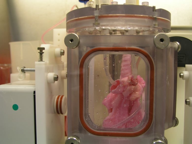
CHAOSMOSIS mAchInes exhibition/performance/discussion/panel/in-situ experiments/art/ science/ techne/ philosophy, 28 September, 2019 in Toronto
An Art/Sci Salon September 2, 2019 announcement (received via email), Note: I have made some formatting changes,
CHAOSMOSIS mAchInes
28 September, 2019
7pm-11pm.
Helen-Gardiner-Phelan Theatre, 2nd floor
University of Toronto. 79 St. George St.A playful co-presentation by the Topological Media Lab (Concordia U-Montreal) and The Digital Dramaturgy Labsquared (U of T-Toronto). This event is part of our collaboration with DDLsquared lab, the Topological Lab and the Leonardo LASER network
7pm-9.30pm, Installation-performances,
9.30pm-11pm, Reception and cash bar, Front and Long Room, Ground floor
Description:
From responsive sculptures to atmosphere-creating machines; from sensorial machines to affective autonomous robots, Chaosmosis mAchInes is an eclectic series of installations and performances reflecting on today’s complex symbiotic relations between humans, machines and the environment.
This will be the first encounter between Montreal-based Topological Media Lab (Concordia University) and the Toronto-based Digital Dramaturgy Labsquared (U of T) to co-present current process-based and experimental works. Both labs have a history of notorious playfulness, conceptual abysmal depth, human-machine interplays, Art&Science speculations (what if?), collaborative messes, and a knack for A/I as in Artistic Intelligence.
Thanks to Nina Czegledy (Laser series, Leonardo network) for inspiring the event and for initiating the collaboration
Visit our Facebook event page
Register through Evenbrite
Supported by
Main sponsor: Centre for Drama, Theatre and Performance Studies, U of T
Sponsors: Computational Arts Program (York U.), Cognitive Science Program (U of T), Knowledge Media Design Institute (U of T), Institute for the History and Philosophy of Science and Technology (IHPST), Fonds de Recherche du Québec – Société et culture (FRQSC), The Centre for Comparative Literature (U of T)
A collaboration between
Laser events, Leonardo networks – Science Artist, Nina Czegledy
ArtsSci Salon – Artistic Director, Roberta Buiani
Digital Dramaturgy Labsquared – Creative Research Director, Antje Budde
Topological Media Lab – Artistic-Research Co-directors, Michael Montanaro | Navid Navab
Project presentations will include:
Topological Media Lab
tangibleFlux φ plenumorphic ∴ chaosmosis
SPIEL
On Air
The Sound That Severs Now from Now
Cloud Chamber (2018) | Caustic Scenography, Responsive Cloud Formation
Liquid Light
Robots: Machine Menagerie
Phaze
Phase
Passing Light
Info projects
Digital Dramaturgy Labsquared
Btw Lf & Dth – interFACING disappearance
Info project
This is a very active September.
ETA September 4, 2019 at 1607 hours PDT: That last comment is even truer than I knew when I published earlier. I missed a Vancouver event, Maker Faire Vancouver will be hosted at Science World on Saturday, September 14. Here’s a little more about it from a Sept. 3, 2019 at Science World at Telus Science World blog posting,
Earlier last month [August 2019?], surgeons at St Paul’s Hospital performed an ankle replacement for a Cloverdale resident using a 3D printed bone. The first procedure of its kind in Western Canada, it saved the patient all of his ten toes — something doctors had originally decided to amputate due to the severity of the motorcycle accident.
Maker Faire Vancouver Co-producer, John Biehler, may not be using his 3D printer for medical breakthroughs, but he does see a subtle connection between his home 3D printer and the Health Canada-approved bone.
“I got into 3D printing to make fun stuff and gadgets,” John says of the box-sized machine that started as a hobby and turned into a side business. “But the fact that the very same technology can have life-changing and life-saving applications is amazing.”
When John showed up to Maker Faire Vancouver seven years ago, opportunities to access this hobby were limited. Armed with a 3D printer he had just finished assembling the night before, John was hoping to meet others in the community with similar interests to build, experiment and create. Much like the increase in accessibility to these portable machines has changed over the years—with universities, libraries and makerspaces making them readily available alongside CNC Machines, laser cutters and more — John says the excitement around crafting and tinkering has skyrocketed as well.
“The kind of technology that inspires people to print a bone or spinal insert all starts at ground zero in places like a Maker Faire where people get exposed to STEAM,” John says …
…
… From 3D printing enthusiasts like John to knitters, metal artists and roboticists, this full one-day event [Maker Faire Vancouver on Saturday, September 14, 2019] will facilitate cross-pollination between hobbyists, small businesses, artists and tinkerers. Described as part science fair, part county fair and part something entirely new, Maker Faire Vancouver hopes to facilitate discovery and what John calls “pure joy moments.”
Hopefully that’s it.