Tag Archives: University of Western Ontario

Mite silk as the basis for a new nanobiomaterial

For the record, this is spider mite silk (I have many posts about spider silk and its possible applications on this blog; just search ‘spider silk’)..

The international collaborative team includes a Canadian university in combination with a Spanish university and a Serbian university. The composition of the team is one I haven’t seen here before. From a December 17, 2020 news item on phys.org (Note: A link has been removed),

An international team of researchers has developed a new nanomaterial from the silk produced by the Tetranychus lintearius mite. This nanomaterial has the ability to penetrate human cells without damaging them and, therefore, has “promising biomedical properties”.

The Nature Scientific Reports journal has published an article by an international scientific team led by Miodrag Grbiç, a researcher from the universities of La Rioja (Spain), Western Ontario (Canada) and Belgrade (Serbia), in its latest issue entitled “The silk of gorse spider mite Tetranychus lintearius represents a novel natural source of nanoparticles and biomaterials.”

In it, researchers from the Murcian Institute for Agricultural and Food Research and Development (IMIDA), the Barcelona Institute of Photonic Sciences, the University of Western Ontario (Canada), the University of Belgrade (Serbia) and the University of La Rioja describe the discovery and characterisation of this mite silk. They also demonstrate its great potential as a source of nanoparticles and biomaterials for medical and technological uses.

A December 17, 2020 Universidad de La Rioja press release (also on EurekAlert), which originated the news item, further explains the research,

The interest of this new material, which is more resistant than steel, ultra flexible, nano-sized, biodegradable, biocompatible and has an excellent ability to penetrate human cells without damaging them, lies in its natural character and its size (a thousand times smaller than human hair), which facilitates cell penetration.

These characteristics are ideal for use in pharmacology and biomedicine since it is biocompatible with organic tissues (stimulates cell proliferation without producing toxicity) and, in principle, biodegradable due to its protein structure (it does not produce residues).

Researcher Miodrag Grbi?, who heads the international group that has researched this mite silk, highlights “its enormous potential for biomedical applications, as thanks to its size it is able to easily penetrate both healthy and cancerous human cells”, which makes it ideal for transporting drugs in cancer therapies, as well as for the development of biosensors to detect pathogens and viruses.

THE ‘RIOJANO BUG’

Tetranychus lintearius is an endemic mite from the European Atlantic coast that feeds exclusively on gorse (Ulex europaeus). It is around 0.3 mm in size, making it smaller than the comma on a keyboard, while the strength of its silk is twice as high as standard spider silk.

It is a very rare species that has only been found so far in the municipality of Valgañón (La Rioja, Spain), in Sierra de la Demanda. It was located thanks to the collaboration of Rosario García, a botanist and former dean of the Faculty of Science and Technology at the University of La Rioja, which is why researchers call it “the Rioja bug” (“El Bicho Riojano”).

The resistance of the silk produced by Tetranychus lintearius is twice that of spider silk, a standard material used for this type of research, and stronger than steel. It also has advantages over the fibres secreted by the silkworm due to its higher Young’s modulus, its electrical charge and its smaller size. These characteristics, along with its lightness, make it a promising natural nanomaterial for technological uses.

This finding is the result of work carried out by the international group of researchers led by Miodrag Grbi?, who sequenced the genome of the red spider Tetranychus urticae in 2011, publishing the results in Nature: https://www.nature.com/articles/nature10640.

Unlike the red spider (Tetranychus urticae), the gorse mite (Tetranychus lintearius) produces a large amount of silk. It has been reared in the laboratories of the Department of Agriculture and Food of the University of La Rioja, under the care of Professor Ignacio Pérez Moreno, allowing research to continue. Red spider silk is difficult to handle and has a lower production rate.

Here’s a link to and a citation for the 2020 paper,

The silk of gorse spider mite Tetranychus lintearius represents a novel natural source of nanoparticles and biomaterials by Antonio Abel Lozano-Pérez, Ana Pagán, Vladimir Zhurov, Stephen D. Hudson, Jeffrey L. Hutter, Valerio Pruneri, Ignacio Pérez-Moreno, Vojislava Grbic’, José Luis Cenis, Miodrag Grbic’ & Salvador Aznar-Cervantes. Scientific Reports volume 10, Article number: 18471 (2020) DOI: https://doi.org/10.1038/s41598-020-74766-7 Published: 28 October 2020

This paper is open access.
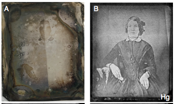
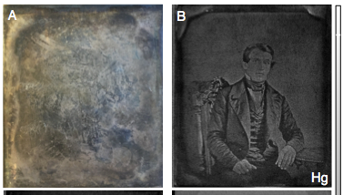
Seeing ghosts: recovering images from dageurreotypes with help from the Canadian Light Source (synchrotron)

A daguerreotype plate with the photograph hidden by the tarnish (left) yet visible when imaged with synchrotron X-rays (right). Courtesy of Madalena Kozachuk.

Amazing, yes? Especially when you consider how devastating the inadvertent destruction of important daguerreotypes in an exhibition of US Civil War photography must have been to the curators and owners of the images. The ‘destruction’ occurred in 2005 and inspired research into the cause of the destruction, which was first covered here in a January 10, 2013 posting and followed up in a November 17, 2015 posting about an exhibit showcasing the results of the research.

A daguerreotype plate with the photograph hidden by the tarnish (left) yet visible when imaged with synchrotron X-rays (right). Courtesy of Madalena Kozachuk.

This latest research into daguerreotypes was performed at the Canadian Light Source (CLS; Saskatoon, Saskatchewan, Canada). Unlike my previous postings, this research was an attempt to retrieve the original image rather than research the reasons for its ‘destruction’. From a June 22, 2018 CLS news release (also on EurekAlert) by Lana Haight and Jeffrey Renaud (Note: Links have been removed),

Art curators will be able to recover images on daguerreotypes, the earliest form of photography that used silver plates, after scientists learned how to use light to see through degradation that has occurred over time.

Research published today [June 22, 2018] in Scientific Reports includes two images from the National Gallery of Canada’s photography research unit that show photographs that were taken, perhaps as early as 1850, but were no longer visible because of tarnish and other damage. The retrieved images, one of a woman and the other of a man, were beyond recognition.

“It’s somewhat haunting because they are anonymous and yet it is striking at the same time,” said Madalena Kozachuk, a PhD student in the Department of Chemistry at Western University [formerly University of Western Ontario] and lead author of the scientific paper.

“The image is totally unexpected because you don’t see it on the plate at all. It’s hidden behind time. But then we see it and we can see such fine details: the eyes, the folds of the clothing, the detailed embroidered patterns of the table cloth.”

The identities of the woman and the man are not known. It’s possible that the plates were produced in the United States, but they could be from Europe.

For the past three years, Kozachuk and an interdisciplinary team of scientists have been exploring how to use synchrotron technology to learn more about chemical changes that damage daguerreotypes.

Invented in 1839, daguerreotype images were created using a highly polished silver-coated copper plate that was sensitive to light when exposed to an iodine vapour. Subjects had to pose without moving for two to three minutes for the image to imprint on the plate, which was then developed as a photograph using a mercury vapour that was heated.

Kozachuk conducts much of her research at the Canadian Light Source and previously published results in scientific journals in 2017 and earlier this year. In those articles, the team members identified the chemical composition of the tarnish and how it changed from one point to another on a daguerreotype.

“We compared degradation that looked like corrosion versus a cloudiness from the residue from products used during the rinsing of the photographs versus degradation from the cover glass. When you look at these degraded photographs, you don’t see one type of degradation,” said Ian Coulthard, a senior scientist at the CLS and one of Kozachuk’s supervisors. He is also a co- author on the research papers.

This preliminary research at the CLS led to today’s [June 22, 2018] paper and the images Kozachuk collected at the Cornell High Energy Synchrotron Source where she was able to analyze the daguerreotypes in their entirety.

Kozachuk used rapid-scanning micro-X-ray fluorescence imaging to analyze the plates, which are about 7.5 cm wide, and identified where mercury was distributed on the plates. With an X-ray beam as small as 10 by 10 microns (a human scalp hair averages 75 microns across) and at an energy most sensitive to mercury absorption, the scan of each daguerreotype took about eight hours.

“Mercury is the major element that contributes to the imagery captured in these photographs. Even though the surface is tarnished, those image particles remain intact. By looking at the mercury, we can retrieve the image in great detail,” said Tsun-Kong (T.K.) Sham, Canada Research Chair in Materials and Synchrotron Radiation at Western University. He also is a co-author of the research and one of Kozachuk’s supervisors.

This is one of the many examples of successful research collaboration between Western University and CLS scientists.

Kozachuk’s research, which is ongoing, will contribute to improving how daguerreotype images are recovered when cleaning is possible and will provide a way to see what’s below the tarnish when cleaning is not possible. She will be back at the CLS this fall to continue her work.

The prospect of improved conservation methods intrigues John P. McElhone, recently retired as the chief of the Conservation and Technical Research branch at the Canadian Photography Institute of the National Gallery of Canada. He provided the daguerreotypes from the institute’s research collection.

“There are a lot of interesting questions that at this stage of our knowledge can only be answered by a sophisticated scientific approach,” said McElhone, another of the co-authors of today’s paper.

“A conservator’s first step is to have a full and complete understanding of what the material isand how it is assembled on a microscopic and even nanoscale level. We want to find out how the chemicals are arranged on the surface and that understanding gives us access to theories about how degradation happens and how that degradation can possibly or possibly not be reversed.”

As the first commercialized photographic process, the daguerreotype is thought to be the first “true” visual representation of history. Unlike painters who could use “poetic licence” in their work, the daguerreotype reflected precisely what was photographed.

Thousands and perhaps millions of daguerreotypes were created over 20 years in the 19th century before the process was replaced. The Canadian Photography Institute collection numbers more than 2,700, not including the daguerreotypes in the institute’s research collection.

By improving the process of restoring these centuries-old images, the scientists are contributing to the historical record. What was thought to be lost that showed the life and times of people from the 19th century can now be found. [emphases mine]

That last sentence seems to be borrowing from a line in the song, Amazing Grace, “I once was lost, but now am found,” from the song’s Wikipedia entry.

Here’s a link to and a citation for the paper,

Recovery of Degraded-Beyond-Recognition 19th Century Daguerreotypes with Rapid High Dynamic Range Elemental X-ray Fluorescence Imaging of Mercury L Emission by Madalena S. Kozachuk, Tsun-Kong Sham, Ronald R. Martin, Andrew J. Nelson, Ian Coulthard, & John P. McElhone. Scientific Reports volume 8, Article number: 9565 (2018) DOI:10.1038/s41598-018-27714 Published online June 22, 2018

This paper is open access. By the way, the research into the ‘destruction’ of the daguerreotypes in the 2005 exhibition? It’s cited in this paper.

At the nanoscale, grapefruit swings from being medication danger to medication enhancer

It’s known that grapefruit, despite its health benefits, can inhibit (or even a pose danger) to a medication’s effectiveness. Most of us have been warned at one time or another to avoid grapefruit juice when downing a pill. So, the news from the University of Louisville (Kentucky; UofL) about grapefruit as part of a drug delivery system seems a little counter-intuitive (from the May 22, 2013 news item on Azonano),

Grapefruits have long been known for their health benefits, and the subtropical fruit may revolutionize how medical therapies like anti-cancer drugs are delivered to specific tumor cells.

University of Louisville researchers have uncovered how to create nanoparticles using natural lipids derived from grapefruit, and have discovered how to use them as drug delivery vehicles. UofL scientists Huang-Ge Zhang, D.V.M., Ph.D., Qilong Wang, Ph.D., and their team today (May 21, 2013), published their findings in Nature Communications.

The May 21, 2013 University of Louisville news release by Julie Heflin, which originated the news item, describes how the nanoparticles are derived and their advantages,

“These nanoparticles, which we’ve named grapefruit-derived nanovectors (GNVs), are derived from an edible plant, and we believe they are less toxic for patients, result in less biohazardous waste for the environment and are much cheaper to produce at large scale than nanoparticles made from synthetic materials,” said Zhang, who holds the Founders Chair in Cancer Research at the Brown Cancer Center.

The researchers demonstrated that GNVs can transport various therapeutic agents, including anti-cancer drugs, DNA/RNA and proteins such as antibodies. Treatment of animals with GNVs seemed to cause less adverse effects than treatment with drugs encapsulated in synthetic lipids.

“Our GNVs can be modified to target specific cells — we can use them like missiles to carry a variety of therapeutic agents for the purpose of destroying diseased cells,” he said. “Furthermore, we can do this at an affordable price.”

The therapeutic potential of grapefruit derived nanoparticles was further validated through a Phase 1 clinical trial for treatment of colon cancer patients. So far, researchers have observed no toxicity in the patients who orally took the anti-inflammatory agent curcumin encapsulated in grapefruit nanoparticles.

The UofL scientists also plan to test whether this technology can be applied in the treatment of inflammation related autoimmune diseases like rheumatoid arthritis.

Here’s a link to and a citation for the researchers’ paper,

Delivery of therapeutic agents by nanoparticles made of grapefruit-derived lipids by Qilong Wang, Xiaoying Zhuang, Jingyao Mu, Zhong-Bin Deng, Hong Jiang, Xiaoyu Xiang, Baomei Wang, Jun Yan, Donald Miller, & Huang-Ge Zhang. Nature Communications 4, 1867 doi:10.1038/ncomms2886 Published 21 May 2013

This paper is behind a paywall.

As for the dangers of grapefruit-medication interactions, ABC (American Broadcasting Corporation) has a Nov. 26, 2012 news item featuring then new research suggesting that even more medications are affected by grapefruit/grapefruit juice than had previously been believed,

It has long been known that grapefruit juice can pose dangerous — and even deadly — risks when taken along with certain medications. Now, experts warn the list of medications that can result in these interactions is longer than many may have believed.

In a new report released Monday in the Canadian Medical Association Journal [CMAJ], researchers at the University of Western Ontario said that while 17 drugs were identified in 2008 as having the potential to cause serious problems when taken with grapefruit, this number has now grown to 43.

So how does a common breakfast fruit cause these problems? Grapefruits contain chemicals called furanocoumarins that interfere with how your body breaks down drugs before they enter the bloodstream. By preventing this normal breakdown of a drug, these chemicals in grapefruit can effectively cause a drug overdose and more severe side-effects.

Among the side effects sometimes seen with grapefruit-induced overdoses are heart rhythm problems, kidney failure, muscle breakdown, difficulty with breathing and blood clots. …

ABC provides a list of drugs that are affected by grapefruit here.

For interested parties, here’s a link to and a citation for the research on grapefruit-medication interactions,

Grapefruit–medication interactions: Forbidden fruit or avoidable consequences? by David G. Bailey, George Dresser, and J. Malcolm O. Arnold. CMAJ March 5, 2013 185:309-316; published ahead of print November 26, 2012,

This paper is behind a paywall.

I have a couple of final comments. (1) It would seem that the grapefruit’s characteristics at the macroscale are not echoed at the nanoscale. (2) Interestingly, the grapefruit nanoparticles (grapefruit nanovectors [GNVs]) are being used to encapsulate curcumin (a constituent of turmeric). I wrote about turmeric and its healing properties in a Dec. 26, 2011 posting, which features a number of links to research in this area.

Is a philosophy of the Higgs and other physics particles a good idea?

Michael  Krämer of the RWTH Aachen University (Germany) muses about philosophy, the Higgs Boson, and more in a Mar. 24, 2013 posting on Jon Butterworth’s Life and Physics blog (Guardian science blogs; Note: A link has been removed),

Many of the great physicists of the 20th century have appreciated the importance of philosophy for science. Einstein, for example, wrote in a letter in 1944:

    I fully agree with you about the significance and educational value of methodology as well as history and philosophy of science. So many people today—and even professional scientists—seem to me like somebody who has seen thousands of trees but has never seen a forest.

At the same time, physics has always played a vital role in shaping ideas in modern philosophy. It appears, however, that we are now faced with the ruins of this beautiful marriage between physics and philosophy. Stephen Hawking has claimed recently that philosophy is “dead” because philosophers have not kept up with science …

Krämer is part of an interdisciplinary (physics and philosophy) project at the LHC (Large Hadron Collider at CERN [European Particle Physics Laboratory]), The Epistemology of the Large Hadron Collider. From the project home page (Note: A link has been removed),

This research collaboration works at the crossroads of physics, philosophy of science, and contemporary history of science. It aims at an epistemological analysis of the recently launched new accelerator experiment at CERN, the Large Hadron Collider (LHC). Central themes are (i) the mechanisms of generating the masses of the particles of the standard model, especially the Higgs-mechanism and the Higgs-particle the LHC has set out to detect; (ii) the ongoing research process with special emphasis on the interaction between a large experiment and a community of theoreticians; and (iii) the implications of an experiment that is characterized by its enormous complexity and the need to be highly selective in data gathering. With the heading “Epistemology of the LHC” the research group intends both a philosophical analysis of the theoretical structures and of the conditions of knowledge production, among them the criteria of acceptance, and a real-time monitoring of the ongoing physical development from the perspective of the history of science. Theresearch group has emerged from a collaboration between a High Energy Working group and the Interdisciplinary Centre for Science and Technology Studies and is based in Wuppertal but also involves external members and collaborators.

Krämer shares some of his ideas and the type of thinking generated when physicists and philosophers collide (I plead guilty to the word play; from Butterworth’s Guardian science blog),

… The relationship between experiment and theory (what impact does theoretical prejudice have on empirical findings?) or the role of models (how can we assess the uncertainty of a simplified representation of reality?) are scientific issues, but also issues from the foundation of philosophy of science. In that sense they are equally important for both fields, and philosophy may add a wider and critical perspective to the scientific discussion. And while not every particle physicist may be concerned with the ontological question of whether particles or fields are the more fundamental objects, our research practice is shaped by philosophical concepts. We do, for example, demand that a physical theory can be tested experimentally and thereby falsified, a criterion that has been emphasized by the philosopher Karl Popper already in 1934. The Higgs mechanism can be falsified, because it predicts how Higgs particles are produced and how they can be detected at the Large Hadron Collider.

On the other hand, some philosophers tell us that falsification is strictly speaking not possible: What if a Higgs property does not agree with the standard theory of particle physics? How do we know it is not influenced by some unknown and thus unaccounted factor, like a mysterious blonde walking past the LHC experiments and triggering the Higgs to decay? (This was an actual argument given in the meeting!)

The meeting Krämer is referring to is this one (from the meeting/conference website),

The first international conference and kick-off meeting of the German Society for Philosophy of Science/Gesellschaft für Wissenschaftsphilosophie (GWP) will take place from 11-14 March 2013 at the University of Hannover under the title:

How Much Philosophy in the Philosophy of Science?

Krämer then highlights some of the discussion that most interested in him (Note: A link has been removed),

… It is very hard for a philosopher to keep up with scientific progress, and how could one integrate various fields without having fully appreciated the essential features of the individual sciences? As Margaret Morrison from the University of Toronto pointed out in her talk, if philosophy steps back too far from the individual sciences, the account becomes too general and isolated from scientific practice. On the other hand, if philosophy is too close to an individual science, it may not be philosophy any longer.

I think philosophy of science should not consider itself primarily as a service to science, but rather identify and answer questions within its own domain. I certainly would not be concerned if my own research went unnoticed by biologists, chemists, or philosophers, as long as it advances particle physics. On the other hand, as Morrison pointed out, science does generate its own philosophical problems, and philosophy may provide some kind of broader perspective for understanding those problems.

It’s well worth reading Krämer’s full post for anyone who’s interested in how physicists (or Krämer) think about the role that philosophy could play (or not) in the field of physics.

The reference to Margaret Morrison from the University of Toronto (U of T) reminded me of the Bubble Chamber blog which is written by U of T historians and philosophers of science. Here’s a July 10, 2012 posting by Mike Thicke about the Higgs Boson and his response to philosopher Wayne Myrvold’s (University of Western Ontario) explanation of the statistics claims being made about the particle at that time,

We can all agree that reasoning and decision making in science is complicated. Scientists reason in many different contexts: in the lab, in their published papers, as career-minded professionals, as interested consumers of science, and as people going about their lives. It’s plausible to think that they reason in different ways in all of these contexts. When we’re discussing their reasoning as scientists, I believe distinguishing between the first three contexts is especially important. While Wayne’s explanation of the statistics behind the Higgs Boson discovery is very interesting, informative, and as far as I can tell correct, I think there are some confusions arising from his failure to make these distinctions.

Thicke does advise reading Myrvold’s July 4, 2012 posting before tackling his riposte.

Canadian soil remediation expert in Australia

Back in my Nov. 4, 2011 posting where I reviewed the third episode in a limited series on nanotechnology, broadcast as a Nature of Things television science programme on  Canadian Broadcasting Corporation stations, I noted Dr. Dennis O’Carroll’s soil remediation work in southern Ontario.

There’s more news about professor O’Carroll, currently visiting Australia, in a June 4, 2012 news item on Nanowerk,

“Toxic contamination of soils is an historical problem,” says Dr Denis O’Carroll, a visiting academic at the University of New South Wales (UNSW) Water Research Lab. “Until the 1970s, people wrongly believed that if we put these toxins into the ground they would simply disappear – that the subsurface would act as a natural filtration unit.”

“The possibility of this waste polluting the environment, and potentially contaminating groundwater sources and remaining there for decades was ignored,” he says.

Far from magically disappearing, chemical contaminants from spilled gas and solvents, when not directly polluting surface waters, seep down into the earth, travelling through microscopic soil cracks, where they accumulate and can eventually reach the groundwater table.

Traditional clean-up methods have focussed on pumping out the contaminated water or flushing out toxins with a specially designed cleansing solution, but these are limited by difficulties in accurately pinpointing and accessing locations where contamination has occurred, says O’Carroll.

His approach is to tackle toxic contaminants with nanotechnology. O’Carroll, who is visiting UNSW from the University of Western Ontario in Canada, has been trialling an innovative new groundwater clean-up technology using metal nanoparticles 500 to 5,000 times narrower than a human hair.

There are more details about O’Carroll’s specific innovations in this field in the June 4, 2012 news item. As well, I published, in its entirety (and with permission), an excellent description of nanotechnology-enabled soil remediation by Joe Martin, a graduate student at the University of Michigan, in my March 30, 2012 posting. Here’s a tidbit from Joe’s article,

… The use of iron oxides to adsorb and immobilize metals and arsenic is not a new concept, but nano-particles offer new advantages. When I wrote “adsorb”, I was not making a spelling error; adsorption is a process by which particles adhere to the surface of another material, but do not penetrate into the interior. This makes surface area, not volume, the important characteristic. Nano-particles provide the maximum surface area-to-weight ratio, maximizing the adsorptive surfaces onto which these elements can attach. These adsorptive processes a very effective at binding and immobilizing metals and arsenic, but they do not allow for the removal of the toxic components. This may be less-than-ideal, but in places like Bangladesh, where arsenic contamination of groundwater poses major health risks, it may be just short of a miracle.

There’s an extensive list with links to further reading and videos on the topic of nanotechnology and site remediation at the end of the March 30, 2012 posting.

Nature of Things’ series: The Nano Revolution (Episode 3); Will Nano Save the Planet?

I’m never thrilled with titles of this ilk, Will Nano Save the Planet? Refreshingly, this episode featured some work being done by Canadian scientists (two of them) although the average Canadian could be forgiven for thinking that it’s the only nanotechnology research taking place in Canada.

It’s a little puzzling that they chose this final episode for a description of the term nanoscale. David Suzuki, the host, mentioned the ridges of skin on your fingers and noted that a nanoparticle is 80,000 times smaller than the distance between the ridges. (If you want a really good description of scale, I recommend listening to Professor Ravi Silva’s audio interview with Alok Jha on the (UK) Guardian’s Oct. 14, 2011 Science Weekly podcast.)

In general, I found the descriptions of the science in this episode were not of the same standard as the previous two, which were very good.

The vignettes, as always, were problematic largely since they were internal monologues of some character who’s grappling with ethical issues and other social impacts of these technologies. Interestingly, men starred in the vignettes where the ‘big’ issues are covered: ethics of health care; longer life; access to energy sources; pollution from nanotechnology-enabled products; etc. The woman who starred in the vignettes from episode one (as I noted in my review) was concerned with cleanliness, tidiness, shopping, and privacy. I guess things don’t change that much in our future, especially in 2050 where nanotechnology protestors are putting up banners, spraypainting, and leafletting (almost as if it were 1968) to express their opposition (in episode three).

There was some interesting work being covered. They profiled Professor Ted Sargent, based at the University of Toronto, who’s doing some exciting work with solar cells (he wants to make them flexible and, even, paintable). His latest breakthrough is mentioned in my Sept. 20, 2011 posting.

Professor Vicki Colvin, Rice University in Texas, is working to purify water. The project is in Mexico and highlights the difficulties when water supplies are contaminated, in this case, with arsenic. (Here in the Pacific Northwest we tend to forget that access to fresh clean water is not easy in many parts of the world.) Colvin and her colleagues are working on a simple solution that can be implemented with some sand, gravel, a tube, and active nanoparticles. (Her work with the Environmental Nanoscience Initiative; a UK/US collaboration was mentioned in my Jan. 28, 2011 posting.)

The third project was focussed on soil remediation and a team from the University of Western Ontario headed by Professor Dennis O’Carroll. I have not come across O’Carroll’s work previously so this was a find for me. As you may or may not know, there are many sites with contaminated soil throughout North America and elsewhere. If successful, O’Carroll’s technique promises to remediate (rehabiltate) the soil without having to move massive amounts of soil and use big  equipment.

This episode featured more discussion about the risks and uncertainties associated with nanotechnology and its use. Unfortunately, I did not recognize the names and (one of my major pet peeves with this series) they either didn’t write out the names on screen or they flashed them briefly which meant that unless I recognized the names it was difficult to find out more about the experts.

I did recognize the mesocosm project at Duke University, which was featured here in my August 15, 2011 posting. The researchers are trying to understand what impact silver nanoparticles have on life. They spray silver nanoparticles in various mesocosms (they look like raised plant beds) and then track what happens to the plant, the soil, and the water supply as the silver nanoparticles cycle through.

There’s work in the UK examining air and the nanoparticles released through the use of internal combustion engines (cars/trucks) as well as our newly engineered nanoparticles. I’m glad to see this material in the episode, perhaps it will finally motivate some public discussion in Canada.

Buckyballs in space

Astronomers are excited! They thought they’d found buckyballs (buckminster fullerenes) in some stars about 15 years ago but that finding still hasn’t been confirmed with laboratory data. Meanwhile, a new team including Jan Cami from the University of Western Ontario (Canada) and the SETI (Search for Extraterrestrial Intelligence) Institute in Mountainview, California recently made an unexpected discovery—buckyballs—while examining a planetary nebula (remains of a star shedding its outer layer of gas and dust as it ages). According to the news item on physorg.com,

“We found what are now the largest molecules known to exist in space,” said astronomer Jan Cami of the University of Western Ontario, Canada, and the SETI Institute in Mountain View, Calif. “We are particularly excited because they have unique properties that make them important players for all sorts of physical and chemical processes going on in space.” Cami has authored a paper about the discovery that will appear online Thursday [July 29, 2010?] in the journal Science.

Buckyballs are made of 60 carbon atoms arranged in three-dimensional, spherical structures. Their alternating patterns of hexagons and pentagons match a typical black-and-white soccer ball. The research team also found the more elongated relative of buckyballs, known as C70, for the first time in space. These molecules consist of 70 carbon atoms and are shaped more like an oval rugby ball. Both types of molecules belong to a class known officially as buckminsterfullerenes, or fullerenes.

You can also find the news item at Nanowerk where an alternative video clip (featuring an interview with Jan Cami discussing buckyballs) to the the silent animation featuring buckyballs and their movement  available on the physorg.com site.

The folks at Rice University must be thrilled since proof of the existence of buckyballs on this planet is strongly associated with discoveries made by scientists at Rice (my May 13, 2010 posting provides a fuller picture of some of the twists and turns associated with that science story).