Tag Archives: US Argonne National Laboratory

Dumbbells at the nanoscale according to researchers at the (US) Argonne National Laboratory

Researchers at the US Dept. of Energy’s Argonne National Laboratory are providing new insight into how nanoparticles ‘grow’. From a Dec. 5, 2014 news item on Nanowerk,

Like snowflakes, nanoparticles come in a wide variety of shapes and sizes. The geometry of a nanoparticle is often as influential as its chemical makeup in determining how it behaves, from its catalytic properties to its potential as a semiconductor component.

Thanks to a new study from the U.S. Department of Energy’s (DOE) Argonne National Laboratory, researchers are closer to understanding the process by which nanoparticles made of more than one material – called heterostructured nanoparticles – form. This process, known as heterogeneous nucleation, is the same mechanism by which beads of condensation form on a windowpane.

The scientists have provided an image which illustrates their findings,

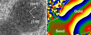
This picture combines a transmission electron microscope image of a nanodumbbell with a gold domain oriented in direction. The seed and gold domains in the dumbbell in the image on the right are identified by geometric phase analysis. Image credit: Soon Gu Kwon.

This picture combines a transmission electron microscope image of a nanodumbbell with a gold domain oriented in direction. The seed and gold domains in the dumbbell in the image on the right are identified by geometric phase analysis. Image credit: Soon Gu Kwon.

A Dec. 4, 2014 Argonne National Laboratory news release by Jared Sagoff, which originated the news item, describes the structures being examined and the reason for doing so,

Heterostructured nanoparticles can be used as catalysts and in advanced energy conversion and storage systems. Typically, these nanoparticles are created from tiny “seeds” of one material, on top of which another material is grown.  In this study, the Argonne researchers noticed that the differences in the atomic arrangements of the two materials have a big impact on the shape of the resulting nanoparticle.

“Before we started this experiment, it wasn’t entirely clear what’s happening at the interface when one material grows on another,” said nanoscientist Elena Shevchenko of Argonne Center for Nanoscale Materials, a DOE Office of Science user facility.

In this study, the researchers observed the formation of a nanoparticle consisting of platinum and gold.  The researchers started with a platinum seed and grew gold around it. Initially, the gold covered the platinum seed’s surface uniformly, creating a type of nanoparticle known as “core-shell.” However, as more gold was deposited, it started to grow unevenly, creating a dumbbell-like structure.

Thanks to state-of-the-art X-ray analysis provided by Argonne’s Advanced Photon Source (APS), a DOE Office of Science user facility, the researchers identified the cause of the dumbbell formation as “lattice mismatch,” in which the spacing between the atoms in the two materials doesn’t align.

“Essentially, you can think of lattice mismatch as having a row of smaller boxes on the bottom layer and larger boxes on the top layer.  When you try to fit the larger boxes into the space for a smaller box, it creates an immense strain,” said Argonne physicist Byeongdu Lee.

While the lattice mismatch is only fractions of a nanometer, the effect accumulates as layer after layer of gold forms on the platinum. The mismatch can be handled by the first two layers of gold atoms – creating the core-shell effect – but afterwards it proves too much to overcome. “The arrangement of atoms is the same in the two materials, but the distance between atoms is different,” said Argonne postdoctoral researcher Soon Gu Kwon. “Eventually, this becomes unstable, and the growth of the gold becomes unevenly distributed.”

As the gold continues to accumulate on one side of the seed nanoparticle, small quantities “slide” down the side of the nanoparticle like grains of sand rolling down the side of a sand hill, creating the dumbbell shape.

The advantage of the Argonne study comes from the researchers’ ability to perform in situ observations of the material in realistic conditions using the APS. “This is the first time anyone has been able to study the kinetics of this heterogeneous nucleation process of nanoparticles in real-time under realistic conditions,” said Argonne physicist Byeongdu Lee. “The combination of two X-ray techniques gave us the ability to observe the material at both the atomic level and the nanoscale, which gave us a good view of how the nanoparticles form and transform.” All conclusions made based on the X-ray studies were further confirmed using atomic-resolution microscopy in the group of Professor Robert Klie of the University of Illinois at Chicago.

This analysis of nanoparticle formation will help to lay the groundwork for the formation of new materials with different and controllable properties, according to Shevchenko. “In order to design materials, you have to understand how these processes happen at a very basic level,” she said.

Here’s a link to and a citation for the paper,

Heterogeneous nucleation and shape transformation of multicomponent metallic nanostructures by Soon Gu Kwon, Galyna Krylova, Patrick J. Phillips, Robert F. Klie, Soma Chattopadhyay, Tomohiro Shibata, Emilio E. Bunel, Yuzi Liu, Vitali B. Prakapenka, Byeongdu Lee, & Elena V. Shevchenko. Nature Materials (2014) doi:10.1038/nmat4115 Published online 02 November 2014

This paper is behind a paywall but there is a free preview via ReadCube Access.

Single layer graphene as a solid lubricant

Graphite (from which graphene springs) has been used as a solid lubricant for many years but it has limitations which researchers at the US Dept. of Energy’s Argonne National Laboratory are attempting to overcome by possibly replacing it with graphene. An Oct. 14, 2014 news item on phys.org describes the research (Note: A link has been removed),

Nanoscientist Anirudha Sumant and his colleagues at Argonne’s Center for Nanoscale Materials and Argonne’s Energy Systems division applied a one-atom-thick layer of graphene, a two-dimensional form of carbon, in between a steel ball and a steel disk. They found that just the single layer of graphene lasted for more than 6,500 “wear cycles,” a dramatic improvement over conventional lubricants like graphite or molybdenum disulfide.

An Oct. 13, 2014 Argonne National Laboratory news release by Jared Sagoff, which originated the news item, provides more information about this research (Note: A link has been removed),

“For comparison,” Sumant said, “conventional lubricants would need about 1,000 layers to last for 1,000 wear cycles. That’s a huge advantage in terms of cost savings with much better performance.”

Graphite has been used as an industrial lubricant for more than 40 years, but not without certain drawbacks, Sumant explained.  “Graphite is limited by the fact that it really works only in humid environments. If you have a dry setting, it’s not going to be nearly as effective,” he said.

This limitation arises from the fact that graphite – unlike graphene – has a three-dimensional structure.  The water molecules in the moist air create slipperiness by weaving themselves in between graphite’s carbon sheets. When there are not enough water molecules in the air, the material loses its slickness.

Molybdenum disulfide, another common lubricant, has the reverse problem, Sumant said. It works in dry environments but not well in wet ones. “Essentially the challenge is to find a single all-purpose lubricant that works well for mechanical systems, no matter where they are,” he said.

Graphene’s two-dimensional structure gives it a significant advantage. “The material is able to bond directly to the surface of the stainless steel ball, making it so perfectly even that even hydrogen atoms are not able to penetrate it,” said Argonne materials scientist Ali Erdemir, a collaborator on the study who tested graphene-coated steel surfaces in his lab.

In a previous study in Materials Today, Sumant and his colleagues showed that a few layers of graphene works equally well in humid and dry environments as a solid lubricant, solving the 40-year-old puzzle of finding a flawless solid lubricant. However, the team wanted to go further and test just a single graphene layer.

While doing so in an environment containing molecules of pure hydrogen, they observed a dramatic improvement in graphene’s operational lifetime. When the graphene monolayer eventually starts to wear away, hydrogen atoms leap in to repair the lattice, like stitching a quilt back together. “Hydrogen can only get into the fabric where there is already an opening,” said Subramanian Sankaranarayanan, an Argonne computational scientist and co-author in this study. This means the graphene layer stays intact longer.

Researchers had previously done experiments to understand the mechanical strength of a single sheet of graphene, but the Argonne study is the first to explain the extraordinary wear resistance of one-atom-thick graphene.

Here’s a link to and a citation for the August 2014 study,

Extraordinary Macroscale Wear Resistance of One Atom Thick Graphene Layer by Diana Berman, Sanket A. Deshmukh, Subramanian K. R. S. Sankaranarayanan, Ali Erdemir, and Anirudha V. Sumant. Advanced Funtional Materials DOI: 10.1002/adfm.201401755 Article first published online: 26 AUG 2014

© 2014 WILEY-VCH Verlag GmbH & Co. KGaA, Weinheim

This article is behind a paywall.

A query into the existence of silicene

There’s some fascinating work on silicene at the Argonne National Laboratory which questions current scientific belief as per a July 25, 2014 news item on Nanowerk (Note: A link has been removed),

Sometimes, scientific findings can shake the foundations of what was once held to be true, causing us to step back and re-examine our basic assumptions.

A recent study (“Silicon Growth at the Two-Dimensional Limit on Ag(111)”) at the U.S. Department of Energy’s Argonne National Laboratory has called into question the existence of silicene, thought to be one of the world’s newest and hottest two-dimensional nanomaterials. The study may have great implications to a multi-billion dollar electronics industry that seeks to revolutionize technology at scales 80,000 times smaller than the human hair.

A July 24, 2014 Argonne National Laboratory news release by Justin H.S. Breaux , which originated the news item, describes both silicene and silicon in preparation for the discussion about whether or not silicene exists,

Silicene was proposed as a two-dimensional sheet of silicon atoms that can be created experimentally by super-heating silicon and evaporating atoms onto a silver platform. Silver is the platform of choice because it will not affect the silicon via chemical bonding nor should alloying occur due to its low solubility. During the heating process, as the silicon atoms fall onto the platform, researchers believed that they were arranging themselves in certain ways to create a single sheet of interlocking atoms.

Silicon, on the other hand, exists in three dimensions and is one of the most common elements on Earth. A metal, semiconductor and insulator, purified silicon is extremely stable and has become essential to the integrated circuits and transistors that run most of our computers.

Both silicene and silicon should react immediately with oxygen, but they react slightly differently. In the case of silicon, oxygen breaks some of the silicon bonds of the first one or two atomic layers to form a layer of silicon-oxygen. This, surprisingly, acts a chemical barrier to prevent the decay of the lower layers.

Because it consists of only one layer of silicon atoms, silicene must be handled in a vacuum. Exposure to any amount of oxygen would completely destroy the sample.

This difference is one of the keys to the researchers’ discovery. After depositing the atoms onto the silver platform, initial tests identified that alloy-like surface phases would form until bulk silicon layers, or “platelets” would precipitate out, which has been mistaken as two-dimensional silicene.

The news release next describes how the scientists solved the puzzle,

“Some of the bulk silicon platelets were more than one layer thick,” said Argonne scientist Nathan Guisinger of Argonne’s Center for Nanoscale Materials. “We determined that if we were dealing with multiple layers of silicon atoms, we could bring it out of our ultra-high vacuum chamber and bring it into air and do some other tests.”

“Everybody assumed the sample would immediately decay as soon as they pulled it out of the chamber,” added Northwestern University graduate student Brian Kiraly, one of the principal authors of the study. “We were the first to actually bring it out and perform major experiments outside of the vacuum.”

Each new series of experiments presented a new set of clues that this was, in fact, not silicene.

By examining and categorizing the top layers of the material, the researchers discovered silicon oxide, a sign of oxidation in the top layers. They were also surprised to find that particles from the silver platform alloyed with the silicon at significant depths.

“We found out that what previous researchers identified as silicene is really just a combination of the silicon and the silver,” said Northwestern graduate student Andrew Mannix.

For their final test, the researchers decided to probe the atomic signature of the material.

Materials are made up of systems of atoms that bond and vibrate in unique ways. Raman spectroscopy allows researchers to measure these bonds and vibrations. Housed within the Center for Nanoscale Materials, a DOE Office of Science User Facility, the spectroscope allows researchers to use light to “shift” the position of one atom in a crystal lattice, which in turn causes a shift in the position of its neighbors. Scientists define a material by measuring how strong or weak these bonds are in relation to the frequency at which the atoms vibrate.

The researchers noticed something oddly familiar when looking at the vibrational signatures and frequencies of their sample. Their sample did not exhibit characteristic vibrations of silicene, but it did match those of silicon.

“Having this many research groups and papers potentially be wrong does not happen often,” says Guisinger. “I hope our research helps guide future studies and convincingly demonstrates that silver is not a good platform if you are trying to grow silicene.”

Here’s an image illustrating the vibrational signatures of what scientists had believed to be silicene,

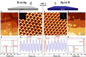
Argonne researchers investigating the properties of silicene (a one-atom thick sheet of silicon atoms) compared scanning tunneling microscope images of atomic silicon growth on silver and atomic silver growth on silicon. The study finds that both growth processes exhibit identical heights and shapes (a, g), indistinguishable honeycomb structures (c, e) and atomic periodicity (d, f). This suggests the growth of bulk silicon on silver, with a silver-induced surface reconstruction, rather than silicene. Courtesy: Argonne National Laboratory

Argonne researchers investigating the properties of silicene (a one-atom thick sheet of silicon atoms) compared scanning tunneling microscope images of atomic silicon growth on silver and atomic silver growth on silicon. The study finds that both growth processes exhibit identical heights and shapes (a, g), indistinguishable honeycomb structures (c, e) and atomic periodicity (d, f). This suggests the growth of bulk silicon on silver, with a silver-induced surface reconstruction, rather than silicene. Courtesy: Argonne National Laboratory

Here’s a link to and a citation for the paper,

Silicon Growth at the Two-Dimensional Limit on Ag(111) by Andrew J. Mannix, Brian Kiraly, Brandon L. Fisher, Mark C. Hersam, and Nathan P. Guisinger. ACS Nano, 2014, 8 (7), pp 7538–7547 DOI: 10.1021/nn503000w Publication Date (Web): July 5, 2014

Copyright © 2014 American Chemical Society

This paper is behind a paywall.

It will be interesting to note what kind of a response the Argonne researchers receive from the scientific community. As for ‘silicene’ items on this blog, there’s a Jan. 14, 2014 posting about work on silicene at the University of Twente (Netherlands). That research was instrumental in helping a student achieve a master’s degree.  While I can describe the Argonne research as fascinating, I imagine the student who got a master’s degree has a different adjective.

Nanotechnology-enabled water resource collaboraton between Israel and Chicago

A June 25, 2013 news item on Azonano describes a collaborative agreement between the University of Chicago and Ben-Gurion University of the Negev (Israel) to work together and fund nanotechnology-enabled solutions for more water in the Middle East and elsewhere,

The University of Chicago and Ben-Gurion University of the Negev will begin funding a series of ambitious research collaborations that apply the latest discoveries in nanotechnology to create new materials and processes for making clean, fresh drinking water more plentiful and less expensive by 2020.

The announcement came June 23 following a meeting in Jerusalem among Israeli President Shimon Peres, Chicago Mayor Rahm Emanuel, University of Chicago President Robert J. Zimmer, Ben-Gurion University President Rivka Carmi and leading scientists in the field. The joint projects will explore innovative solutions at the water-energy nexus, developing more efficient ways of using water to produce energy and using energy to treat and deliver clean water.

There are more details in the June 23, 2013 University of Chicago news release, which originated the news item (Note: Links have been removed),

The University of Chicago also brings to the effort two powerful research partners already committed to clean water research: the Argonne National Laboratory in Lemont, Ill., and the Marine Biological Laboratory in Woods Hole, Mass.

“We feel it is critical to bring outstanding scientists together to address water resource challenges that are being felt around the world, and will only become more acute over time,” said Zimmer. “Our purification challenges in the Great Lakes region right now are different from some of the scarcity issues some of our colleagues at Ben-Gurion are addressing, but our combined experience will be a tremendous asset in turning early-stage technologies into innovative solutions that may have applications far beyond local issues.”

“Clean, plentiful water is a strategic issue in the Middle East and the world at large, and a central research focus of our university for more than three decades,” said Carmi. “We believe that this partnership will enhance state-of-the-art science in both universities, while having a profound effect on the sustainable availability of clean water to people around the globe.”

The first wave of research proposals include fabricating new materials tailored to remove contaminants, bacteria, viruses and salt from drinking water at a fraction of the cost of current technologies; biological engineering that will help plants maximize their own drought-resistance mechanisms; and polymers that can change the water retention properties of soil in agriculture.

UChicago, BGU and Argonne have jointly committed more than $1 million in seed money over the next two years to support inaugural projects, with the first projects getting under way this fall.

One proposed project would attempt to devise multi-functional and anti-fouling membranes for water purification. These membranes, engineered at the molecular level, could be switched or tuned to remove a wide range of biological and chemical contaminants and prevent the formation of membrane-fouling bacterial films. Keeping those membranes free of fouling would extend their useful lives and decrease energy usage while reducing the operational cost of purifying water.

Another proposal focuses on developing polymers for soil infusion or seed coatings to promote water retention. Such polymers conjure visions of smart landscapes that can substantially promote agricultural growth while reducing irrigation needs.

Officials from both the U.S. and Israel hailed the collaboration as an example of the potential for collaborative innovation that can improve quality of life and boost economic vitality.

You can read more about the University of Chicago’s March 8, 2013 memorandum of understanding with the Ben-Gurion University of the Negev in this March 19,2013 University of Chicago news article by Steve Koppes.

Sidenote: In early May 2013, internationally renowned physicist Stephen Hawking participated in an ‘academic’ boycott of Israel over its position on Palestine. The May 9, 2013 article, Stephen Hawking: Furore deepens over Israel boycott, by Harriet Sherwood, Matthew Kalman, and Sam Jones for the Guardian newspaper reveals some of the content of Hawking’s letter to the organizers and his reasons for participating in the boycott,

Hawking, a world-renowned scientist and bestselling author who has had motor neurone disease for 50 years, cancelled his appearance at the high-profile Presidential Conference, which is personally sponsored by Israel’s president, Shimon Peres, after a barrage of appeals from Palestinian academics.

The full text of the letter [from Hawking], dated 3 May, said: “I accepted the invitation to the Presidential Conference with the intention that this would not only allow me to express my opinion on the prospects for a peace settlement but also because it would allow me to lecture on the West Bank. However, I have received a number of emails from Palestinian academics. They are unanimous that I should respect the boycott. In view of this, I must withdraw from the conference. Had I attended, I would have stated my opinion that the policy of the present Israeli government is likely to lead to disaster.”

But Palestinians welcomed Hawking’s decision. “Palestinians deeply appreciate Stephen Hawking’s support for an academic boycott of Israel,” said Omar Barghouti, a founding member of the Boycott, Divestment and Sanctions movement. “We think this will rekindle the kind of interest among international academics in academic boycotts that was present in the struggle against apartheid in South Africa.”

Steve Caplan in a May 13, 2013 piece (Occam’s Corner hosted by the Guardian) explained why he profoundly disagreed with Hawking’s position (Note: Links have been removed),

My respect for Hawking as a scientist and person of enormous courage has made my dismay at his recent decision all the greater. In these very virtual pages I have previously opined on the folly of imposing an academic boycott on Israel. The UK, which sports many of the supporters of this policy – dubiously known as the Boycott Divestment and Sanctions (BDS) – also appears to be particularly fertile ground for anti-Semitism. To what degree British anti-Semitism, the anti-Israel BDS lobby and legitimate criticism of Israel’s policies are related is an inordinately complex question, but it is clear that anti-Semitism plays a role among some BDS supporters.

The decision by Hawking to join the boycotters of Israel and Israeli academics is particularly ironic in light of the fact that the conference is being hosted in honor of the 90th birthday of Israel’s president, Shimon Peres. More than any other Israeli leader, Peres has been committed to negotiations and comprehensive peace with the Palestinians, and he was awarded the Nobel Peace Prize for his efforts. At 90, despite his figurehead position, Peres remains steadfastly optimistic in his relentless goal of a fair two-state solution for Israel and the Palestinians.

Caplan’s summary of how the ‘Palestine problem’ was created and how we got to the current state of affairs is one of most the clear-headed I’ve seen,

Pinning the blame on one side with a propaganda machine and a sleeve full of slogans is easy to do, but there is nothing simple or straightforward about the Israeli-Palestinian conflict. From the very birth of the State of Israel in 1948, the mode by which the Palestinian refugee problem was created has been debated intensely by historians. There is little question that a combination of intimidation by Israelis and acquiescence of the refugees to calls by Palestinian and Arab leaders to flee (and return with the victorious Arab armies) were the major causes of Palestinian uprooting.

To what degree was each side responsible? The Palestinians and Arab countries initiated the war in 1948, vetoing by force the United Nations Partition Plan to divide the country between Israelis and Palestinians – in an attempt to prevent any Jewish state from arising. And at the time, Israelis doubtlessly showed little concern at the growing numbers of Palestinians who fled or were forced from their homes. And later, after the Six-Day War in 1967, the Israelis displayed poor judgment (that unfortunately continues to this day) in allowing her citizens to build settlements in these conquered territories.

Both sides have suffered from poor leadership over the years.

Caplan also discusses the relationship between Israel’s government and its academics as he explains why he is opposed academic boycotts,

… in any case, Israeli academics and scientists are neither government mouthpieces nor puppets. There have frequently been serious disagreements between the government and the universities in Israel, highlighting the independence of Israel’s academic institutions. One such example is the Israeli government’s decision last year to upgrade the status of a college built in Ariel – a town inside the West Bank – to that of a university. This was vehemently opposed by Israel’s institutions of higher learning (and by perhaps 50% of the general population).

A second example is the unsuccessful attempt by the Israeli government to shut down Ben-Gurion University’s Department of Politics and Government – which was attacked for its leftist views. The rallying opposition and petition by Israeli academics across the country who warned of the danger to academic freedom helped prevent the department’s closure.

You’ll note the reference to Ben-Gurion University in that last paragraph excerpted from Caplan’s piece, which brings this posting back to where it started, collaboration between two universities to come up with solutions that address problems with access to water. In the end, I am inclined to agree with Caplan that we need to open up and maintain the lines of communication.

ETA June 27, 2013: There is no hint in the University of Chicago news releases that these water projects will benefit any parties other than Israel and the US but it is tempting to hope that this work might also have an impact in Palestine given its current water crisis there as described in a June 26, 2013 news item in the World Bulletin (Note: Links have been removed),

A tiny wedge of land jammed between Israel, Egypt and the Mediterranean sea, the Gaza Strip is heading inexorably into a water crisis that the United Nations says could make the Palestinian enclave unliveable in just a few years.

With 90-95 percent of the territory’s only aquifer contaminated by sewage, chemicals and seawater, neighbourhood desalination facilities and their public taps are a lifesaver for some of Gaza’s 1.6 million residents.

But these small-scale projects provide water for only about 20 percent of the population, forcing many more residents in the impoverished Gaza Strip to buy bottled water at a premium.

“There is a crisis. There is a serious deficit in the water resources in Gaza and there is a serious deterioration in the water quality,” said Rebhi El Sheikh, deputy chairman of the Palestinian Water Authority (PWA).

A NASA study of satellite data released this year showed that between 2003 and 2009 the region lost 144 cubic km of stored freshwater – equivalent to the amount of water held in the Dead Sea – making an already bad situation much worse.

But the situation in Gaza is particularly acute, with the United Nations warning that its sole aquifer might be unusable by 2016, with the damage potentially irreversible by 2020.

H/T June 26, 2013 Reuters news item.
Only for the truly obsessed: a movie featuring gold nanocrystal vibrations

Folks at the London Centre for Nanotechnology (at the University College of London) have released a film made with a pioneering 3D imaging technique that shows how gold nanocrystals vibrate. From the May 23, 2013 news release on EurekAlert,

A billon-frames-per-second film has captured the vibrations of gold nanocrystals in stunning detail for the first time.

The film, which was made using 3D imaging pioneered at the London Centre for Nanotechnology (LCN) at UCL [University College of London], reveals important information about the composition of gold. The findings are published in the journal Science.

Jesse Clark, from the LCN and lead author of the paper said: “Just as the sound quality of a musical instrument can provide great detail about its construction, so too can the vibrations seen in materials provide important information about their composition and functions.”

“It is absolutely amazing that we are able to capture snapshots of these nanoscale motions and create movies of these processes. This information is crucial to understanding the response of materials after perturbation. “

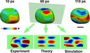
Caption: The acoustic phonons can be visualized on the surface as regions of contraction (blue) and expansion (red). Also shown are two-dimensional images comparing the experimental results with theory and molecular dynamics simulation. The scale bar is 100 nanometers. Credit: Jesse Clark/UCL

Caption: The acoustic phonons can be visualized on the surface as regions of contraction (blue) and expansion (red). Also shown are two-dimensional images comparing the experimental results with theory and molecular dynamics simulation. The scale bar is 100 nanometers. Credit: Jesse Clark/UCL

Here are more details from the news release,

Scientists found that the vibrations were unusual because they start off at exactly the same moment everywhere inside the crystal. It was previously expected that the effects of the excitation would travel across the gold nanocrystal at the speed of sound, but they were found to be much faster, i.e., supersonic.

The new images support theoretical models for light interaction with metals, where energy is first transferred to electrons, which are able to short-circuit the much slower motion of the atoms.

The team carried out the experiments at the SLAC National Accelerator Laboratory using a revolutionary X-ray laser called the “Linac Coherent Light Source”. The pulses of X-rays are extremely short (measured in femtoseconds, or quadrillionths of a second), meaning they are able to freeze all motion of the atoms in any sample, leaving only the electrons still moving.

However, the X-ray pulses are intense enough that the team was able to take single snapshots of the vibrations of the gold nanocrystals they were examining. The vibration was started with a short pulse of infrared light.

The real keeners can watch the movie if they click on the link to the May 23, 2013 news release on EurekAlert.

The team developing this movie was international in scope (from the news release),

The research team included contributors from UCL, University of Oxford, SLAC, Argonne National Laboratory [US] and LaTrobe University, Australia.

Gold nanoparticle self-assembly visualization at the Argonne National Laboratory (US)

There’s a Mar. 13, 2013 news item on phys.org which seems to have been written by someone who’s very technical,

The self-assembly of gold nanoparticles (Au NPs) coated with specific organic ions in water was observed by Center for Nanoscale Materials staff in the Nanobio Interfaces, Electronic & Magnetic Materials & Devices, and Nanophotonics groups at the Argonne National Laboratory using in situ transmission electron microscopy (TEM) equipped with a liquid cell. The Au NPs formed one-dimensional chains within a few minutes.

The originating March 2013 article is on an Argonne National Laboratory’s Center for Nanoscale Materials page,

The self-assembly of NPs attracts intense attention for its potential application in the fabrication of hybrid systems with collective properties from different types of materials. The observations provided here clearly elucidate the complex mechanism of charged NP self-assembly processes. They also paint a cautionary tale on using TEM in situ cells to imitate self-assembly processes in actual solution environments. [emphasis mine]

The hydrated electrons formed in radiolysis of water decrease the overall positive charge of cetyltrimethylammonium (CTA)-coated Au NPs. The NPs also were coated with negative citrate ions. (With citrate alone, however, the Au NPs remained steady in the liquid cell regardless of electron-beam intensity). The anisotropic attractive interactions, including dipolar and Van der Waals interactions, overcome the repulsion among the NPs and induce the assembly of NPs. The spatial segregation of different sizes of NPs as a result of electric field gradients within the cell was observed as well.

I’m not sure why the observations paint a cautionary tale. Perhaps a reader could enlighten me?

The researchers also provided an image,

Cetyltrimethylammonium-ion-coated gold nanoparticles before (top) and after (bottom) 500 seconds of electron-beam exposure inside a TEM liquid cell at 200 kV. Scale bar: 100 nm. [downloaded from http://nano.anl.gov/news/highlights/2013_gold_nanoparticles.html]

Cetyltrimethylammonium-ion-coated gold nanoparticles before (top) and after (bottom) 500 seconds of electron-beam exposure inside a TEM liquid cell at 200 kV. Scale bar: 100 nm. [downloaded from http://nano.anl.gov/news/highlights/2013_gold_nanoparticles.html]

For anyone who can understand the technical explanations, here’s a citation and a link to the research paper,

In Situ Visualization of Self-Assembly of Charged Gold Nanoparticles by Yuzi Liu, Xiao-Min Lin, Yugang Sun, and Tijana Rajh. J. Am. Chem. Soc., 2013, 135 (10), pp 3764–3767 DOI: 10.1021/ja312620e Publication Date (Web): February 22, 2013

Copyright © 2013 American Chemical Society

The paper is behind a paywall.


Picasso, paint, and the hard x-ray nanoprobe

There’s the paint you put on your walls and there’s the paint you put on your body and there’s the paint artists use for their works of art. Well, it turns out that a very well known artist used common house paint to create some of his masterpieces,

Among the Picasso paintings in the Art Institute of Chicago collection, The Red Armchair is the most emblematic of his Ripolin usage and is the painting that was examined with APS X-rays at Argonne National Laboratory. To view a larger version of the image, click on it. Courtesy Art Institute of Chicago, Gift of Mr. and Mrs. Daniel Saidenberg (AIC 1957.72) © Estate of Pablo Picasso / Artists Rights Society (ARS), New York [downloaded from http://www.anl.gov/articles/high-energy-x-rays-shine-light-mystery-picasso-s-paints]

Among the Picasso paintings in the Art Institute of Chicago collection, The Red Armchair is the most emblematic of his Ripolin usage and is the painting that was examined with APS X-rays at Argonne National Laboratory. To view a larger version of the image, click on it. Courtesy Art Institute of Chicago, Gift of Mr. and Mrs. Daniel Saidenberg (AIC 1957.72) © Estate of Pablo Picasso / Artists Rights Society (ARS), New York [downloaded from http://www.anl.gov/articles/high-energy-x-rays-shine-light-mystery-picasso-s-paints]

The Art Institute of Chicago teamed with the US Argonne National Laboratory to solve a decades-long mystery as to what kind of paint Picasso used. From the Feb. 8, 2013 news item on Azonano,

The Art Institute of Chicago teamed up with Argonne National Laboratory to unravel a decades-long debate among art scholars about what kind of paint Picasso used to create his masterpieces.

The results published last month in the journal Applied Physics A: Materials Science & Processing adds significant weight to the widely held theory that Picasso was one of the first master painters to use common house paint rather than traditional artists’ paint. That switch in painting material gave birth to a new style of art marked by canvasses covered in glossy images with marbling, muted edges, and occasional errant paint drips but devoid of brush marks. Fast-drying enamel house paint enabled this dramatic departure from the slow-drying heavily blended oil paintings that dominated the art world up until Picasso’s time.

The key to decoding this long-standing mystery was the development of a unique high-energy X-ray instrument, called the hard X-ray nanoprobe, at the U.S. Department of Energy’s Advanced Photon Source (APS) X-ray facility and the Center for Nanoscale Materials, both housed at Argonne. The nanoprobe is designed to advance the development of high-performance materials and sustainable energies by giving scientists a close up view of the type and arraignment of chemical elements in material.

At that submicroscopic level is where science and art crossed paths.

The Argonne National Laboratory Feb. 6, 2013 news release by Tona Kunz, which originated the news item,  provides more  technical detail,

Volker Rose, a physicist at Argonne, uses the nanoprobe at the APS [Advanced Photon Source]/CNM [Center for Nanoscale Materials] to study zinc oxide, a key chemical used in wide-band-gap semiconductors. White paint contains the same chemical in varying amounts, depending on the type and brand of paint, which makes it a valuable clue for learning about Picasso’s work.

By comparing decades-old paint samples collected through e-Bay purchases with samples from Picasso paintings, scientists were able to learn that the chemical makeup of paint used by Picasso matched the chemical makeup of the first commercial house paint, Ripolin. Scientists also learned about the correlation of the spacing of impurities at the nanoscale in zinc oxide, offering important clues to how zinc oxide could be modified to improve performance in a variety of products, including sensors for radiation detection, LEDs and energy-saving windows as well as liquid-crystal displays for computers, TVs and instrument panels.

“Everything that we learn about how materials are structured and how chemicals react at the nanolevel can help us in our quest to design a better and more sustainable future,” Rose said.

Physicists weren’t the first to investigate the question,

Many art conservators and historians have tried over the years to use traditional optical and electron microscopes to determine whether Picasso or one of his contemporaries was the first to break with the cultural tradition of professional painters using expensive paints designed specifically for their craft. Those art world detectives all failed, because traditional tools wouldn’t let them see deeply enough into the layers of paint or with enough resolution to distinguish between store-bought enamel paint and techniques designed to mimic its appearance.

“Appearances can deceive, so this is where art can benefit from scientific research,” said Francesca Casadio, senior conservator scientist at the Art Institute of Chicago, and co-lead author on the result publication. “We needed to reverse-engineer the paint so that we could figure out if there was a fingerprint that we could then go look for in the pictures around the world that are suspected to be painted with Ripolin, the first commercial brand of house paint.”

Just as criminals leave a signature at a crime scene, each batch of paint has a chemical signature determined by its ingredients and impurities from the area and time period it was made. These signatures can’t be imitated and lie in the nanoscale range.

Yet until now, it was difficult to differentiate the chemical components of the paint pigments from the chemical components in the binders, fillers, other additives and contaminates that were mixed in with the pigments or layered on top of them. Only the nanoprobe at the APS /CNM can distinguish that level of detail: elemental composition and nanoscale distribution of elements within individualized submicrometeric pigment particles.

“The nanoprobe at the APS and CNM allowed unprecedented visualization of information about chemical composition within a singe grain of paint pigment, significantly reducing doubt that Picasso used common house paint in some of his most famous works,” said Rose, co-lead author on the result publication titled “High-Resolution Fluorescence Mapping of Impurities in the Historical Zinc Oxide Pigments: Hard X-ray Nanoprobe Applications to the Paints of Pablo Picasso.”

The nanoprobe’s high spatial resolution and micro-focusing abilities gave it the unique ability to identify individual chemical elements and distinguish between the size of paint particles crushed by hand in artists’ studios and those crushed even smaller by manufacturing equipment. The nanoprobe peered deeper than previous similar paint studies limited to a one-micrometer viewing resolution. The nanoprobe gave scientists an unprecedented look at 30-nanometer-wide particles of paint and impurities from the paint manufacturing process. For comparison, a typical sheet of copier paper is 100,000 nanometers thick.

Using the nanoprobe, scientists were able to determine that Picasso used enamel paint to create in 1931 The Red Armchair, on display at the Art Institute of Chicago. They were also able to determine the paint brand and from what manufacturing region the paint originated.

X-ray analysis of white paints produced under the Ripolin brand and used in artists’ traditional tube paints revealed that both contained nearly contaminate-free zinc oxide pigment. However, artists’ tube paints contained more fillers of other white-colored pigments than did the Ripolin, which was mostly pure zinc oxide.

Casaido [sic] views this type of chemical characterization of paints as a having a much wider application than just the study of Picasso’s paintings. By studying the chemical composition of art materials, she said, historians can learn about trade movements in ancient times, better determine the time period a piece was created, and even learn about the artist themselves through their choice of materials.

Perhaps not so coincidentally, the Art Institute of Chicago is celebrating the 100 year relationship between Picasso and Chicago, excerpted from their Jan. 14, 2013 news release,

THE ART INSTITUTE HONORS 100-YEAR RELATIONSHIP BETWEEN PICASSO AND CHICAGO WITH LANDMARK MUSEUM–WIDE CELEBRATION

First Large-Scale Picasso Exhibition Presented by the Art Institute in 30 Years Commemorates Centennial Anniversary of the Armory Show

Picasso and Chicago on View Exclusively at the Art Institute February 20–May 12, 2013

This winter, the Art Institute of Chicago celebrates the unique relationship between Chicago and one of the preeminent artists of the 20th century—Pablo Picasso—with special presentations, singular paintings on loan from the Philadelphia Museum of Art, and programs throughout the museum befitting the artist’s unparalleled range and influence. The centerpiece of this celebration is the major exhibition Picasso and Chicago, on view from February 20 through May 12, 2013 in the Art Institute’s Regenstein Hall, which features more than 250 works selected from the museum’s own exceptional holdings and from private collections throughout Chicago. Representing Picasso’s innovations in nearly every media—paintings, sculpture, prints, drawings, and ceramics—the works not only tell the story of Picasso’s artistic development but also the city’s great interest in and support for the artist since the Armory Show of 1913, a signal event in the history of modern art.

The Art Institute of Chicago, Francesca Casadio, and art conservation (specifically in regard to Winslow Homer) were mentioned here in an April 11, 2011 posting.