Tag Archives: US National Institute of Biomedical Imaging and Bioengineering (NIBIB)

Injectable bandages for internal bleeding and hydrogel for the brain

This injectable bandage could be a gamechanger (as they say) if it can be taken beyond the ‘in vitro’ (i.e., petri dish) testing stage. A May 22, 2018 news item on Nanowerk makes the announcement (Note: A link has been removed),

While several products are available to quickly seal surface wounds, rapidly stopping fatal internal bleeding has proven more difficult. Now researchers from the Department of Biomedical Engineering at Texas A&M University are developing an injectable hydrogel bandage that could save lives in emergencies such as penetrating shrapnel wounds on the battlefield (Acta Biomaterialia, “Nanoengineered injectable hydrogels for wound healing application”).

A May 22, 2018 US National Institute of Biomedical Engineering and Bioengiineering news release, which originated the news item, provides more detail (Note: Links have been removed),

The researchers combined a hydrogel base (a water-swollen polymer) and nanoparticles that interact with the body’s natural blood-clotting mechanism. “The hydrogel expands to rapidly fill puncture wounds and stop blood loss,” explained Akhilesh Gaharwar, Ph.D., assistant professor and senior investigator on the work. “The surface of the nanoparticles attracts blood platelets that become activated and start the natural clotting cascade of the body.”

Enhanced clotting when the nanoparticles were added to the hydrogel was confirmed by standard laboratory blood clotting tests. Clotting time was reduced from eight minutes to six minutes when the hydrogel was introduced into the mixture. When nanoparticles were added, clotting time was significantly reduced, to less than three minutes.

In addition to the rapid clotting mechanism of the hydrogel composite, the engineers took advantage of special properties of the nanoparticle component. They found they could use the electric charge of the nanoparticles to add growth factors that efficiently adhered to the particles. “Stopping fatal bleeding rapidly was the goal of our work,” said Gaharwar. “However, we found that we could attach growth factors to the nanoparticles. This was an added bonus because the growth factors act to begin the body’s natural wound healing process—the next step needed after bleeding has stopped.”

The researchers were able to attach vascular endothelial growth factor (VEGF) to the nanoparticles. They tested the hydrogel/nanoparticle/VEGF combination in a cell culture test that mimics the wound healing process. The test uses a petri dish with a layer of endothelial cells on the surface that create a solid skin-like sheet. The sheet is then scratched down the center creating a rip or hole in the sheet that resembles a wound.

When the hydrogel containing VEGF bound to the nanoparticles was added to the damaged endothelial cell wound, the cells were induced to grow back and fill-in the scratched region—essentially mimicking the healing of a wound.

“Our laboratory experiments have verified the effectiveness of the hydrogel for initiating both blood clotting and wound healing,” said Gaharwar. “We are anxious to begin tests in animals with the hope of testing and eventual use in humans where we believe our formulation has great potential to have a significant impact on saving lives in critical situations.”

The work was funded by grant EB023454 from the National Institute of Biomedical Imaging and Bioengineering (NIBIB), and the National Science Foundation. The results were reported in the February issue of the journal Acta Biomaterialia.

The paper was published back in April 2018 and there was an April 2, 2018 Texas A&M University news release on EurekAlert making the announcement (and providing a few unique details),

A penetrating injury from shrapnel is a serious obstacle in overcoming battlefield wounds that can ultimately lead to death.Given the high mortality rates due to hemorrhaging, there is an unmet need to quickly self-administer materials that prevent fatality due to excessive blood loss.

With a gelling agent commonly used in preparing pastries, researchers from the Inspired Nanomaterials and Tissue Engineering Laboratory have successfully fabricated an injectable bandage to stop bleeding and promote wound healing.

In a recent article “Nanoengineered Injectable Hydrogels for Wound Healing Application” published in Acta Biomaterialia, Dr. Akhilesh K. Gaharwar, assistant professor in the Department of Biomedical Engineering at Texas A&M University, uses kappa-carrageenan and nanosilicates to form injectable hydrogels to promote hemostasis (the process to stop bleeding) and facilitate wound healing via a controlled release of therapeutics.

“Injectable hydrogels are promising materials for achieving hemostasis in case of internal injuries and bleeding, as these biomaterials can be introduced into a wound site using minimally invasive approaches,” said Gaharwar. “An ideal injectable bandage should solidify after injection in the wound area and promote a natural clotting cascade. In addition, the injectable bandage should initiate wound healing response after achieving hemostasis.”

The study uses a commonly used thickening agent known as kappa-carrageenan, obtained from seaweed, to design injectable hydrogels. Hydrogels are a 3-D water swollen polymer network, similar to Jell-O, simulating the structure of human tissues.

When kappa-carrageenan is mixed with clay-based nanoparticles, injectable gelatin is obtained. The charged characteristics of clay-based nanoparticles provide hemostatic ability to the hydrogels. Specifically, plasma protein and platelets form blood adsorption on the gel surface and trigger a blood clotting cascade.

“Interestingly, we also found that these injectable bandages can show a prolonged release of therapeutics that can be used to heal the wound” said Giriraj Lokhande, a graduate student in Gaharwar’s lab and first author of the paper. “The negative surface charge of nanoparticles enabled electrostatic interactions with therapeutics thus resulting in the slow release of therapeutics.”
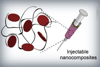
Nanoparticles that promote blood clotting and wound healing (red discs), attached to the wound-filling hydrogel component (black) form a nanocomposite hydrogel. The gel is designed to be self-administered to stop bleeding and begin wound-healing in emergency situations. Credit: Lokhande, et al. 1

Here’s a link to and a citation for the paper,

Nanoengineered injectable hydrogels for wound healing application by Giriraj Lokhande, James K. Carrow, Teena Thakur, Janet R. Xavier, Madasamy Parani, Kayla J. Bayless, Akhilesh K. Gaharwar. Acta Biomaterialia Volume 70, 1 April 2018, Pages 35-47
https://doi.org/10.1016/j.actbio.2018.01.045

This paper is behind a paywall.

Hydrogel and the brain

It’s been an interesting week for hydrogels. On May 21, 2018 there was a news item on ScienceDaily about a bioengineered hydrogel which stimulated brain tissue growth after a stroke (mouse model),

In a first-of-its-kind finding, a new stroke-healing gel helped regrow neurons and blood vessels in mice with stroke-damaged brains, UCLA researchers report in the May 21 issue of Nature Materials.

“We tested this in laboratory mice to determine if it would repair the brain in a model of stroke, and lead to recovery,” said Dr. S. Thomas Carmichael, Professor and Chair of neurology at UCLA. “This study indicated that new brain tissue can be regenerated in what was previously just an inactive brain scar after stroke.”

The brain has a limited capacity for recovery after stroke and other diseases. Unlike some other organs in the body, such as the liver or skin, the brain does not regenerate new connections, blood vessels or new tissue structures. Tissue that dies in the brain from stroke is absorbed, leaving a cavity, devoid of blood vessels, neurons or axons, the thin nerve fibers that project from neurons.

After 16 weeks, stroke cavities in mice contained regenerated brain tissue, including new neural networks — a result that had not been seen before. The mice with new neurons showed improved motor behavior, though the exact mechanism wasn’t clear.

Remarkable stuff.

3D microtopographic scaffolds for transplantation and generation of reprogrammed human neurons

Should this technology prove successful once they start testing on people, the stated goal is to use it for the treatment of human neurodegenerative disorders such as Parkinson’s disease.  But, I can’t help wondering if they might also consider constructing an artificial brain.

Getting back to the 3D scaffolds for neurons, a March 17, 2016 US National Institutes of Health (NIH) news release (also on EurekAlert), makes the announcement,

National Institutes of Health-funded scientists have developed a 3D micro-scaffold technology that promotes reprogramming of stem cells into neurons, and supports growth of neuronal connections capable of transmitting electrical signals. The injection of these networks of functioning human neural cells — compared to injecting individual cells — dramatically improved their survival following transplantation into mouse brains. This is a promising new platform that could make transplantation of neurons a viable treatment for a broad range of human neurodegenerative disorders.

Previously, transplantation of neurons to treat neurodegenerative disorders, such as Parkinson’s disease, had very limited success due to poor survival of neurons that were injected as a solution of individual cells. The new research is supported by the National Institute of Biomedical Imaging and Bioengineering (NIBIB), part of NIH.

“Working together, the stem cell biologists and the biomaterials experts developed a system capable of shuttling neural cells through the demanding journey of transplantation and engraftment into host brain tissue,” said Rosemarie Hunziker, Ph.D., director of the NIBIB Program in Tissue Engineering and Regenerative Medicine. “This exciting work was made possible by the close collaboration of experts in a wide range of disciplines.”

The research was performed by researchers from Rutgers University, Piscataway, New Jersey, departments of Biomedical Engineering, Neuroscience and Cell Biology, Chemical and Biochemical Engineering, and the Child Health Institute; Stanford University School of Medicine’s Institute of Stem Cell Biology and Regenerative Medicine, Stanford, California; the Human Genetics Institute of New Jersey, Piscataway; and the New Jersey Center for Biomaterials, Piscataway. The results are reported in the March 17, 2016 issue of Nature Communications.

The researchers experimented in creating scaffolds made of different types of polymer fibers, and of varying thickness and density. They ultimately created a web of relatively thick fibers using a polymer that stem cells successfully adhered to. The stem cells used were human induced pluripotent stem cells (iPSCs), which can be readily generated from adult cell types such as skin cells. The iPSCs were induced to differentiate into neural cells by introducing the protein NeuroD1 into the cells.

The space between the polymer fibers turned out to be critical. “If the scaffolds were too dense, the stem cell-derived neurons were unable to integrate into the scaffold, whereas if they are too sparse then the network organization tends to be poor,” explained Prabhas Moghe, Ph.D., distinguished professor of biomedical engineering & chemical engineering at Rutgers University and co-senior author of the paper. “The optimal pore size was one that was large enough for the cells to populate the scaffold but small enough that the differentiating neurons sensed the presence of their neighbors and produced outgrowths resulting in cell-to-cell contact. This contact enhances cell survival and development into functional neurons able to transmit an electrical signal across the developing neural network.”

To test the viability of neuron-seeded scaffolds when transplanted, the researchers created micro-scaffolds that were small enough for injection into mouse brain tissue using a standard hypodermic needle. They injected scaffolds carrying the human neurons into brain slices from mice and compared them to human neurons injected as individual, dissociated cells.

The neurons on the scaffolds had dramatically increased cell-survival compared with the individual cell suspensions. The scaffolds also promoted improved neuronal outgrowth and electrical activity. Neurons injected individually in suspension resulted in very few cells surviving the transplant procedure.

Human neurons on scaffolds compared to neurons in solution were then tested when injected into the brains of live mice. Similar to the results in the brain slices, the survival rate of neurons on the scaffold network was increased nearly 40-fold compared to injected isolated cells. A critical finding was that the neurons on the micro-scaffolds expressed proteins that are involved in the growth and maturation of neural synapses–a good indication that the transplanted neurons were capable of functionally integrating into the host brain tissue.

The success of the study gives this interdisciplinary group reason to believe that their combined areas of expertise have resulted in a system with much promise for eventual treatment of human neurodegenerative disorders. In fact, they are now refining their system for specific use as an eventual transplant therapy for Parkinson’s disease. The plan is to develop methods to differentiate the stem cells into neurons that produce dopamine, the specific neuron type that degenerates in individuals with Parkinson’s disease. The work also will include fine-tuning the scaffold materials, mechanics and dimensions to optimize the survival and function of dopamine-producing neurons, and finding the best mouse models of the disease to test this Parkinson’s-specific therapy.

Here’s a link to and a citation for the paper,

Generation and transplantation of reprogrammed human neurons in the brain using 3D microtopographic scaffolds by Aaron L. Carlson, Neal K. Bennett, Nicola L. Francis, Apoorva Halikere, Stephen Clarke, Jennifer C. Moore, Ronald P. Hart, Kenneth Paradiso, Marius Wernig, Joachim Kohn, Zhiping P. Pang, & Prabhas V. Moghe. Nature Communications 7, Article number: 10862  doi:10.1038/ncomms10862 Published 17 March 2016

This paper is open access.

Finding a way to prevent sunscreens from penetrating the skin

While nanosunscreens have been singled out for their possible impact on our health, the fact is many sunscreens contain dangerous ingredients penetrating the skin. A Dec. 14, 2015 news item on ScienceDaily describes some research into getting sunscreens to stay on the skin surface avoiding penetration,

A new sunscreen has been developed that encapsulates the UV-blocking compounds inside bio-adhesive nanoparticles, which adhere to the skin well, but do not penetrate beyond the skin’s surface. These properties resulted in highly effective UV protection in a mouse model, without the adverse effects observed with commercial sunscreens, including penetration into the bloodstream and generation of reactive oxygen species, which can damage DNA and lead to cancer.

A US National Institute of Biomedical Imaging and Bioengineering (NIBIB) Dec. 14, 2015 news release, which originated the news item, expands on the theme (Note: Links have been removed),

Commercial sunscreens use compounds that effectively filter out damaging UV light. However, there is concern that these agents have a variety of harmful effects due to penetration past the surface skin. For example, these products have been found in human breast tissue and urine and are known to disrupt the normal function of some hormones. Also, the exposure of the UV filters to light can produce toxic reactive oxygen species that are destructive to cells and tissues and can cause tumors through DNA damage.

“This work applies a novel bioengineering idea to a little known but significant health problem, adds Jessica Tucker, Ph.D., Director of the NIBIB Program in Delivery Systems and Devices for Drugs and Biologics. “While we are all familiar with the benefits of sunscreen, the potential toxicities from sunscreen due to penetration into the body and creation of DNA-damaging agents are not well known. Bioengineering sunscreen to inhibit penetration and keep any DNA-damaging compounds isolated in the nanoparticle and away from the skin is a great example of how a sophisticated technology can be used to solve a problem affecting the health of millions of people.”

Bioengineers and dermatologists at Yale University in New Haven, Connecticut combined their expertise in nanoparticle-based drug delivery and the molecular and cellular characteristics of the skin to address these potential health hazards of current commercial sunscreens.

The news release then goes on to provide some technical details,

The group encapsulated a commonly used sunscreen, padimate O (PO), inside a nanoparticle (a very small molecule often used to transport drugs and other agents into the body). PO is related to the better-known sunscreen PABA.

The bioadhesive nanoparticle containing the sunscreen PO was tested on pigs for penetration into the skin. A control group of pigs received the PO alone, not encapsulated in a nanoparticle. The PO penetrated beyond the surface layers of skin where it could potentially enter the bloodstream through blood vessels that are in the deeper skin layers. However, the PO inside the nanoparticle remained on the surface of the skin and did not penetrate into deeper layers.

Because the bioadhesive nanoparticles, or BNPs are larger than skin pores it was somewhat expected that they could not enter the body by that route. However, skin is full of hair follicles that are larger than BNPs and so could be a way for migration into the body. Surprisingly, BNPs did not pass through the hair follicle openings either. Tests indicated that the adhesive properties of the BNPs caused them to stick to the skin surface, unable to move through the hair follicles.

Further testing showed that the BNPs were water resistant and remained on the skin for a day or more, yet were easily removed by towel wiping. They also disappeared in several days through natural exfoliation of the surface skin.

BNPs enhance the effect of sunscreen

An important test was whether the BNP-encapsulated sunscreen retained its UV filtering properties. The researchers used a mouse model to test whether PO blocked sunburn when encapsulated in the BNPs. The BNP formulation successfully provided the same amount of UV protection as the commercial products applied directly to the skin of the hairless mouse model. Surprisingly, this was achieved even though the BNPs carried only a fraction (5%) of the amount of commercial sunblock applied to the mice.

Finally, the encapsulated sunscreen was tested for the formation of damaging oxygen-carrying molecules known as reactive oxygen species, (ROS) when exposed to UV light. The researchers hypothesized that any ROS created by the sunscreen’s interaction with UV would stay contained inside the BNP, unable to damage surrounding tissue. Following exposure to UV light, no damaging ROS were detected outside of the nanoparticle, indicating that any harmful agents that were formed remained inside of the nanoparticle, unable to make contact with the skin.

“We are extremely pleased with the properties and performance of our BNP formulation,” says senior author Mark Saltzman, Ph.D., Yale School of Engineering and Applied Science. “The sunscreen loaded BNPs combine the best properties of an effective sunscreen with a safety profile that alleviates the potential toxicities of the actual sunscreen product because it is encapsulated and literally never touches the skin.” Adds co-senior author, Michael Girardi, M.D. “Our nanoparticles performed as expected, however, these are preclinical findings. We are now in a position to assess the effects on human skin.”

So, all of this work has been done on animal models, which means that human clinical trials are the likely next step. As we wait, here’s a link to and a citation for this group’s paper,

A sunblock based on bioadhesive nanoparticles by Yang Deng, Asiri Ediriwickrema, Fan Yang, Julia Lewis, Michael Girardi, & W. Mark Saltzman. Nature Materials 14, 1278–1285 (2015) doi:10.1038/nmat4422 Published online 28 September 2015

This paper is behind a paywall.