Tag Archives: US National Institutes of Health (NIH)

Brain surgery with no scalpel or incisions

A December 3, 2021 news item on ScienceDaily announces some very exciting work from the University of Virginia UVA) and Stanford University,

University of Virginia School of Medicine researchers have developed a noninvasive way to remove faulty brain circuits that could allow doctors to treat debilitating neurological diseases without the need for conventional brain surgery.

The UVA team, together with colleagues at Stanford University, indicate that the approach, if successfully translated to the operating room, could revolutionize the treatment of some of the most challenging and complex neurological diseases, including epilepsy, movement disorders and more. The approach uses low-intensity focused ultrasound waves combined with microbubbles to briefly penetrate the brain’s natural defenses and allow the targeted delivery of a neurotoxin. This neurotoxin kills the culprit brain cells while sparing other healthy cells and preserving the surrounding brain architecture.

A November 22, 2021 University of Virginia news release (also on EurekAlert but published on December 3, 2021), which originated the news item, offers technical details (Note: Links have been removed),

“This novel surgical strategy has the potential to supplant existing neurosurgical procedures used for the treatment of neurological disorders that don’t respond to medication,” said researcher Kevin S. Lee of UVA’s Departments of Neuroscience and Neurosurgery and the Center for Brain Immunology and Glia, or BIG. “This unique approach eliminates the diseased brain cells, spares adjacent healthy cells and achieves these outcomes without even having to cut into the scalp.”

The Power of PING

The new approach, called “PING,” has already demonstrated exciting potential in laboratory studies. For instance, one of the promising applications for PING could be for the surgical treatment of epilepsies that do not respond to medication. Approximately a third of patients with epilepsy do not respond to anti-seizure drugs, and surgery can reduce or eliminate seizures for some of them. Lee and his team, along with their collaborators at Stanford, have shown that PING can reduce or eliminate seizures in two research models of epilepsy. The findings raise the possibility of treating epilepsy in a carefully targeted and noninvasive manner without the need for traditional brain surgery. 

Another important potential advantage of PING is that it could encourage the surgical treatment of appropriate patients with epilepsy who are reluctant to undergo conventional invasive or ablative surgery.

In a scientific paper newly published in the Journal of Neurosurgery, Lee and his collaborators detail the ability of PING to focally eliminate neurons in a brain region, while sparing non-target cells in the same area. In contrast, currently available surgical approaches damage all cells in a treated brain region. 

A key advantage of the approach is its incredible precision. PING harnesses the power of magnetic-resonance imaging to let scientists peer inside the skull so that they can precisely guide sound waves to open the body’s natural blood-brain barrier exactly where needed. This barrier is designed to keep harmful cells and molecules out of the brain, but it also prevents the delivery of potentially beneficial treatments.

The UVA group’s new paper concludes that PING allows the delivery of a highly targeted neurotoxin, cleanly wiping out problematic neurons, a type of brain cell, without causing collateral damage. 

Another key advantage of the precision of this approach is that it can be used on irregularly shaped targets in areas that would be extremely difficult or impossible to reach through regular brain surgery. “If this strategy translates to the clinic,” the researchers write in their new paper, “the noninvasive nature and specificity of the procedure could positively influence both physician referrals for, and patient confidence in, surgery for medically intractable neurological disorders.”

“Our hope is that the PING strategy will become a key element in the next generation of very precise, noninvasive, neurosurgical approaches to treat major neurological disorders,” said Lee, who is part of the UVA Brain Institute.

About the Research

Lee’s groundbreaking research has been supported by the National Institutes of Health, the Chester Fund and the Charlottesville-based Focused Ultrasound Foundation. The work is part of an expansive effort at UVA Health to explore the potential of scalpel-free focused ultrasound to treat complex diseases throughout the body.

UVA’s pioneering research has already paved the way for the federal Food and Drug Administration to approve focused ultrasound for the treatment of essential tremor, a common movement disorder, and Parkinson’s disease symptoms. Research is underway on its potential applications for many more conditions, including breast cancer and glioblastoma, a deadly form of brain tumor. Learn more about UVA’s focused ultrasound research.

The research team included Yi Wang, Matthew J. Anzivino, Yanrong Zhang, Edward H. Bertram, James Woznak, Alexander L. Klibanov, Erik Dumont and Max Wintermark. 

An application to patent the PING procedure has been submitted by members of the research group. 

The research was funded by the National Institutes of Health, grants R01 NS102194 and R01 CA217953-01; the Chester Fund; and the Focused Ultrasound Foundation.

To keep up with the latest medical research news from UVA, subscribe to the Making of Medicine blog at http://makingofmedicine.virginia.edu.

Here’s a link to and a citation for the paper,

Noninvasive disconnection of targeted neuronal circuitry sparing axons of passage and nonneuronal cells by Yi Wang, Matthew J. Anzivino, Yanrong Zhang, Edward H. Bertram, James Woznak, Alexander L. Klibanov, Erik Dumont, Max Wintermark, and Kevin S. Lee. Journal of Neurosurgery DOI: https://doi.org/10.3171/2021.7.JNS21123 Online Publication Date: 19 Nov 2021

This paper is behind a paywall.

Nanopore-tal enables cells to talk to computers?

An August 25, 2021 news item on ScienceDaily announced research that will allow more direct communication between cells and computers,

Genetically encoded reporter proteins have been a mainstay of biotechnology research, allowing scientists to track gene expression, understand intracellular processes and debug engineered genetic circuits.

But conventional reporting schemes that rely on fluorescence and other optical approaches come with practical limitations that could cast a shadow over the field’s future progress. Now, researchers at the University of Washington and Microsoft have created a “nanopore-tal” into what is happening inside these complex biological systems, allowing scientists to see reporter proteins in a whole new light.

The team introduced a new class of reporter proteins that can be directly read by a commercially available nanopore sensing device. The new system ― dubbed “Nanopore-addressable protein Tags Engineered as Reporters” or “NanoporeTERs” ― can detect multiple protein expression levels from bacterial and human cell cultures far beyond the capacity of existing techniques.

An August 12, 2021 University of Washington news release (also on EurekAlert but published August 24, 2021), which originated the news item, provides more detail (Note: Links have been removed),

“NanoporeTERs offer a new and richer lexicon for engineered cells to express themselves and shed new light on the factors they are designed to track. They can tell us a lot more about what is happening in their environment all at once,” said co-lead author Nicolas Cardozo, a doctoral student with the UW Molecular Engineering and Sciences Institute. “We’re essentially making it possible for these cells to ‘talk’ to computers about what’s happening in their surroundings at a new level of detail, scale and efficiency that will enable deeper analysis than what we could do before.”

For conventional labeling methods, researchers can track only a few optical reporter proteins, such as green fluorescent protein, simultaneously because of their overlapping spectral properties. For example, it’s difficult to distinguish between more than three different colors of fluorescent proteins at once. In contrast, NanoporeTERs were designed to carry distinct protein “barcodes” composed of strings of amino acids that, when used in combination, allow at least ten times more multiplexing possibilities. 

These synthetic proteins are secreted outside of a cell into the surrounding environment, where researchers can collect and analyze them using a commercially available nanopore array. Here, the team used the Oxford Nanopore Technologies MinION device. 

The researchers engineered the NanoporeTER proteins with charged “tails” so that they can be pulled into the nanopore sensors by an electric field. Then the team uses machine learning to classify the electrical signals for each NanoporeTER barcode in order to determine each protein’s output levels.

“This is a fundamentally new interface between cells and computers,” said senior author Jeff Nivala, a UW research assistant professor in the Paul G. Allen School of Computer Science & Engineering. “One analogy I like to make is that fluorescent protein reporters are like lighthouses, and NanoporeTERs are like messages in a bottle. 

“Lighthouses are really useful for communicating a physical location, as you can literally see where the signal is coming from, but it’s hard to pack more information into that kind of signal. A message in a bottle, on the other hand, can pack a lot of information into a very small vessel, and you can send many of them off to another location to be read. You might lose sight of the precise physical location where the messages were sent, but for many applications that’s not going to be an issue.”

As a proof of concept, the team developed a library of more than 20 distinct NanoporeTERs tags. But the potential is significantly greater, according to co-lead author Karen Zhang, now a doctoral student in the UC Berkeley-UCSF bioengineering graduate program.

“We are currently working to scale up the number of NanoporeTERs to hundreds, thousands, maybe even millions more,” said Zhang, who graduated this year from the UW with bachelor’s degrees in both biochemistry and microbiology. “The more we have, the more things we can track.

“We’re particularly excited about the potential in single-cell proteomics, but this could also be a game-changer in terms of our ability to do multiplexed biosensing to diagnose disease and even target therapeutics to specific areas inside the body. And debugging complicated genetic circuit designs would become a whole lot easier and much less time-consuming if we could measure the performance of all the components in parallel instead of by trial and error.”

These researchers have made novel use of the MinION device before, when they developed a molecular tagging system to replace conventional inventory control methods. That system relied on barcodes comprising synthetic strands of DNA that could be decoded on demand using the portable reader. 

This time, the team went a step farther.

“This is the first paper to show how a commercial nanopore sensor device can be repurposed for applications other than the DNA and RNA sequencing for which they were originally designed,” said co-author Kathryn Doroschak, a computational biologist at Adaptive Biotechnologies who completed this work as a doctoral student at the Allen School. “This is exciting as a precursor for nanopore technology becoming more accessible and ubiquitous in the future. You can already plug a nanopore device into your cell phone. I could envision someday having a choice of ‘molecular apps’ that will be relatively inexpensive and widely available outside of traditional genomics.”

Additional co-authors of the paper are Aerilynn Nguyen at Northeastern University and Zoheb Siddiqui at Amazon, both former UW undergraduate students; Nicholas Bogard at Patch Biosciences, a former UW postdoctoral research associate; Luis Ceze, an Allen School professor; and Karin Strauss, an Allen School affiliate professor and a senior principal research manager at Microsoft. This research was funded by the National Science Foundation, the National Institutes of Health and a sponsored research agreement from Oxford Nanopore Technologies. 

Here’s a link to and a citation for the paper,

Multiplexed direct detection of barcoded protein reporters on a nanopore array by Nicolas Cardozo, Karen Zhang, Kathryn Doroschak, Aerilynn Nguyen, Zoheb Siddiqui, Nicholas Bogard, Karin Strauss, Luis Ceze & Jeff Nivala. Nature Biotechnology (2021) DOI: https://doi.org/10.1038/s41587-021-01002-6 Published: 12 August 2021

This paper is behind a paywall.

There’s no ‘I’ in team: coaching scientists to work together

While it’s true enough in English where you don’t spell the word team with the letter ‘I’, that’s not the case in French where the word is ‘equipe’. it makes me wonder how many other languages in the world have an ‘I’ in team.

Moving on. This English language saying is true enough in its way but there is no team unless you have a group of ‘I’s’ and the trick is getting them to work together as a July 18, 2019 Northwestern University news release (received via email) about a new online training tool notes,

Coaching scientists to play well together

Free tool shows how to avoid fights over data and authorship conflicts   

‘You stole my idea’ or ‘I’m not getting credit for my work’ are common disputes
Only tool validated by research to help scientists collaborate smoothly
Many NSF [US National Science Foundation] and NIH [US National Institutes of Health] grants now require applicants to show readiness for team science
Scientists can’t do it on their own

CHICAGO — When scientists from different disciplines collaborate – as is increasingly necessary to confront the complexity of challenging research problems – interpersonal tussles often arise. One scientist may accuse another of stealing her ideas. Or, a researcher may feel he is not getting credit for his work or doesn’t have access to important data. 
 
“Interdisciplinary team science is now the state of the art across all branches of science and engineering,” said Bonnie Spring, professor of preventive medicine at Northwestern University Feinberg School of Medicine. “But very few scientists have been trained to work with others outside of their own disciplinary silo.”
 
The skill is critical because many National Institute[s] of Health and National Science Foundationgrants require applicants to show readiness for team science.
 
A free, online training tool developed by Northwestern — teamscience.net — has been been proven to help scientists develop skills to work with other scientists outside their own discipline. 
 
A new study led by Spring showed scientists who completed the program’s modules – called COALESCE – significantly boosted their knowledge about team science and increased their self-confidence about being able to successfully work in scientific teams. Most people who completed one or more modules (84%) said that the experience of taking the modules was very likely to positively impact their future research.
 
The study will be published July 18 [2019] in the Journal of Clinical and Translational Science.
 
There are few training resources to teach scientists how to collaborate, and the ones that are available don’t have evidence of their effectiveness. Teamscience.net is the only free, validated-by-research tool available to anyone at any time. 
 
Almost 1,000 of the COALESCE users opted voluntarily to respond to questions about the learning modules, providing information about how taking each module influenced team science knowledge, skills and attitudes.
 
‘You stole my idea’
 
The most common area of dispute among collaborating scientists is authorship concerns, such as accusations that one person stole ideas from another or that a contributor was not getting credit for his or her work, the study authors said. Other disputes arise around access to and analysis of data, utilization of materials or resources and the general direction of the research itself. Underlying all of these issues is a common failure to prepare for working collaboratively with other scientists. 
 
“Preparing in advance before starting to collaborate, often through the creation of a formal collaboration agreement document, is the best way to head off these types of disputes,” said Angela Pfammatter, assistant professor of preventive medicine at Feinberg and a coauthor on the paper.
  
Spring suggested “having scientists discuss their expectations of one another and the collaboration to prevent acrimonious conflicts.” 
 
Skills to play well together
 
These skills are critical to a successful scientific team, the authors said: 

The ability to choose team members who have the right mix of expertise, temperament and accessibility to round out a team. 
The ability to anticipate what could go wrong and to develop contingency plans in advance. 
The ability to manage conflict within the team 

The teamscience.net modules help scientists acquire these skills by letting them interact with different problem scenarios that can arise in team-based research. Scientists can try out different solutions and learn from mistakes in a safe, online environment. 
 
More than 16,000 people have accessed the resource in the past six years.  Demand for team science training is expected to increase as interdisciplinary teams set out to tackle some of science’s most challenging problems. 
 
Other Northwestern authors on the paper are Ekaterina Klyachko, Phillip Rak, H. Gene McFadden, Juned Siddique and Leland Bardsley. 
 
Funding support for COALESCE is from the National Institutes of Health, National Center for Advancing Translational Sciences grants 3UL1RR025741 and UL1TR001422 and its Office of Behavioral and Social Sciences Research.

i once got caught here on this blog between two warring scientists. My August 24, 2015 posting was a pretty standard one for me. Initially, it was one of my more minimalistic pieces with a copy of the text from a university news release announcing the research and a link to the academic paper. I can’t remember if the problem was which scientist was listed first and which was listed last but one of them took exception and contacted me explaining how it was wrong. (Note: These decisions are not made by me.) I did my best to fix whatever the problem was and then the other scientist contacted me. After the dust settled, I ended up with a dog’s breakfast for my posting and a new policy.

Getting back to COALESCE: I wish the Northwestern University researchers all the best as they look for ways to help scientists work together more smoothly and cooperatively.

Here’s a link to and a citation for the paper,

Online, cross-disciplinary team science training for health and medical professionals: Evaluation of COALESCE (teamscience.net) by Bonnie Spring, Ekaterina A. Klyachko, Phillip W. Rak, H. Gene McFadden, Donald Hedeker, Juned Siddique, Leland R. Bardsley, and Angela Fidler Pfammatter. Jurnal of Clinical and Translational Science DOI: https://doi.org/10.1017/cts.2019.383 Published online by Cambridge University Press: 18 July 2019

This paper is open access.

Editing the genome with CRISPR ((clustered regularly interspaced short palindromic repeats)-carrying nanoparticles

MIT (Massachusetts Institute of Technology) researchers have developed a new nonviral means of delivering CRISPR ((clustered regularly interspaced short palindromic repeats)-CAS9 gene therapy according to a November 13, 2017 news item on Nanowerk,

In a new study, MIT researchers have developed nanoparticles that can deliver the CRISPR genome-editing system and specifically modify genes in mice. The team used nanoparticles to carry the CRISPR components, eliminating the need to use viruses for delivery.

Using the new delivery technique, the researchers were able to cut out certain genes in about 80 percent of liver cells, the best success rate ever achieved with CRISPR in adult animals.

In a new study, MIT researchers have developed nanoparticles that can deliver the CRISPR genome-editing system and specifically modify genes, eliminating the need to use viruses for delivery. Image: MIT News

A November 13, 2017 MIT news release (also on EurekAlert), which originated the news item, provides more details about the research and a good description of and comparison between using a viral system and using a nanoparticle-based system to deliver CRISPR-CAS9,

“What’s really exciting here is that we’ve shown you can make a nanoparticle that can be used to permanently and specifically edit the DNA in the liver of an adult animal,” says Daniel Anderson, an associate professor in MIT’s Department of Chemical Engineering and a member of MIT’s Koch Institute for Integrative Cancer Research and Institute for Medical Engineering and Science (IMES).

One of the genes targeted in this study, known as Pcsk9, regulates cholesterol levels. Mutations in the human version of the gene are associated with a rare disorder called dominant familial hypercholesterolemia, and the FDA recently approved two antibody drugs that inhibit Pcsk9. However these antibodies need to be taken regularly, and for the rest of the patient’s life, to provide therapy. The new nanoparticles permanently edit the gene following a single treatment, and the technique also offers promise for treating other liver disorders, according to the MIT team.

Anderson is the senior author of the study, which appears in the Nov. 13 [2017] issue of Nature Biotechnology. The paper’s lead author is Koch Institute research scientist Hao Yin. Other authors include David H. Koch Institute Professor Robert Langer of MIT, professors Victor Koteliansky and Timofei Zatsepin of the Skolkovo Institute of Science and Technology [Russia], and Professor Wen Xue of the University of Massachusetts Medical School.

Targeting disease

Many scientists are trying to develop safe and efficient ways to deliver the components needed for CRISPR, which consists of a DNA-cutting enzyme called Cas9 and a short RNA that guides the enzyme to a specific area of the genome, directing Cas9 where to make its cut.

In most cases, researchers rely on viruses to carry the gene for Cas9, as well as the RNA guide strand. In 2014, Anderson, Yin, and their colleagues developed a nonviral delivery system in the first-ever demonstration of curing a disease (the liver disorder tyrosinemia) with CRISPR in an adult animal. However, this type of delivery requires a high-pressure injection, a method that can also cause some damage to the liver.

Later, the researchers showed they could deliver the components without the high-pressure injection by packaging messenger RNA (mRNA) encoding Cas9 into a nanoparticle instead of a virus. Using this approach, in which the guide RNA was still delivered by a virus, the researchers were able to edit the target gene in about 6 percent of hepatocytes, which is enough to treat tyrosinemia.

While that delivery technique holds promise, in some situations it would be better to have a completely nonviral delivery system, Anderson says. One consideration is that once a particular virus is used, the patient will develop antibodies to it, so it couldn’t be used again. Also, some patients have pre-existing antibodies to the viruses being tested as CRISPR delivery vehicles.

In the new Nature Biotechnology paper, the researchers came up with a system that delivers both Cas9 and the RNA guide using nanoparticles, with no need for viruses. To deliver the guide RNAs, they first had to chemically modify the RNA to protect it from enzymes in the body that would normally break it down before it could reach its destination.

The researchers analyzed the structure of the complex formed by Cas9 and the RNA guide, or sgRNA, to figure out which sections of the guide RNA strand could be chemically modified without interfering with the binding of the two molecules. Based on this analysis, they created and tested many possible combinations of modifications.

“We used the structure of the Cas9 and sgRNA complex as a guide and did tests to figure out we can modify as much as 70 percent of the guide RNA,” Yin says. “We could heavily modify it and not affect the binding of sgRNA and Cas9, and this enhanced modification really enhances activity.”

Reprogramming the liver

The researchers packaged these modified RNA guides (which they call enhanced sgRNA) into lipid nanoparticles, which they had previously used to deliver other types of RNA to the liver, and injected them into mice along with nanoparticles containing mRNA that encodes Cas9.

They experimented with knocking out a few different genes expressed by hepatocytes, but focused most of their attention on the cholesterol-regulating Pcsk9 gene. The researchers were able to eliminate this gene in more than 80 percent of liver cells, and the Pcsk9 protein was undetectable in these mice. They also found a 35 percent drop in the total cholesterol levels of the treated mice.

The researchers are now working on identifying other liver diseases that might benefit from this approach, and advancing these approaches toward use in patients.

“I think having a fully synthetic nanoparticle that can specifically turn genes off could be a powerful tool not just for Pcsk9 but for other diseases as well,” Anderson says. “The liver is a really important organ and also is a source of disease for many people. If you can reprogram the DNA of your liver while you’re still using it, we think there are many diseases that could be addressed.”

“We are very excited to see this new application of nanotechnology open new avenues for gene editing,” Langer adds.

The research was funded by the National Institutes of Health (NIH), the Russian Scientific Fund, the Skoltech Center, and the Koch Institute Support (core) Grant from the National Cancer Institute.

Here’s a link to and a citation for the paper,

Structure-guided chemical modification of guide RNA enables potent non-viral in vivo genome editing by Hao Yin, Chun-Qing Song, Sneha Suresh, Qiongqiong Wu, Stephen Walsh, Luke Hyunsik Rhym, Esther Mintzer, Mehmet Fatih Bolukbasi, Lihua Julie Zhu, Kevin Kauffman, Haiwei Mou, Alicia Oberholzer, Junmei Ding, Suet-Yan Kwan, Roman L Bogorad, Timofei Zatsepin, Victor Koteliansky, Scot A Wolfe, Wen Xue, Robert Langer, & Daniel G Anderson. Nature Biotechnology doi:10.1038/nbt.4005 Published online: 13 November 2017

This paper is behind a paywall.

CRISPR and editing the germline in the US (part 3 of 3): public discussions and pop culture

After giving a basic explanation of the technology and some of the controversies in part 1 and offering more detail about the technology and about the possibility of designer babies in part 2; this part covers public discussion, a call for one and the suggestion that one is taking place in popular culture.

But a discussion does need to happen

In a move that is either an exquisite coincidence or has been carefully orchestrated (I vote for the latter), researchers from the University of Wisconsin-Madison have released a study about attitudes in the US to human genome editing. From an Aug. 11, 2017 University of Wisconsin-Madison news release (also on EurekAllert),

In early August 2017, an international team of scientists announced they had successfully edited the DNA of human embryos. As people process the political, moral and regulatory issues of the technology — which nudges us closer to nonfiction than science fiction — researchers at the University of Wisconsin-Madison and Temple University show the time is now to involve the American public in discussions about human genome editing.

In a study published Aug. 11 in the journal Science, the researchers assessed what people in the United States think about the uses of human genome editing and how their attitudes may drive public discussion. They found a public divided on its uses but united in the importance of moving conversations forward.

“There are several pathways we can go down with gene editing,” says UW-Madison’s Dietram Scheufele, lead author of the study and member of a National Academy of Sciences committee that compiled a report focused on human gene editing earlier this year. “Our study takes an exhaustive look at all of those possible pathways forward and asks where the public stands on each one of them.”

Compared to previous studies on public attitudes about the technology, the new study takes a more nuanced approach, examining public opinion about the use of gene editing for disease therapy versus for human enhancement, and about editing that becomes hereditary versus editing that does not.

The research team, which included Scheufele and Dominique Brossard — both professors of life sciences communication — along with Michael Xenos, professor of communication arts, first surveyed study participants about the use of editing to treat disease (therapy) versus for enhancement (creating so-called “designer babies”). While about two-thirds of respondents expressed at least some support for therapeutic editing, only one-third expressed support for using the technology for enhancement.

Diving even deeper, researchers looked into public attitudes about gene editing on specific cell types — somatic or germline — either for therapy or enhancement. Somatic cells are non-reproductive, so edits made in those cells do not affect future generations. Germline cells, however, are heritable, and changes made in these cells would be passed on to children.

Public support of therapeutic editing was high both in cells that would be inherited and those that would not, with 65 percent of respondents supporting therapy in germline cells and 64 percent supporting therapy in somatic cells. When considering enhancement editing, however, support depended more upon whether the changes would affect future generations. Only 26 percent of people surveyed supported enhancement editing in heritable germline cells and 39 percent supported enhancement of somatic cells that would not be passed on to children.

“A majority of people are saying that germline enhancement is where the technology crosses that invisible line and becomes unacceptable,” says Scheufele. “When it comes to therapy, the public is more open, and that may partly be reflective of how severe some of those genetically inherited diseases are. The potential treatments for those diseases are something the public at least is willing to consider.”

Beyond questions of support, researchers also wanted to understand what was driving public opinions. They found that two factors were related to respondents’ attitudes toward gene editing as well as their attitudes toward the public’s role in its emergence: the level of religious guidance in their lives, and factual knowledge about the technology.

Those with a high level of religious guidance in their daily lives had lower support for human genome editing than those with low religious guidance. Additionally, those with high knowledge of the technology were more supportive of it than those with less knowledge.

While respondents with high religious guidance and those with high knowledge differed on their support for the technology, both groups highly supported public engagement in its development and use. These results suggest broad agreement that the public should be involved in questions of political, regulatory and moral aspects of human genome editing.

“The public may be split along lines of religiosity or knowledge with regard to what they think about the technology and scientific community, but they are united in the idea that this is an issue that requires public involvement,” says Scheufele. “Our findings show very nicely that the public is ready for these discussions and that the time to have the discussions is now, before the science is fully ready and while we have time to carefully think through different options regarding how we want to move forward.”

Here’s a  link to and a citation for the paper,

U.S. attitudes on human genome editing by Dietram A. Scheufele, Michael A. Xenos, Emily L. Howell, Kathleen M. Rose, Dominique Brossard1, and Bruce W. Hardy. Science 11 Aug 2017: Vol. 357, Issue 6351, pp. 553-554 DOI: 10.1126/science.aan3708

This paper is behind a paywall.

A couple of final comments

Briefly, I notice that there’s no mention of the ethics of patenting this technology in the news release about the study.

Moving on, it seems surprising that the first team to engage in germline editing in the US is in Oregon; I would have expected the work to come from Massachusetts, California, or Illinois where a lot of bleeding edge medical research is performed. However, given the dearth of financial support from federal funding institutions, it seems likely that only an outsider would dare to engage i the research. Given the timing, Mitalipov’s work was already well underway before the recent about-face from the US National Academy of Sciences (Note: Kaiser’s Feb. 14, 2017 article does note that for some the recent recommendations do not represent any change).

As for discussion on issues such as editing of the germline, I’ve often noted here that popular culture (including advertising with the science fiction and other dramas laid in various media) often provides an informal forum for discussion. Joelle Renstrom in an Aug. 13, 2017 article for slate.com writes that Orphan Black (a BBC America series featuring clones) opened up a series of questions about science and ethics in the guise of a thriller about clones. She offers a précis of the first four seasons (Note: A link has been removed),

If you stopped watching a few seasons back, here’s a brief synopsis of how the mysteries wrap up. Neolution, an organization that seeks to control human evolution through genetic modification, began Project Leda, the cloning program, for two primary reasons: to see whether they could and to experiment with mutations that might allow people (i.e., themselves) to live longer. Neolution partnered with biotech companies such as Dyad, using its big pharma reach and deep pockets to harvest people’s genetic information and to conduct individual and germline (that is, genetic alterations passed down through generations) experiments, including infertility treatments that result in horrifying birth defects and body modification, such as tail-growing.

She then provides the article’s thesis (Note: Links have been removed),

Orphan Black demonstrates Carl Sagan’s warning of a time when “awesome technological powers are in the hands of a very few.” Neolutionists do whatever they want, pausing only to consider whether they’re missing an opportunity to exploit. Their hubris is straight out of Victor Frankenstein’s playbook. Frankenstein wonders whether he ought to first reanimate something “of simpler organisation” than a human, but starting small means waiting for glory. Orphan Black’s evil scientists embody this belief: if they’re going to play God, then they’ll control not just their own destinies, but the clones’ and, ultimately, all of humanity’s. Any sacrifices along the way are for the greater good—reasoning that culminates in Westmoreland’s eugenics fantasy to genetically sterilize 99 percent of the population he doesn’t enhance.

Orphan Black uses sci-fi tropes to explore real-world plausibility. Neolution shares similarities with transhumanism, the belief that humans should use science and technology to take control of their own evolution. While some transhumanists dabble in body modifications, such as microchip implants or night-vision eye drops, others seek to end suffering by curing human illness and aging. But even these goals can be seen as selfish, as access to disease-eradicating or life-extending technologies would be limited to the wealthy. Westmoreland’s goal to “sell Neolution to the 1 percent” seems frighteningly plausible—transhumanists, who statistically tend to be white, well-educated, and male, and their associated organizations raise and spend massive sums of money to help fulfill their goals. …

On Orphan Black, denial of choice is tantamount to imprisonment. That the clones have to earn autonomy underscores the need for ethics in science, especially when it comes to genetics. The show’s message here is timely given the rise of gene-editing techniques such as CRISPR. Recently, the National Academy of Sciences gave germline gene editing the green light, just one year after academy scientists from around the world argued it would be “irresponsible to proceed” without further exploring the implications. Scientists in the United Kingdom and China have already begun human genetic engineering and American scientists recently genetically engineered a human embryo for the first time. The possibility of Project Leda isn’t farfetched. Orphan Black warns us that money, power, and fear of death can corrupt both people and science. Once that happens, loss of humanity—of both the scientists and the subjects—is inevitable.

In Carl Sagan’s dark vision of the future, “people have lost the ability to set their own agendas or knowledgeably question those in authority.” This describes the plight of the clones at the outset of Orphan Black, but as the series continues, they challenge this paradigm by approaching science and scientists with skepticism, ingenuity, and grit. …

I hope there are discussions such as those Scheufele and Brossard are advocating but it might be worth considering that there is already some discussion underway, as informal as it is.

-30-

Part 1: CRISPR and editing the germline in the US (part 1 of 3): In the beginning

Part 2: CRISPR and editing the germline in the US (part 2 of 3): ‘designer babies’?

CRISPR and editing the germline in the US (part 2 of 3): ‘designer babies’?

Having included an explanation of CRISPR-CAS9 technology along with the news about the first US team to edit the germline and bits and pieces about ethics and a patent fight (part 1), this part hones in on the details of the work and worries about ‘designer babies’.

The interest flurry

I found three articles addressing the research and all three concur that despite some of the early reporting, this is not the beginning of a ‘designer baby’ generation.

First up was Nick Thieme in a July 28, 2017 article for Slate,

MIT Technology Review reported Thursday that a team of researchers from Portland, Oregon were the first team of U.S.-based scientists to successfully create a genetically modified human embryo. The researchers, led by Shoukhrat Mitalipov of Oregon Health and Science University, changed the DNA of—in MIT Technology Review’s words—“many tens” of genetically-diseased embryos by injecting the host egg with CRISPR, a DNA-based gene editing tool first discovered in bacteria, at the time of fertilization. CRISPR-Cas9, as the full editing system is called, allows scientists to change genes accurately and efficiently. As has happened with research elsewhere, the CRISPR-edited embryos weren’t implanted—they were kept sustained for only a couple of days.

In addition to being the first American team to complete this feat, the researchers also improved upon the work of the three Chinese research teams that beat them to editing embryos with CRISPR: Mitalipov’s team increased the proportion of embryonic cells that received the intended genetic changes, addressing an issue called “mosaicism,” which is when an embryo is comprised of cells with different genetic makeups. Increasing that proportion is essential to CRISPR work in eliminating inherited diseases, to ensure that the CRISPR therapy has the intended result. The Oregon team also reduced the number of genetic errors introduced by CRISPR, reducing the likelihood that a patient would develop cancer elsewhere in the body.

Separate from the scientific advancements, it’s a big deal that this work happened in a country with such intense politicization of embryo research. …

But there are a great number of obstacles between the current research and the future of genetically editing all children to be 12-foot-tall Einsteins.

Ed Yong in an Aug. 2, 2017 article for The Atlantic offered a comprehensive overview of the research and its implications (unusually for Yong, there seems to be mildly condescending note but it’s worth ignoring for the wealth of information in the article; Note: Links have been removed),

… the full details of the experiment, which are released today, show that the study is scientifically important but much less of a social inflection point than has been suggested. “This has been widely reported as the dawn of the era of the designer baby, making it probably the fifth or sixth time people have reported that dawn,” says Alta Charo, an expert on law and bioethics at the University of Wisconsin-Madison. “And it’s not.”

Given the persistent confusion around CRISPR and its implications, I’ve laid out exactly what the team did, and what it means.

Who did the experiments?

Shoukhrat Mitalipov is a Kazakhstani-born cell biologist with a history of breakthroughs—and controversy—in the stem cell field. He was the scientist to clone monkeys. He was the first to create human embryos by cloning adult cells—a move that could provide patients with an easy supply of personalized stem cells. He also pioneered a technique for creating embryos with genetic material from three biological parents, as a way of preventing a group of debilitating inherited diseases.

Although MIT Tech Review name-checked Mitalipov alone, the paper splits credit for the research between five collaborating teams—four based in the United States, and one in South Korea.

What did they actually do?

The project effectively began with an elevator conversation between Mitalipov and his colleague Sanjiv Kaul. Mitalipov explained that he wanted to use CRISPR to correct a disease-causing gene in human embryos, and was trying to figure out which disease to focus on. Kaul, a cardiologist, told him about hypertrophic cardiomyopathy (HCM)—an inherited heart disease that’s commonly caused by mutations in a gene called MYBPC3. HCM is surprisingly common, affecting 1 in 500 adults. Many of them lead normal lives, but in some, the walls of their hearts can thicken and suddenly fail. For that reason, HCM is the commonest cause of sudden death in athletes. “There really is no treatment,” says Kaul. “A number of drugs are being evaluated but they are all experimental,” and they merely treat the symptoms. The team wanted to prevent HCM entirely by removing the underlying mutation.

They collected sperm from a man with HCM and used CRISPR to change his mutant gene into its normal healthy version, while simultaneously using the sperm to fertilize eggs that had been donated by female volunteers. In this way, they created embryos that were completely free of the mutation. The procedure was effective, and avoided some of the critical problems that have plagued past attempts to use CRISPR in human embryos.

Wait, other human embryos have been edited before?

There have been three attempts in China. The first two—in 2015 and 2016—used non-viable embryos that could never have resulted in a live birth. The third—announced this March—was the first to use viable embryos that could theoretically have been implanted in a womb. All of these studies showed that CRISPR gene-editing, for all its hype, is still in its infancy.

The editing was imprecise. CRISPR is heralded for its precision, allowing scientists to edit particular genes of choice. But in practice, some of the Chinese researchers found worrying levels of off-target mutations, where CRISPR mistakenly cut other parts of the genome.

The editing was inefficient. The first Chinese team only managed to successfully edit a disease gene in 4 out of 86 embryos, and the second team fared even worse.

The editing was incomplete. Even in the successful cases, each embryo had a mix of modified and unmodified cells. This pattern, known as mosaicism, poses serious safety problems if gene-editing were ever to be used in practice. Doctors could end up implanting women with embryos that they thought were free of a disease-causing mutation, but were only partially free. The resulting person would still have many tissues and organs that carry those mutations, and might go on to develop symptoms.

What did the American team do differently?

The Chinese teams all used CRISPR to edit embryos at early stages of their development. By contrast, the Oregon researchers delivered the CRISPR components at the earliest possible point—minutes before fertilization. That neatly avoids the problem of mosaicism by ensuring that an embryo is edited from the very moment it is created. The team did this with 54 embryos and successfully edited the mutant MYBPC3 gene in 72 percent of them. In the other 28 percent, the editing didn’t work—a high failure rate, but far lower than in previous attempts. Better still, the team found no evidence of off-target mutations.

This is a big deal. Many scientists assumed that they’d have to do something more convoluted to avoid mosaicism. They’d have to collect a patient’s cells, which they’d revert into stem cells, which they’d use to make sperm or eggs, which they’d edit using CRISPR. “That’s a lot of extra steps, with more risks,” says Alta Charo. “If it’s possible to edit the embryo itself, that’s a real advance.” Perhaps for that reason, this is the first study to edit human embryos that was published in a top-tier scientific journal—Nature, which rejected some of the earlier Chinese papers.

Is this kind of research even legal?

Yes. In Western Europe, 15 countries out of 22 ban any attempts to change the human germ line—a term referring to sperm, eggs, and other cells that can transmit genetic information to future generations. No such stance exists in the United States but Congress has banned the Food and Drug Administration from considering research applications that make such modifications. Separately, federal agencies like the National Institutes of Health are banned from funding research that ultimately destroys human embryos. But the Oregon team used non-federal money from their institutions, and donations from several small non-profits. No taxpayer money went into their work. [emphasis mine]

Why would you want to edit embryos at all?

Partly to learn more about ourselves. By using CRISPR to manipulate the genes of embryos, scientists can learn more about the earliest stages of human development, and about problems like infertility and miscarriages. That’s why biologist Kathy Niakan from the Crick Institute in London recently secured a license from a British regulator to use CRISPR on human embryos.

Isn’t this a slippery slope toward making designer babies?

In terms of avoiding genetic diseases, it’s not conceptually different from PGD, which is already widely used. The bigger worry is that gene-editing could be used to make people stronger, smarter, or taller, paving the way for a new eugenics, and widening the already substantial gaps between the wealthy and poor. But many geneticists believe that such a future is fundamentally unlikely because complex traits like height and intelligence are the work of hundreds or thousands of genes, each of which have a tiny effect. The prospect of editing them all is implausible. And since genes are so thoroughly interconnected, it may be impossible to edit one particular trait without also affecting many others.

“There’s the worry that this could be used for enhancement, so society has to draw a line,” says Mitalipov. “But this is pretty complex technology and it wouldn’t be hard to regulate it.”

Does this discovery have any social importance at all?

“It’s not so much about designer babies as it is about geographical location,” says Charo. “It’s happening in the United States, and everything here around embryo research has high sensitivity.” She and others worry that the early report about the study, before the actual details were available for scrutiny, could lead to unnecessary panic. “Panic reactions often lead to panic-driven policy … which is usually bad policy,” wrote Greely [bioethicist Hank Greely].

As I understand it, despite the change in stance, there is no federal funding available for the research performed by Mitalipov and his team.

Finally, University College London (UCL) scientists Joyce Harper and Helen O’Neill wrote about CRISPR, the Oregon team’s work, and the possibilities in an Aug. 3, 2017 essay for The Conversation (Note: Links have been removed),

The genome editing tool used, CRISPR-Cas9, has transformed the field of biology in the short time since its discovery in that it not only promises, but delivers. CRISPR has surpassed all previous efforts to engineer cells and alter genomes at a fraction of the time and cost.

The technology, which works like molecular scissors to cut and paste DNA, is a natural defence system that bacteria use to fend off harmful infections. This system has the ability to recognise invading virus DNA, cut it and integrate this cut sequence into its own genome – allowing the bacterium to render itself immune to future infections of viruses with similar DNA. It is this ability to recognise and cut DNA that has allowed scientists to use it to target and edit specific DNA regions.

When this technology is applied to “germ cells” – the sperm and eggs – or embryos, it changes the germline. That means that any alterations made would be permanent and passed down to future generations. This makes it more ethically complex, but there are strict regulations around human germline genome editing, which is predominantly illegal. The UK received a licence in 2016 to carry out CRISPR on human embryos for research into early development. But edited embryos are not allowed to be inserted into the uterus and develop into a fetus in any country.

Germline genome editing came into the global spotlight when Chinese scientists announced in 2015 that they had used CRISPR to edit non-viable human embryos – cells that could never result in a live birth. They did this to modify the gene responsible for the blood disorder β-thalassaemia. While it was met with some success, it received a lot of criticism because of the premature use of this technology in human embryos. The results showed a high number of potentially dangerous, off-target mutations created in the procedure.

Impressive results

The new study, published in Nature, is different because it deals with viable human embryos and shows that the genome editing can be carried out safely – without creating harmful mutations. The team used CRISPR to correct a mutation in the gene MYBPC3, which accounts for approximately 40% of the myocardial disease hypertrophic cardiomyopathy. This is a dominant disease, so an affected individual only needs one abnormal copy of the gene to be affected.

The researchers used sperm from a patient carrying one copy of the MYBPC3 mutation to create 54 embryos. They edited them using CRISPR-Cas9 to correct the mutation. Without genome editing, approximately 50% of the embryos would carry the patients’ normal gene and 50% would carry his abnormal gene.

After genome editing, the aim would be for 100% of embryos to be normal. In the first round of the experiments, they found that 66.7% of embryos – 36 out of 54 – were normal after being injected with CRIPSR. Of the remaining 18 embryos, five had remained unchanged, suggesting editing had not worked. In 13 embryos, only a portion of cells had been edited.

The level of efficiency is affected by the type of CRISPR machinery used and, critically, the timing in which it is put into the embryo. The researchers therefore also tried injecting the sperm and the CRISPR-Cas9 complex into the egg at the same time, which resulted in more promising results. This was done for 75 mature donated human eggs using a common IVF technique called intracytoplasmic sperm injection. This time, impressively, 72.4% of embryos were normal as a result. The approach also lowered the number of embryos containing a mixture of edited and unedited cells (these embryos are called mosaics).

Finally, the team injected a further 22 embryos which were grown into blastocyst – a later stage of embryo development. These were sequenced and the researchers found that the editing had indeed worked. Importantly, they could show that the level of off-target mutations was low.

A brave new world?

So does this mean we finally have a cure for debilitating, heritable diseases? It’s important to remember that the study did not achieve a 100% success rate. Even the researchers themselves stress that further research is needed in order to fully understand the potential and limitations of the technique.

In our view, it is unlikely that genome editing would be used to treat the majority of inherited conditions anytime soon. We still can’t be sure how a child with a genetically altered genome will develop over a lifetime, so it seems unlikely that couples carrying a genetic disease would embark on gene editing rather than undergoing already available tests – such as preimplantation genetic diagnosis or prenatal diagnosis – where the embryos or fetus are tested for genetic faults.

-30-

As might be expected there is now a call for public discussion about the ethics about this kind of work. See Part 3.

For anyone who started in the middle of this series, here’s Part 1 featuring an introduction to the technology and some of the issues.
CRISPR and editing the germline in the US (part 1 of 3): In the beginning

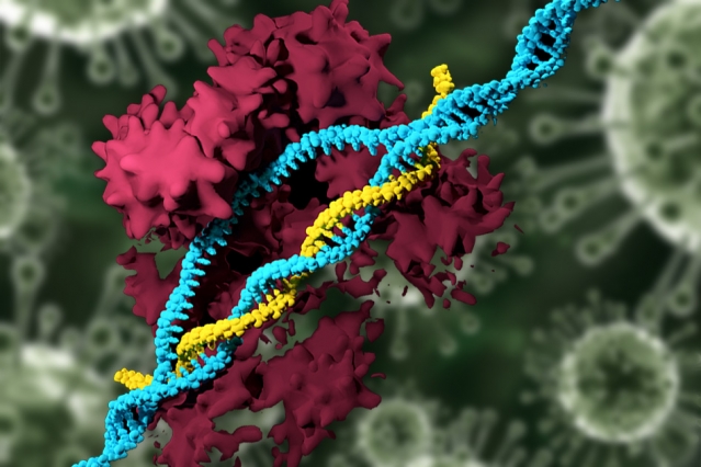
There’s been a minor flurry of interest in CRISPR (Clustered Regularly Interspaced Short Palindromic Repeats; also known as CRISPR-CAS9), a gene-editing technique, since a team in Oregon announced a paper describing their work editing the germline. Since I’ve been following the CRISPR-CAS9 story for a while this seems like a good juncture for a more in-depth look at the topic. In this first part I’m including an introduction to CRISPR, some information about the latest US work, and some previous writing about ethics issues raised when Chinese scientists first announced their work editing germlines in 2015 and during the patent dispute between the University of California at Berkeley and Harvard University’s Broad Institute.

Introduction to CRISPR

I’ve been searching for a good description of CRISPR and this helped to clear up some questions for me (Thank you to MIT Review),

For anyone who’s been reading about science for a while, this upbeat approach to explaining how a particular technology will solve all sorts of problems will seem quite familiar. It’s not the most hyperbolic piece I’ve seen but it barely mentions any problems associated with research (for some of the problems see: ‘The interest flurry’ later in part 2).

Oregon team

Steve Connor’s July 26, 2017 article for the MIT (Massachusetts Institute of Technology) Technology Review breaks the news (Note: Links have been removed),

The first known attempt at creating genetically modified human embryos in the United States has been carried out by a team of researchers in Portland, Oregon, MIT Technology Review has learned.

The effort, led by Shoukhrat Mitalipov of Oregon Health and Science University, involved changing the DNA of a large number of one-cell embryos with the gene-editing technique CRISPR, according to people familiar with the scientific results.

Until now, American scientists have watched with a combination of awe, envy, and some alarm as scientists elsewhere were first to explore the controversial practice. To date, three previous reports of editing human embryos were all published by scientists in China.

Now Mitalipov is believed to have broken new ground both in the number of embryos experimented upon and by demonstrating that it is possible to safely and efficiently correct defective genes that cause inherited diseases.

Although none of the embryos were allowed to develop for more than a few days—and there was never any intention of implanting them into a womb—the experiments are a milestone on what may prove to be an inevitable journey toward the birth of the first genetically modified humans.

In altering the DNA code of human embryos, the objective of scientists is to show that they can eradicate or correct genes that cause inherited disease, like the blood condition beta-thalassemia. The process is termed “germline engineering” because any genetically modified child would then pass the changes on to subsequent generations via their own germ cells—the egg and sperm.

Some critics say germline experiments could open the floodgates to a brave new world of “designer babies” engineered with genetic enhancements—a prospect bitterly opposed by a range of religious organizations, civil society groups, and biotech companies.

The U.S. intelligence community last year called CRISPR a potential “weapon of mass destruction.”

Here’s a link to a citation for the groundbreaking paper,

Correction of a pathogenic gene mutation in human embryos by Hong Ma, Nuria Marti-Gutierrez, Sang-Wook Park, Jun Wu, Yeonmi Lee, Keiichiro Suzuki, Amy Koski, Dongmei Ji, Tomonari Hayama, Riffat Ahmed, Hayley Darby, Crystal Van Dyken, Ying Li, Eunju Kang, A.-Reum Park, Daesik Kim, Sang-Tae Kim, Jianhui Gong, Ying Gu, Xun Xu, David Battaglia, Sacha A. Krieg, David M. Lee, Diana H. Wu, Don P. Wolf, Stephen B. Heitner, Juan Carlos Izpisua Belmonte, Paula Amato, Jin-Soo Kim, Sanjiv Kaul, & Shoukhrat Mitalipov. Nature (2017) doi:10.1038/nature23305 Published online 02 August 2017

This paper appears to be open access.

CRISPR Issues: ethics and patents

In my May 14, 2015 posting I mentioned a ‘moratorium’ on germline research, the Chinese research paper, and the stance taken by the US National Institutes of Health (NIH),

The CRISPR technology has reignited a discussion about ethical and moral issues of human genetic engineering some of which is reviewed in an April 7, 2015 posting about a moratorium by Sheila Jasanoff, J. Benjamin Hurlbut and Krishanu Saha for the Guardian science blogs (Note: A link has been removed),

On April 3, 2015, a group of prominent biologists and ethicists writing in Science called for a moratorium on germline gene engineering; modifications to the human genome that will be passed on to future generations. The moratorium would apply to a technology called CRISPR/Cas9, which enables the removal of undesirable genes, insertion of desirable ones, and the broad recoding of nearly any DNA sequence.

Such modifications could affect every cell in an adult human being, including germ cells, and therefore be passed down through the generations. Many organisms across the range of biological complexity have already been edited in this way to generate designer bacteria, plants and primates. There is little reason to believe the same could not be done with human eggs, sperm and embryos. Now that the technology to engineer human germlines is here, the advocates for a moratorium declared, it is time to chart a prudent path forward. They recommend four actions: a hold on clinical applications; creation of expert forums; transparent research; and a globally representative group to recommend policy approaches.

The authors go on to review precedents and reasons for the moratorium while suggesting we need better ways for citizens to engage with and debate these issues,

An effective moratorium must be grounded in the principle that the power to modify the human genome demands serious engagement not only from scientists and ethicists but from all citizens. We need a more complex architecture for public deliberation, built on the recognition that we, as citizens, have a duty to participate in shaping our biotechnological futures, just as governments have a duty to empower us to participate in that process. Decisions such as whether or not to edit human genes should not be left to elite and invisible experts, whether in universities, ad hoc commissions, or parliamentary advisory committees. Nor should public deliberation be temporally limited by the span of a moratorium or narrowed to topics that experts deem reasonable to debate.

I recommend reading the post in its entirety as there are nuances that are best appreciated in the entirety of the piece.

Shortly after this essay was published, Chinese scientists announced they had genetically modified (nonviable) human embryos. From an April 22, 2015 article by David Cyranoski and Sara Reardon in Nature where the research and some of the ethical issues discussed,

In a world first, Chinese scientists have reported editing the genomes of human embryos. The results are published1 in the online journal Protein & Cell and confirm widespread rumours that such experiments had been conducted — rumours that sparked a high-profile debate last month2, 3 about the ethical implications of such work.

In the paper, researchers led by Junjiu Huang, a gene-function researcher at Sun Yat-sen University in Guangzhou, tried to head off such concerns by using ‘non-viable’ embryos, which cannot result in a live birth, that were obtained from local fertility clinics. The team attempted to modify the gene responsible for β-thalassaemia, a potentially fatal blood disorder, using a gene-editing technique known as CRISPR/Cas9. The researchers say that their results reveal serious obstacles to using the method in medical applications.

“I believe this is the first report of CRISPR/Cas9 applied to human pre-implantation embryos and as such the study is a landmark, as well as a cautionary tale,” says George Daley, a stem-cell biologist at Harvard Medical School in Boston, Massachusetts. “Their study should be a stern warning to any practitioner who thinks the technology is ready for testing to eradicate disease genes.”

….

Huang says that the paper was rejected by Nature and Science, in part because of ethical objections; both journals declined to comment on the claim. (Nature’s news team is editorially independent of its research editorial team.)

He adds that critics of the paper have noted that the low efficiencies and high number of off-target mutations could be specific to the abnormal embryos used in the study. Huang acknowledges the critique, but because there are no examples of gene editing in normal embryos he says that there is no way to know if the technique operates differently in them.

Still, he maintains that the embryos allow for a more meaningful model — and one closer to a normal human embryo — than an animal model or one using adult human cells. “We wanted to show our data to the world so people know what really happened with this model, rather than just talking about what would happen without data,” he says.

This, too, is a good and thoughtful read.

There was an official response in the US to the publication of this research, from an April 29, 2015 post by David Bruggeman on his Pasco Phronesis blog (Note: Links have been removed),

In light of Chinese researchers reporting their efforts to edit the genes of ‘non-viable’ human embryos, the National Institutes of Health (NIH) Director Francis Collins issued a statement (H/T Carl Zimmer).

“NIH will not fund any use of gene-editing technologies in human embryos. The concept of altering the human germline in embryos for clinical purposes has been debated over many years from many different perspectives, and has been viewed almost universally as a line that should not be crossed. Advances in technology have given us an elegant new way of carrying out genome editing, but the strong arguments against engaging in this activity remain. These include the serious and unquantifiable safety issues, ethical issues presented by altering the germline in a way that affects the next generation without their consent, and a current lack of compelling medical applications justifying the use of CRISPR/Cas9 in embryos.” …

The US has modified its stance according to a February 14, 2017 article by Jocelyn Kaiser for Science Magazine (Note: Links have been removed),

Editing the DNA of a human embryo to prevent a disease in a baby could be ethically allowable one day—but only in rare circumstances and with safeguards in place, says a widely anticipated report released today.

The report from an international committee convened by the U.S. National Academy of Sciences (NAS) and the National Academy of Medicine in Washington, D.C., concludes that such a clinical trial “might be permitted, but only following much more research” on risks and benefits, and “only for compelling reasons and under strict oversight.” Those situations could be limited to couples who both have a serious genetic disease and for whom embryo editing is “really the last reasonable option” if they want to have a healthy biological child, says committee co-chair Alta Charo, a bioethicist at the University of Wisconsin in Madison.

Some researchers are pleased with the report, saying it is consistent with previous conclusions that safely altering the DNA of human eggs, sperm, or early embryos—known as germline editing—to create a baby could be possible eventually. “They have closed the door to the vast majority of germline applications and left it open for a very small, well-defined subset. That’s not unreasonable in my opinion,” says genome researcher Eric Lander of the Broad Institute in Cambridge, Massachusetts. Lander was among the organizers of an international summit at NAS in December 2015 who called for more discussion before proceeding with embryo editing.

But others see the report as lowering the bar for such experiments because it does not explicitly say they should be prohibited for now. “It changes the tone to an affirmative position in the absence of the broad public debate this report calls for,” says Edward Lanphier, chairman of the DNA editing company Sangamo Therapeutics in Richmond, California. Two years ago, he co-authored a Nature commentary calling for a moratorium on clinical embryo editing.

One advocacy group opposed to embryo editing goes further. “We’re very disappointed with the report. It’s really a pretty dramatic shift from the existing and widespread agreement globally that human germline editing should be prohibited,” says Marcy Darnovsky, executive director of the Center for Genetics and Society in Berkeley, California.

Interestingly, this change of stance occurred just prior to a CRISPR patent decision (from my March 15, 2017 posting),

I have written about the CRISPR patent tussle (Harvard & MIT’s [Massachusetts Institute of Technology] Broad Institute vs the University of California at Berkeley) previously in a Jan. 6, 2015 posting and in a more detailed May 14, 2015 posting. I also mentioned (in a Jan. 17, 2017 posting) CRISPR and its patent issues in the context of a posting about a Slate.com series on Frankenstein and the novel’s applicability to our own time. This patent fight is being bitterly fought as fortunes are at stake.

It seems a decision has been made regarding the CRISPR patent claims. From a Feb. 17, 2017 article by Charmaine Distor for The Science Times,

After an intense court battle, the US Patent and Trademark Office (USPTO) released its ruling on February 15 [2017]. The rights for the CRISPR-Cas9 gene editing technology was handed over to the Broad Institute of Harvard University and the Massachusetts Institute of Technology (MIT).

According to an article in Nature, the said court battle was between the Broad Institute and the University of California. The two institutions are fighting over the intellectual property right for the CRISPR patent. The case between the two started when the patent was first awarded to the Broad Institute despite having the University of California apply first for the CRISPR patent.

Heidi Ledford’s Feb. 17, 2017 article for Nature provides more insight into the situation (Note: Links have been removed),

It [USPTO] ruled that the Broad Institute of Harvard and MIT in Cambridge could keep its patents on using CRISPR–Cas9 in eukaryotic cells. That was a blow to the University of California in Berkeley, which had filed its own patents and had hoped to have the Broad’s thrown out.

The fight goes back to 2012, when Jennifer Doudna at Berkeley, Emmanuelle Charpentier, then at the University of Vienna, and their colleagues outlined how CRISPR–Cas9 could be used to precisely cut isolated DNA1. In 2013, Feng Zhang at the Broad and his colleagues — and other teams — showed2 how it could be adapted to edit DNA in eukaryotic cells such as plants, livestock and humans.

Berkeley filed for a patent earlier, but the USPTO granted the Broad’s patents first — and this week upheld them. There are high stakes involved in the ruling. The holder of key patents could make millions of dollars from CRISPR–Cas9’s applications in industry: already, the technique has sped up genetic research, and scientists are using it to develop disease-resistant livestock and treatments for human diseases.

….

I also noted this eyebrow-lifting statistic,  “As for Ledford’s 3rd point, there are an estimated 763 patent families (groups of related patents) claiming CAS9 leading to the distinct possibility that the Broad Institute will be fighting many patent claims in the future.)

-30-

Part 2 covers three critical responses to the reporting and between them describe the technology in more detail and the possibility of ‘designer babies’.  CRISPR and editing the germline in the US (part 2 of 3): ‘designer babies’?

Part 3 is all about public discussion or, rather, the lack of and need for according to a couple of social scientists. Informally, there is some discussion via pop culture and Joelle Renstrom notes although she is focused on the larger issues touched on by the television series, Orphan Black and as I touch on in my final comments. CRISPR and editing the germline in the US (part 3 of 3): public discussions and pop culture

Titanium dioxide nanoparticles have subtle effects on oxidative stress genes?

There’s research from the Georgia Institute of Technology (Georgia Tech; US) suggesting that titanium dioxide nanoparticles may have long term side effects. From a May 10, 2016 news item on ScienceDaily,

A nanoparticle commonly used in food, cosmetics, sunscreen and other products can have subtle effects on the activity of genes expressing enzymes that address oxidative stress inside two types of cells. While the titanium dioxide (TiO2) nanoparticles are considered non-toxic because they don’t kill cells at low concentrations, these cellular effects could add to concerns about long-term exposure to the nanomaterial.

A May 9, 2016 Georgia Tech news release on Newswire (also on EurekAlert), which originated the news item, describes the research in more detail,

Researchers at the Georgia Institute of Technology used high-throughput screening techniques to study the effects of titanium dioxide nanoparticles on the expression of 84 genes related to cellular oxidative stress. Their work found that six genes, four of them from a single gene family, were affected by a 24-hour exposure to the nanoparticles.

The effect was seen in two different kinds of cells exposed to the nanoparticles: human HeLa* cancer cells commonly used in research, and a line of monkey kidney cells. Polystyrene nanoparticles similar in size and surface electrical charge to the titanium dioxide nanoparticles did not produce a similar effect on gene expression.

“This is important because every standard measure of cell health shows that cells are not affected by these titanium dioxide nanoparticles,” said Christine Payne, an associate professor in Georgia Tech’s School of Chemistry and Biochemistry. “Our results show that there is a more subtle change in oxidative stress that could be damaging to cells or lead to long-term changes. This suggests that other nanoparticles should be screened for similar low-level effects.”

The research was reported online May 6 in the Journal of Physical Chemistry C. The work was supported by the National Institutes of Health (NIH) through the HERCULES Center at Emory University, and by a Vasser Woolley Fellowship.

Titanium dioxide nanoparticles help make powdered donuts white, protect skin from the sun’s rays and reflect light in painted surfaces. In concentrations commonly used, they are considered non-toxic, though several other studies have raised concern about potential effects on gene expression that may not directly impact the short-term health of cells.

To determine whether the nanoparticles could affect genes involved in managing oxidative stress in cells, Payne and colleague Melissa Kemp – an associate professor in the Wallace H. Coulter Department of Biomedical Engineering at Georgia Tech and Emory University – designed a study to broadly evaluate the nanoparticle’s impact on the two cell lines.

Working with graduate students Sabiha Runa and Dipesh Khanal, they separately incubated HeLa cells and monkey kidney cells with titanium oxide at levels 100 times less than the minimum concentration known to initiate effects on cell health. After incubating the cells for 24 hours with the TiO2, the cells were lysed and their contents analyzed using both PCR and Western Blot techniques to study the expression of 84 genes associated with the cells’ ability to address oxidative processes.

Payne and Kemp were surprised to find changes in the expression of six genes, including four from the peroxiredoxin family of enzymes that helps cells degrade hydrogen peroxide, a byproduct of cellular oxidation processes. Too much hydrogen peroxide can create oxidative stress which can damage DNA and other molecules.

The effect measured was significant – changes of about 50 percent in enzyme expression compared to cells that had not been incubated with nanoparticles. The tests were conducted in triplicate and produced similar results each time.

“One thing that was really surprising was that this whole family of proteins was affected, though some were up-regulated and some were down-regulated,” Kemp said. “These were all related proteins, so the question is why they would respond differently to the presence of the nanoparticles.”

The researchers aren’t sure how the nanoparticles bind with the cells, but they suspect it may involve the protein corona that surrounds the particles. The corona is made up of serum proteins that normally serve as food for the cells, but adsorb to the nanoparticles in the culture medium. The corona proteins have a protective effect on the cells, but may also serve as a way for the nanoparticles to bind to cell receptors.

Titanium dioxide is well known for its photo-catalytic effects under ultraviolet light, but the researchers don’t think that’s in play here because their culturing was done in ambient light – or in the dark. The individual nanoparticles had diameters of about 21 nanometers, but in cell culture formed much larger aggregates.

In future work, Payne and Kemp hope to learn more about the interaction, including where the enzyme-producing proteins are located in the cells. For that, they may use HyPer-Tau, a reporter protein they developed to track the location of hydrogen peroxide within cells.

The research suggests a re-evaluation may be necessary for other nanoparticles that could create subtle effects even though they’ve been deemed safe.

“Earlier work had suggested that nanoparticles can lead to oxidative stress, but nobody had really looked at this level and at so many different proteins at the same time,” Payne said. “Our research looked at such low concentrations that it does raise questions about what else might be affected. We looked specifically at oxidative stress, but there may be other genes that are affected, too.”

Those subtle differences may matter when they’re added to other factors.

“Oxidative stress is implicated in all kinds of inflammatory and immune responses,” Kemp noted. “While the titanium dioxide alone may just be modulating the expression levels of this family of proteins, if that is happening at the same time you have other types of oxidative stress for different reasons, then you may have a cumulative effect.”

*HeLa cells are named for Henrietta Lacks who unknowingly donated her immortal cell line to medical research. You can find more about the  story on the Oprah Winfrey website, which features an excerpt from the Rebecca Skloot book “The Immortal Life of Henrietta Lacks.” By the way, on May 2, 2016 it was announced that Oprah Winfrey would star in a movie for HBO as Henrietta Lacks’ daughter in an adaptation of the Rebecca Skloot book. You can read more about the proposed production in a May 3, 2016 article by Benjamin Lee for the Guardian.

Getting back to titanium dioxide nanoparticles and their possible long term effects, here’s a link to and a citation for the Georgia Tech team’s paper,

TiO2 Nanoparticles Alter the Expression of Peroxiredoxin Antioxidant Genes by Sabiha Runa, Dipesh Khanal, Melissa L. Kemp‡, and Christine K. Payne. J. Phys. Chem. C, Article ASAP DOI: 10.1021/acs.jpcc.6b01939 Publication Date (Web): April 21, 2016

Copyright © 2016 American Chemical Society

This paper is behind a paywall.

University of Toronto (Canada) researchers and lab-grown heart and liver tissue (person-on-a-chip)

Usually called ‘human-on-a-chip’, a team at the University of Toronto have developed a two-organ ‘person on a chip’ according to a March 7, 2016 news item on phys.org (Note: Links have been removed),

Researchers at U of T [University of Toronto] Engineering have developed a new way of growing realistic human tissues outside the body. Their “person-on-a-chip” technology, called AngioChip, is a powerful platform for discovering and testing new drugs, and could eventually be used to repair or replace damaged organs.

Professor Milica Radisic (IBBME, ChemE), graduate student Boyang Zhang and the rest of the team are among those research groups around the world racing to find ways to grow human tissues in the lab, under conditions that mimic a real person’s body. They have developed unique methods for manufacturing small, intricate scaffolds for individual cells to grow on. These artificial environments produce cells and tissues that resemble the real thing more closely than those grown lying flat in a petri dish.

The team’s recent creations have included BiowireTM—an innovative method of growing heart cells around a silk suture—as well as a scaffold for heart cells that snaps together like sheets of Velcro. But AngioChip takes tissue engineering to a whole new level. “It’s a fully three-dimensional structure complete with internal blood vessels,” says Radisic. “It behaves just like vasculature, and around it there is a lattice for other cells to attach and grow.” …

A March 7, 2016 University of Toronto news release (also on EurekAlert), which originated the news item, provides more detail about the AngioChip,

Zhang built the scaffold out of POMaC, a polymer that is both biodegradable and biocompatible. The scaffold is built out of a series of thin layers, stamped with a pattern of channels that are each about 50 to 100 micrometres wide. The layers, which resemble the computer microchips, are then stacked into a 3D structure of synthetic blood vessels. As each layer is added, UV light is used to cross-link the polymer and bond it to the layer below.

When the structure is finished, it is bathed in a liquid containing living cells. The cells quickly attach to the inside and outside of the channels and begin growing just as they would in the human body.

“Previously, people could only do this using devices that squish the cells between sheets of silicone and glass,” says Radisic. “You needed several pumps and vacuum lines to run just one chip. Our system runs in a normal cell culture dish, and there are no pumps; we use pressure heads to perfuse media through the vasculature. The wells are open, so you can easily access the tissue.”

Using the platform, the team has built model versions of both heart and liver tissues that function like the real thing. “Our liver actually produced urea and metabolized drugs,” says Radisic. They can connect the blood vessels of the two artificial organs, thereby modelling not just the organs themselves, but the interactions between them. They’ve even injected white blood cells into the vessels and watched as they squeezed through gaps in the vessel wall to reach the tissue on the other side, just as they do in the human body.

The news release also mentions potential markets and the work that needs to be accomplished before AngioChip is available for purchase,

AngioChip has great potential in the field of pharmaceutical testing. Current drug-testing methods, such as animal testing and controlled clinical trials, are costly and fraught with ethical concerns. Testing on lab-grown human tissues would provide a realistic model at a fraction of the cost, but this area of research is still in its infancy. “In the last few years, it has become possible to order cultures of human cells for testing, but they’re grown on a plate, a two-dimensional environment,” says Radisic. “They don’t capture all the functional hallmarks of a real heart muscle, for example.”

A more realistic platform like AngioChip could enable drug companies to detect dangerous side effects and interactions between organ compartments long before their products reach the market, saving countless lives. It could also be used to understand and validate the effectiveness of current drugs and even to screen libraries of chemical compounds to discover new drugs. Through TARA Biosystems Inc., a spin-off company co-founded by Radisic, the team is already working on commercializing the technology.

In future, Radisic envisions her lab-grown tissues being implanted into the body to repair organs damaged by disease. Because the cells used to seed the platform can come from anyone, the new tissues could be genetically identical to the intended host, reducing the risk of organ rejection. Even in its current form, the team has shown that the AngioChip can be implanted into a living animal, its artificial blood vessels connected to a real circulatory system. The polymer scaffolding itself simply biodegrades after several months.

The team still has much work to do. Each AngioChip is currently made by hand; if the platform is to be used industrially, the team will need to develop high-throughput manufacturing methods to create many copies at once. Still, the potential is obvious. “It really is multifunctional, and solves many problems in the tissue engineering space,” says Radisic. “It’s truly next-generation.”

Here’s a link to and a citation for the paper,

Biodegradable scaffold with built-in vasculature for organ-on-a-chip engineering and direct surgical anastomosis by Boyang Zhang, Miles Montgomery, M. Dean Chamberlain, Shinichiro Ogawa, Anastasia Korolj, Aric Pahnke, Laura A. Wells, Stéphane Massé, Jihye Kim, Lewis Reis, Abdul Momen, Sara S. Nunes, Aaron R. Wheeler, Kumaraswamy Nanthakumar, Gordon Keller, Michael V. Sefton, & Milica Radisic. Nature Materials (2016) doi:10.1038/nmat4570 Published online 07 March 2016

This paper is behind a paywall.

The researchers have made two images illustrating their work available. There’s this still image,

These tiny polymer scaffolds contain channels that are about 100 micrometres wide, about the same diameter as a human hair. When seeded with cells, the channels act as artificial blood vessels. By mimicking tissues in the human heart and other organs, these scaffolds provide a new way to test drugs for potentially dangerous side effects. (Image: Tyler Irving/Boyang Zhang/Kevin Soobrian)

These tiny polymer scaffolds contain channels that are about 100 micrometres wide, about the same diameter as a human hair. When seeded with cells, the channels act as artificial blood vessels. By mimicking tissues in the human heart and other organs, these scaffolds provide a new way to test drugs for potentially dangerous side effects. (Image: Tyler Irving/Boyang Zhang/Kevin Soobrian)

Perhaps more intriguing is this one,

UofT_AngioChipMoving

When seeded with heart cells, the flexible polymer scaffold contracts with a regular rhythm, just like real heart tissue. (Image: Boyang Zhang)

I have mentioned ‘human-on-a-chip’ projects many times here and as the news release writer notes, there is an international race. My July 1, 2015 posting (cross-posted from the June 30, 2015 posting [Testing times: the future of animal alternatives] on the International Innovation blog [a CORDIS-listed project dissemination partner for FP7 and H2020 projects]) notes a couple of those projects,

Organ-on-a-chip projects use stem cells to create human tissues that replicate the functions of human organs. Discussions about human-on-a-chip activities – a phrase used to describe 10 interlinked organ chips – were a highlight of the 9th World Congress on Alternatives to Animal Testing held in Prague, Czech Republic, last year. One project highlighted at the event was a joint US National Institutes of Health (NIH), US Food and Drug Administration (FDA) and US Defense Advanced Research Projects Agency (DARPA) project led by Dan Tagle that claimed it would develop functioning human-on-a-chip by 2017. However, he and his team were surprisingly close-mouthed and provided few details making it difficult to assess how close they are to achieving their goal.

By contrast, Uwe Marx – Leader of the ‘Multi-Organ-Chip’ programme in the Institute of Biotechnology at the Technical University of Berlin and Scientific Founder of TissUse, a human-on-a-chip start-up company – claims to have sold two-organ chips. He also claims to have successfully developed a four-organ chip and that he is on his way to building a human-on-a-chip. Though these chips remain to be seen, if they are, they will integrate microfluidics, cultured cells and materials patterned at the nanoscale to mimic various organs, and will allow chemical testing in an environment that somewhat mirrors a human.

As for where the University of Toronto efforts fit into the race, I don’t know for sure. It’s the first time I’ve come across a reference to liver tissue producing urea but I believe there’s at least one other team in China which has achieved a three-dimensional, more lifelike aspect for liver tissue in my Jan. 29, 2016 posting ‘Constructing a liver’.