Tag Archives: Weiwei Zhong

Gently measuring electrical signals in small animals with nano-SPEARs

This work comes from Rice University (Texas, US) according to an April 17, 2017 news item on Nanowerk,

Microscopic probes developed at Rice University have simplified the process of measuring electrical activity in individual cells of small living animals. The technique allows a single animal like a worm to be tested again and again and could revolutionize data-gathering for disease characterization and drug interactions.

The Rice lab of electrical and computer engineer Jacob Robinson has invented “nanoscale suspended electrode arrays” — aka nano-SPEARs — to give researchers access to electrophysiological signals from the cells of small animals without injuring them. Nano-SPEARs replace glass pipette electrodes that must be aligned by hand each time they are used.”

An April 17, 2017 Rice University news release (also on EurekAltert), which originated the news item, details the work,

“One of the experimental bottlenecks in studying synaptic behavior and degenerative diseases that affect the synapse is performing electrical measurements at those synapses,” Robinson said. “We set out to study large groups of animals under lots of different conditions to screen drugs or test different genetic factors that relate to errors in signaling at those synapses.”

Robinson’s early work at Rice focused on high-quality, high-throughput electrical characterization of individual cells. The new platform adapts the concept to probe the surface cells of nematodes, worms that make up 80 percent of all animals on Earth.

Most of what is known about muscle activity and synaptic transmission in the worms comes from the few studies that successfully used manually aligned glass pipettes to measure electrical activity from individual cells, Robinson said. However, this patch clamp technique requires time-consuming and invasive surgery that could negatively affect the data that is gathered from small research animals.

The platform developed by Robinson’s team works something like a toll booth for traveling worms. As each animal passes through a narrow channel, it is temporarily immobilized and pressed against one or several nano-SPEARS that penetrate its body-wall muscle and record electrical activity from nearby cells. That animal is then released, the next is captured and measured, and so on. Robinson said the device proved much faster to use than traditional electrophysiological cell measurement techniques.

The nano-SPEARs are created using standard thin-film deposition procedures and electron-beam or photolithography and can be made from less than 200 nanometers to more than 5 microns thick, depending on the size of animal to be tested. Because the nano-SPEARs can be fabricated on either silicon or glass, the technique easily combines with fluorescence microscopy, Robinson said.

The animals suitable for probing with a nano-SPEAR can be as large as several millimeters, like hydra, cousins of the jellyfish and the subject of an upcoming study. But nematodes known as Caenorhabditis elegans were practical for several reasons: First, Robinson said, they’re small enough to be compatible with microfluidic devices and nanowire electrodes. Second, there were a lot of them down the hall at the lab of Rice colleague Weiwei Zhong, who studies nematodes as transparent, easily manipulated models for signaling pathways that are common to all animals.

“I used to shy away from measuring electrophysiology because the conventional method of patch clamping is so technically challenging,” said Zhong, an assistant professor of biochemistry and cell biology and co-author of the paper. “Only a few graduate students or postdocs can do it. With Jacob’s device, even an undergraduate student can measure electrophysiology.”

“This meshes nicely with the high-throughput phenotyping she does,” Robinson said. “She can now correlate locomotive phenotypes with activity at the muscle cells. We believe that will be useful to study degenerative diseases centered around neuromuscular junctions.”

In fact, the labs have begun doing so. “We are now using this setup to profile worms with neurodegenerative disease models such as Parkinson’s and screen for drugs that reduce the symptoms,” Zhong said. “This would not be possible using the conventional method.”

Initial tests on C. elegans models for amyotrophic lateral sclerosis and Parkinson’s disease revealed for the first time clear differences in electrophysiological responses between the two, the researchers reported.

Testing the efficacy of drugs will be helped by the new ability to study small animals for long periods. “What we can do, for the first time, is look at electrical activity over a long period of time and discover interesting patterns of behavior,” Robinson said.

Some worms were studied for up to an hour, and others were tested on multiple days, said lead author Daniel Gonzales, a Rice graduate student in Robinson’s lab who took charge of herding nematodes through the microfluidic devices.

“It was in some way easier than working with isolated cells because the worms are larger and fairly sturdy,” Gonzales said. “With cells, if there’s too much pressure, they die. If they hit a wall, they die. But worms are really sturdy, so it was just a matter of getting them up against the electrodes and keeping them there.”

The team constructed microfluidic arrays with multiple channels that allowed testing of many nematodes at once. In comparison with patch-clamping techniques that limit labs to studying about one animal per hour, Robinson said his team measured as many as 16 nematodes per hour.

“Because this is a silicon-based technology, making arrays and producing recording chambers in high numbers becomes a real possibility,” he said.
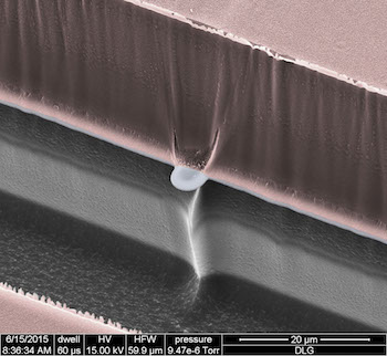
A scanning electron micrograph shows a nano-SPEAR suspended midway between layers of silicon (grey) and photoresist material (pink) that form a recording chamber for immobilized nematodes. The high-throughput technology developed at Rice University can be adapted for other small animals and could enhance data-gathering for disease characterization and drug interactions. Courtesy of the Robinson Lab

Here’s a link to and a citation for the paper,

Scalable electrophysiology in intact small animals with nanoscale suspended electrode arrays by Daniel L. Gonzales, Krishna N. Badhiwala, Daniel G. Vercosa, Benjamin W. Avants, Zheng Liu, Weiwei Zhong, & Jacob T. Robinson. Nature Nanotechnology (2017) doi:10.1038/nnano.2017.55 Published online 17 April 2017

This paper is behind a paywall.
Cute, adorable roundworms help measure nanoparticle toxicity

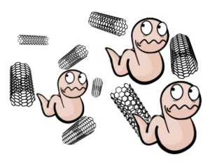
Caption: Low-cost experiments to test the toxicity of nanomaterials focused on populations of roundworms. Rice University scientists were able to test 20 nanomaterials in a short time, and see their method as a way to determine which nanomaterials should undergo more extensive testing. Credit: Zhong Lab/Rice University

Caption: Low-cost experiments to test the toxicity of nanomaterials focused on populations of roundworms. Rice University scientists were able to test 20 nanomaterials in a short time, and see their method as a way to determine which nanomaterials should undergo more extensive testing.
Credit: Zhong Lab/Rice University

Until now, ‘cute’ and ‘adorable’ are not words I would have associated with worms of any kind or with Rice University, for that matter. It’s amazing what a single image can do, eh?

A Feb. 3, 2015 news item on Azonano describes how roundworms have been used in research investigating the toxicity of various kinds of nanoparticles,

The lowly roundworm is the star of an ambitious Rice University project to measure the toxicity of nanoparticles.

The low-cost, high-throughput study by Rice scientists Weiwei Zhong and Qilin Li measures the effects of many types of nanoparticles not only on individual organisms but also on entire populations.

A Feb. 2, 2015 Rice University news release (also on EurekAlert), which originated the news item, provides more details about the research,

The Rice researchers tested 20 types of nanoparticles and determined that five, including the carbon-60 molecules (“buckyballs”) discovered at Rice in 1985, showed little to no toxicity.

Others were moderately or highly toxic to Caenorhabditis elegans, several generations of which the researchers observed to see the particles’ effects on their health.

The results were published by the American Chemical Society journal Environmental Sciences and Technology. They are also available on the researchers’ open-source website.

“Nanoparticles are basically new materials, and we don’t know much about what they will do to human health and the health of the ecosystem,” said Li, an associate professor of civil and environmental engineering and of materials science and nanoengineering. “There have been a lot of publications showing certain nanomaterials are more toxic than others. So before we make more products that incorporate these nanomaterials, it’s important that we understand we’re not putting anything toxic into the environment or into consumer products.

“The question is, How much cost can we bear?” she said. “It’s a long and expensive process to do a thorough toxicological study of any chemical, not just nanomaterials.” She said that due to the large variety of nanomaterials being produced at high speed and at such a large scale, there is “an urgent need for high-throughput screening techniques to prioritize which to study more extensively.”

Rice’s pilot study proves it is possible to gather a lot of toxicity data at low cost, said Zhong, an assistant professor of biosciences, who has performed extensive studies on C. elegans, particularly on their gene networks. Materials alone for each assay, including the worms and the bacteria they consumed and the culture media, cost about 50 cents, she said.

The researchers used four assays to see how worms react to nanoparticles: fitness, movement, growth and lifespan. The most sensitive assay of toxicity was fitness. In this test, the researchers mixed the nanoparticles in solutions with the bacteria that worms consume. Measuring how much bacteria they ate over time served as a measure of the worms’ “fitness.”

“If the worms’ health is affected by the nanoparticles, they reproduce less and eat less,” Zhong said. “In the fitness assay, we monitor the worms for a week. That is long enough for us to monitor toxicity effects accumulated through three generations of worms.” C. elegans has a life cycle of about three days, and since each can produce many offspring, a population that started at 50 would number more than 10,000 after a week. Such a large number of tested animals also enabled the fitness assay to be highly sensitive.

The researchers’ “QuantWorm” system allowed fast monitoring of worm fitness, movement, growth and lifespan. In fact, monitoring the worms was probably the least time-intensive part of the project. Each nanomaterial required specific preparation to make sure it was soluble and could be delivered to the worms along with the bacteria. The chemical properties of each nanomaterial also needed to be characterized in detail.

The researchers studied a representative sampling of three classes of nanoparticles: metal, metal oxides and carbon-based. “We did not do polymeric nanoparticles because the type of polymers you can possibly have is endless,” Li explained.

They examined the toxicity of each nanoparticle at four concentrations. Their results showed C-60 fullerenes, fullerol (a fullerene derivative), titanium dioxide, titanium dioxide-decorated nanotubes and cerium dioxide were the least damaging to worm populations.

Their “fitness” assay confirmed dose-dependent toxicity for carbon black, single- and multiwalled carbon nanotubes, graphene, graphene oxide, gold nanoparticles and fumed silicon dioxide.

They also determined the degree to which surface chemistry affected the toxicity of some particles. While amine-functionalized multiwalled nanotubes proved highly toxic, hydroxylated nanotubes had the least toxicity, with significant differences in fitness, body length and lifespan.

A complete and interactive toxicity chart for all of the tested materials is available online.

Zhong said the method could prove its worth as a rapid way for drug or other companies to narrow the range of nanoparticles they wish to put through more expensive, dedicated toxicology testing.

“Next, we hope to add environmental variables to the assays, for example, to mimic ultraviolet exposure or river water conditions in the solution to see how they affect toxicity,” she said. “We also want to study the biological mechanism by which some particles are toxic to worms.”

Here’s a citation for the paper and links to the paper and to the researchers’ website,

A multi-endpoint, high-throughput study of nanomaterial toxicity in Caenorhabditis elegans by Sang-Kyu Jung, Xiaolei Qu, Boanerges Aleman-Meza, Tianxiao Wang, Celeste Riepe, Zheng Liu, Qilin Li, and Weiwei Zhong. Environ. Sci. Technol., Just Accepted Manuscript DOI: 10.1021/es5056462 Publication Date (Web): January 22, 2015
Copyright © 2015 American Chemical Society

Nanomaterial effects on C. elegans

Home | Download | Tutorial | About

This heat map indicates whether a measurement for the nanomaterial-exposed worms is higher (yellow), or lower (blue) than the control worms. Black indicates no effects from nanomaterial exposure.

Clicking on colored blocks to see detailed experimental data.

The published paper is open access but you need an American Chemical Society site registration to access it. The researchers’ site is open access.