Tag Archives: wound-healing

Wound healing without sutures

Whoever wrote this Technion-Israel Institute of Technology November 28, 2021 press release (also on EurekAlert) they seem to have had a lot of fun doing it,

“Sutures? That’s practically medieval!”

It is a staple of science fiction to mock sutures as outdated. The technique has, after all, been in use for at least 5,000 years. Surely medicine should have advanced since ancient Egypt. Professor Hossam Haick from the Wolfson Department of Chemical Engineering at the Technion has finally turned science fiction into reality. His lab succeeded in creating a smart sutureless dressing that binds the wound together, wards off infection, and reports on the wound’s condition directly to the doctors’ computers. Their study was published in Advanced Materials.

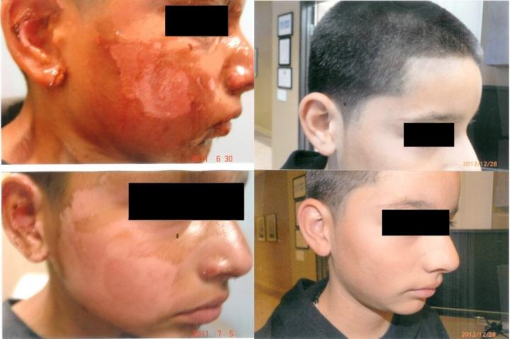
Current surgical procedures entail the surgeon cutting the human body, doing what needs to be done, and sewing the wound shut – an invasive procedure that damages surrounding healthy tissue. Some sutures degrade by themselves – or should degrade – as the wound heals. Others need to be manually removed. Dressing is then applied over the wound and medical personnel monitor the wound by removing the dressing to allow observation for signs of infection like swelling, redness, and heat. This procedure is painful to the patient, and disruptive to healing, but it is unavoidable. Working with these methods also mean that infection is often discovered late, since it takes time for visible signs to appear, and more time for the inspection to come round and see them. In developed countries, with good sanitation available, about 20% of patients develop infections post-surgery, necessitating additional treatment and extending the time to recovery. The figure and consequences are much worse in developing countries.

How will it work with Prof. Haick’s new dressing?

Prior to beginning a procedure, the dressing – which is very much like a smart band-aid – developed by Prof. Haick’s lab will be applied to the site of the planned incision. The incision will then be made through it. Following the surgery, the two ends of the wound will be brought together, and within three seconds the dressing will bind itself together, holding the wound closed, similarly to sutures. From then, the dressing will be continuously monitoring the wound, tracking the healing process, checking for signs of infection like changes in temperature, pH, and glucose levels, and report to the medical personnel’s smartphones or other devices. The dressing will also itself release antibiotics onto the wound area, preventing infection.

“I was watching a movie on futuristic robotics with my kids late one night,” said Prof. Haick, “and I thought, what if we could really make self-repairing sensors?”

Most people discard their late-night cinema-inspired ideas. Not Prof. Haick, who, the very next day after his Eureka moment, was researching and making plans. The first publication about a self-healing sensor came in 2015 (read more about it on the Technion website here). At that time, the sensor needed almost 24 hours to repair itself. By 2020, sensors were healing in under a minute (read about the study by Muhammad Khatib, a student in Prof. Haick’s lab here), but while it had multiple applications, it was not yet biocompatible, that is, not usable in contact with skin and blood. Creating a polymer that would be both biocompatible and self-healing was the next step, and one that was achieved by postdoctoral fellow Dr. Ning Tang.

The new polymer is structured like a molecular zipper, made from sulfur and nitrogen: the surgeon’s scalpel opens it; then pressed together, it closes and holds fast. Integrated carbon nanotubes provide electric conductivity and the integration of the sensor array. In experiments, wounds closed with the smart dressing healed as fast as those closed with sutures and showed reduced rates of infection.

“It’s a new approach to wound treatment,” said Prof. Haick. “We introduce the advances of the fourth industrial revolution – smart interconnected devices, into the day-to-day treatment of patients.”

Prof. Haick is the head of the Laboratory for Nanomaterial-based Devices (LNBD) and the Dean of Undergraduate Studies at the Technion. Dr. Ning Tang was a postdoctoral fellow in Prof. Haick’s laboratory and conducted this study as part of his fellowship. He has now been appointed an associate professor in Shanghai Jiao Tong University.

Here’s a link to and a citation for the paper,

Highly Efficient Self-Healing Multifunctional Dressing with Antibacterial Activity for Sutureless Wound Closure and Infected Wound Monitoring by Ning Tang, Rongjun Zhang, Youbin Zheng, Jing Wang, Muhammad Khatib, Xue Jiang, Cheng Zhou, Rawan Omar, Walaa Saliba, Weiwei Wu, Miaomiao Yuan, Daxiang Cui, Hossam Haick. DOI: https://doi.org/10.1002/adma.202106842 First published: 05 November 2021

This paper is behind a paywall.

I usually like to have three links to a news/press release and in my searches for a third source for this press release, I stumbled onto the technioncanada.org website. They seemed to have scooped everyone including Technion as they have a November 25, 2021posting of the press release.

Shape-conforming hydrogel and the body’s own healing mechanisms

A June 11, 2018 news item on ScienceDaily announces a development of interest to people with diabetes or those who treat them,

A simple scrape or sore might not cause alarm for most people. But for diabetic patients, an untreated scratch can turn into an open wound that could potentially lead to a limb amputation or even death.

A Northwestern University team has developed a new device, called a regenerative bandage, that quickly heals these painful, hard-to-treat sores without using drugs. During head-to-head tests, Northwestern’s bandage healed diabetic wounds 33 percent faster than one of the most popular bandages currently on the market.

A June 11, 2018 Northwestern University news release by Amanda Morris, which originated the news item, provides more detail,

“The novelty is that we identified a segment of a protein in skin that is important to wound healing, made the segment and incorporated it into an antioxidant molecule that self-aggregates at body temperature to create a scaffold that facilitates the body’s ability to regenerate tissue at the wound site,” said Northwestern’s Guillermo Ameer, who led the study. “With this newer approach, we’re not releasing drugs or outside factors to accelerate healing. And it works very well.”

Because the bandage leverages the body’s own healing power without releasing drugs or biologics, it faces fewer regulatory hurdles. This means patients could see it on the market much sooner.

The research was published today, June 11 [2018], in the Proceedings of the National Academy of Sciences. Although Ameer’s laboratory is specifically interested in diabetes applications, the bandage can be used to heal all types of open wounds.

An expert in biomaterials and regenerative engineering, Ameer is the Daniel Hale Williams Professor of Biomedical Engineering in the McCormick School of Engineering, professor of surgery in the Feinberg School of Medicine and director of Northwestern’s new Center for Advanced Regenerative Engineering (CARE).

The difference between a sore in a physically healthy person versus a diabetic patient? Diabetes can cause nerve damage that leads to numbness in the extremities. People with diabetes, therefore, might experience something as simple as a blister or small scratch that goes unnoticed and untreated because they cannot feel it to know it’s there. As high glucose levels also thicken capillary walls, blood circulation slows, making it more difficult for these wounds to heal. It’s a perfect storm for a small nick to become a limb-threatening — or life-threatening — wound.

The secret behind Ameer’s regenerative bandage is laminin, a protein found in most of the body’s tissues including the skin. Laminin sends signals to cells, encouraging them to differentiate, migrate and adhere to one another. Ameer’s team identified a segment of laminin — 12 amino acids in length — called A5G81 that is critical for the wound-healing process.

“This particular sequence caught our eye because it activates cellular receptors to get cells to adhere, migrate and proliferate,” Ameer said. “Then we cut up the sequence to find the minimum size that we needed for it to work.”

By using such a small fragment of laminin rather than the entire protein, it can be easily synthesized in the laboratory — making it more reproducible while keeping manufacturing costs low. Ameer’s team incorporated A5G81 into an antioxidant hydrogel bandage that it previously developed in the laboratory.

The bandage’s antioxidant nature counters inflammation. And the hydrogel is thermally responsive: It is a liquid when applied to the wound bed, then rapidly solidifies into a gel when exposed to body temperature. This phase change allows it to conform to the exact shape of the wound — a property that helped it out-perform other bandages on the market.

“Wounds have irregular shapes and depths. Our liquid can fill any shape and then stay in place,” Ameer said. “Other bandages are mostly based on collagen films or sponges that can move around and shift away from the wound site.”

Patients also must change bandages often, which can rip off the healing tissue and re-injure the site. Ameer’s bandage, however, can be rinsed off with cool saline, so the regenerating tissue remains undisturbed.

Not only will the lack of drugs or biologics make the bandage move to market faster, it also increases the bandage’s safety. So far, Ameer’s team has not noticed any adverse side effects in animal models. This is a stark difference from another product on the market, which contains a growth factor linked to cancer.

“It is not acceptable for patients who are trying to heal an open sore to have to deal with an increased risk of cancer,” Ameer said.

Next, Ameer’s team will continue to investigate the bandage in a larger pre-clinical model.

Here’s a link to and a citation for the paper,

Potent laminin-inspired antioxidant regenerative dressing accelerates wound healing in diabetes by Yunxiao Zhu, Zdravka Cankova, Marta Iwanaszko, Sheridan Lichtor, Milan Mrksich, and Guillermo A. Ameer. PNAS [Proceedings of the National Academy of Science] June 11, 2018. 201804262; published ahead of print June 11, 2018. https://doi.org/10.1073/pnas.1804262115

This paper is behind a paywall.

An artificial enzyme uses light to kill bacteria

An April 4, 2018 news item on ScienceDaily announces a light-based approach to killing bacteria,

Researchers from RMIT University [Australia] have developed a new artificial enzyme that uses light to kill bacteria.

The artificial enzymes could one day be used in the fight against infections, and to keep high-risk public spaces like hospitals free of bacteria like E. coli and Golden Staph.

E. coli can cause dysentery and gastroenteritis, while Golden Staph is the major cause of hospital-acquired secondary infections and chronic wound infections.

Made from tiny nanorods — 1000 times smaller than the thickness of the human hair — the “NanoZymes” use visible light to create highly reactive oxygen species that rapidly break down and kill bacteria.

Lead researcher, Professor Vipul Bansal who is an Australian Future Fellow and Director of RMIT’s Sir Ian Potter NanoBioSensing Facility, said the new NanoZymes offer a major cutting edge over nature’s ability to kill bacteria.

Dead bacteria made beautiful,

Caption: A 3-D rendering of dead bacteria after it has come into contact with the NanoZymes.
Credit: Dr. Chaitali Dekiwadia/ RMIT Microscopy and Microanalysis Facility

An April 5, 2018 RMIT University press release (also on EurekAlert but dated April 4, 2018), which originated the news item, expands on the theme,

“For a number of years we have been attempting to develop artificial enzymes that can fight bacteria, while also offering opportunities to control bacterial infections using external ‘triggers’ and ‘stimuli’,” Bansal said. “Now we have finally cracked it.

“Our NanoZymes are artificial enzymes that combine light with moisture to cause a biochemical reaction that produces OH radicals and breaks down bacteria. Nature’s antibacterial activity does not respond to external triggers such as light.

“We have shown that when shined upon with a flash of white light, the activity of our NanoZymes increases by over 20 times, forming holes in bacterial cells and killing them efficiently.

“This next generation of nanomaterials are likely to offer new opportunities in bacteria free surfaces and controlling spread of infections in public hospitals.”

The NanoZymes work in a solution that mimics the fluid in a wound. This solution could be sprayed onto surfaces.

The NanoZymes are also produced as powders to mix with paints, ceramics and other consumer products. This could mean bacteria-free walls and surfaces in hospitals.

Public toilets — places with high levels of bacteria, and in particular E. coli — are also a prime location for the NanoZymes, and the researchers believe their new technology may even have the potential to create self-cleaning toilet bowls.

While the NanoZymes currently use visible light from torches or similar light sources, in the future they could be activated by sunlight.

The researchers have shown that the NanoZymes work in a lab environment. The team is now evaluating the long-term performance of the NanoZymes in consumer products.

“The next step will be to validate the bacteria killing and wound healing ability of these NanoZymes outside of the lab,” Bansal said.

“This NanoZyme technology has huge potential, and we are seeking interest from appropriate industries for joint product development.”

Here’s a link to and a citation for the paper,

Visible-Light-Triggered Reactive-Oxygen-Species-Mediated Antibacterial Activity of Peroxidase-Mimic CuO Nanorods by Md. Nurul Karim, Mandeep Singh, Pabudi Weerathunge, Pengju Bian, Rongkun Zheng, Chaitali Dekiwadia, Taimur Ahmed, Sumeet Walia, Enrico Della Gaspera, Sanjay Singh, Rajesh Ramanathan, and Vipul Bansal. ACS Appl. Nano Mater., Article ASAP DOI: 10.1021/acsanm.8b00153 Publication Date (Web): March 6, 2018

Copyright © 2018 American Chemical Society

This paper is open access.

‘Origami organs’ for tissue engineering

This is a different approach to tissue engineering and its the consequence of a serendipitous accident.  From an Aug. 7, 2017 Northwestern University news release (also on EurekAlert),

Northwestern Medicine scientists and engineers have invented a range of bioactive “tissue papers” made of materials derived from organs that are thin and flexible enough to even fold into an origami bird. The new biomaterials can potentially be used to support natural hormone production in young cancer patients and aid wound healing.

The tissue papers are made from structural proteins excreted by cells that give organs their form and structure. The proteins are combined with a polymer to make the material pliable.

In the study, individual types of tissue papers were made from ovarian, uterine, kidney, liver, muscle or heart proteins obtained by processing pig and cow organs. Each tissue paper had specific cellular properties of the organ from which it was made.

The article describing the tissue paper and its function will be published Aug. 7 in the journal Advanced Functional Materials.

“This new class of biomaterials has potential for tissue engineering and regenerative medicine as well as drug discovery and therapeutics,” corresponding author Ramille Shah said. “It’s versatile and surgically friendly.”

Shah is an assistant professor of surgery at the Feinberg School of Medicine and an assistant professor of materials science and engineering at McCormick School of Engineering. She also is a member of the Simpson Querrey Institute for BioNanotechnology.

For wound healing, Shah thinks the tissue paper could provide support and the cell signaling needed to help regenerate tissue to prevent scarring and accelerate healing.

The tissue papers are made from natural organs or tissues. The cells are removed, leaving the natural structural proteins – known as the extracellular matrix – that then are dried into a powder and processed into the tissue papers. Each type of paper contains residual biochemicals and protein architecture from its original organ that can stimulate cells to behave in a certain way.

In the lab of reproductive scientist Teresa Woodruff, the tissue paper made from a bovine ovary was used to grow ovarian follicles when they were cultured in vitro. The follicles (eggs and hormone-producing cells) grown on the tissue paper produced hormones necessary for proper function and maturation.

“This could provide another option to restore normal hormone function to young cancer patients who often lose their hormone function as a result of chemotherapy and radiation,” Woodruff, a study coauthor, said.

A strip of the ovarian paper with the follicles could be implanted under the arm to restore hormone production for cancer patients or even women in menopause.

Woodruff is the director of the Oncofertility Consortium and the Thomas J. Watkins Memorial Professor of Obstetrics and Gynecology at Feinberg.

In addition, the tissue paper made from various organs separately supported the growth of adult human stem cells. Scientists placed human bone marrow stem cells on the tissue paper, and all the stem cells attached and multiplied over four weeks.

“That’s a good sign that the paper supports human stem cell growth,” said first author Adam Jakus, who developed the tissue papers. “It’s an indicator that once we start using tissue paper in animal models it will be biocompatible.”

The tissue papers feel and behave much like standard office paper when they are dry, Jakus said. Jakus simply stacks them in a refrigerator or a freezer. He even playfully folded them into an origami bird.

“Even when wet, the tissue papers maintain their mechanical properties and can be rolled, folded, cut and sutured to tissue,” he said.

Jakus was a Hartwell postdoctoral fellow in Shah’s lab for the study and is now chief technology officer and cofounder of the startup company Dimension Inx, LLC, which was also cofounded by Shah. The company will develop, produce and sell 3-D printable materials primarily for medical applications. The Intellectual Property is owned by Northwestern University and will be licensed to Dimension Inx.

An Accidental Spill Sparked Invention

An accidental spill of 3-D printing ink in Shah’s lab by Jakus sparked the invention of the tissue paper. Jakus was attempting to make a 3-D printable ovary ink similar to the other 3-D printable materials he previously developed to repair and regenerate bone, muscle and nerve tissue. When he went to wipe up the spill, the ovary ink had already formed a dry sheet.

“When I tried to pick it up, it felt strong,” Jakus said. “I knew right then I could make large amounts of bioactive materials from other organs. The light bulb went on in my head. I could do this with other organs.”

“It is really amazing that meat and animal by-products like a kidney, liver, heart and uterus can be transformed into paper-like biomaterials that can potentially regenerate and restore function to tissues and organs,” Jakus said. “I’ll never look at a steak or pork tenderloin the same way again.”

For those who like their news in a video,

As someone who once made baklava, that does not look like filo pastry, where an individual sheet is quite thin and rips easily. Enough said.

Here’s a link to and a citation for the paper,

“Tissue Papers” from Organ-Specific Decellularized Extracellular Matrices by Adam E. Jakus, Monica M. Laronda, Alexandra S. Rashedi, Christina M. Robinson, Chris Lee, Sumanas W. Jordan, Kyle E. Orwig, Teresa K. Woodruff, and Ramille N. Shah. Advnaced Functional Materials DOI: 10.1002/adfm.201700992 Version of Record online: 7 AUG 2017

© 2017 WILEY-VCH Verlag GmbH & Co. KGaA, Weinheim

This paper is behind a paywall.

Curcumin gel for burns and scalds

The curcumin debate continues (see my  Jan. 26, 2017 posting titled: Curcumin: a scientific literature review concludes health benefits may be overstated for more about that). In the meantime, scientists at the University of California at Los Angeles’ (UCLA) David Geffen School of Medicine found that curcumin gel might be effective as a treatment for burns. From a March 14, 2017 Pensoft Publishers news release on EurekAlert (Note: Links have been removed),

What is the effect of Topical Curcumin Gel for treating burns and scalds? In a recent research paper, published in the open access journal BioDiscovery, Dr. Madalene Heng, Clinical Professor of Dermatology at the David Geffen School of Medicine, stresses that use of topical curcumin gel for treating skin problems, like burns and scalds, is very different, and appears to work more effectively, when compared to taking curcumin tablets by mouth for other conditions.

“Curcumin gel appears to work much better when used on the skin because the gel preparation allows curcumin to penetrate the skin, inhibit phosphorylase kinase and reduce inflammation,” explains Dr Heng.

In this report, use of curcumin after burns and scalds were found to reduce the severity of the injury, lessen pain and inflammation, and improve healing with less than expected scarring, or even no scarring, of the affected skin. Dr. Heng reports her experience using curcumin gel on such injuries using three examples of patients treated after burns and scalds, and provides a detailed explanation why topical curcumin may work on such injuries.

Curcumin is an ingredient found in the common spice turmeric. Turmeric has been used as a spice for centuries in many Eastern countries and gives well known dishes, such as curry, their typical yellow-gold color. The spice has also been used for cosmetic and medical purposes for just as long in these countries.

In recent years, the medicinal value of curcumin has been the subject of intense scientific studies, with publication numbering in the thousands, looking into the possible beneficial effects of this natural product on many kinds of affliction in humans.

This study published reports that topical curcumin gel applied soon after mild to moderate burns and scalds appears to be remarkably effective in relieving symptoms and improved healing of the affected skin.

“When taken by mouth, curcumin is very poorly absorbed into the body, and may not work as well,” notes Dr. Heng. “Nonetheless, our tests have shown that when the substance is used in a topical gel, the effect is notable.”

The author of the study believes that the effectiveness of curcumin gel on the skin – or topical curcumin – is related to its potent anti-inflammatory activity. Based on studies that she has done both in the laboratory and in patients over 25 years, the key to curcumin’s effectiveness on burns and scalds is that it is a natural inhibitor of an enzyme called phosphorylase kinase.

This enzyme in humans has many important functions, including its involvement in wound healing. Wound healing is the vital process that enables healing of tissues after injury. The process goes through a sequence of acute and chronic inflammatory events, during which there is redness, swelling, pain and then healing, often with scarring in the case of burns and scalds of the skin. The sequence is started by the release of phosphorylase kinase about 5 mins after injury, which activates over 200 genes that are involved in wound healing.

Dr. Heng uses curcumin gel for burns, scalds and other skin conditions as complementary treatment, in addition to standard treatment usually recommended for such conditions.

Caption: These are results from 5 days upon application of curcumin gel to burns, and results after 6 weeks. Credit: Dr. Madalene Heng

Here’s a link to and a citation for the paper,

Phosphorylase Kinase Inhibition Therapy in Burns and Scalds by Madalene Heng. BioDiscovery 20: e11207 (24 Feb 2017) https://doi.org/10.3897/biodiscovery.20.e1120

This paper is in an open access journal.

Antibiotic synthetic spider silk

I have a couple of questions, what is ‘click’ chemistry and how does a chance meeting lead to a five-year, interdisciplinary research project on synthetic spider silk? From a Jan. 4, 2017 news item on ScienceDaily,

A chance meeting between a spider expert and a chemist has led to the development of antibiotic synthetic spider silk.

After five years’ work an interdisciplinary team of scientists at The University of Nottingham has developed a technique to produce chemically functionalised spider silk that can be tailored to applications used in drug delivery, regenerative medicine and wound healing.

The Nottingham research team has shown for the first time how ‘click-chemistry’ can be used to attach molecules, such as antibiotics or fluorescent dyes, to artificially produced spider silk synthesised by E.coli bacteria. The research, funded by the Biotechnology and Biological Sciences Research Council (BBSRC) is published today in the online journal Advanced Materials.

A Jan. 3, 2016 University of Nottingham press release (also on EurekAlert), which originated the news item, provides a few more details about ‘click’ chemistry (not enough for me) and more information about the research,

The chosen molecules can be ‘clicked’ into place in soluble silk protein before it has been turned into fibres, or after the fibres have been formed. This means that the process can be easily controlled and more than one type of molecule can be used to ‘decorate’ individual silk strands.

Nottingham breakthrough

In a laboratory in the Centre of Biomolecular Sciences, Professor Neil Thomas from the School of Chemistry in collaboration with Dr Sara Goodacre from the School of Life Sciences, has led a team of BBSRC DTP-funded PhD students starting with David Harvey who was then joined by Victor Tudorica, Leah Ashley and Tom Coekin. They have developed and diversified this new approach to functionalising ‘recombinant’ — artificial — spider silk with a wide range of small molecules.

They have shown that when these ‘silk’ fibres are ‘decorated’ with the antibiotic levofloxacin it is slowly released from the silk, retaining its anti-bacterial activity for at least five days.

Neil Thomas, a Professor of Medicinal and Biological Chemistry, said: “Our technique allows the rapid generation of biocompatible, mono or multi-functionalised silk structures for use in a wide range of applications. These will be particularly useful in the fields of tissue engineering and biomedicine.”

Remarkable qualities of spider silk

Spider silk is strong, biocompatible and biodegradable. It is a protein-based material that does not appear to cause a strong immune, allergic or inflammatory reaction. With the recent development of recombinant spider silk, the race has been on to find ways of harnessing its remarkable qualities.

The Nottingham research team has shown that their technique can be used to create a biodegradable mesh which can do two jobs at once. It can replace the extra cellular matrix that our own cells generate, to accelerate growth of the new tissue. It can also be used for the slow release of antibiotics.

Professor Thomas said: “There is the possibility of using the silk in advanced dressings for the treatment of slow-healing wounds such as diabetic ulcers. Using our technique infection could be prevented over weeks or months by the controlled release of antibiotics. At the same time tissue regeneration is accelerated by silk fibres functioning as a temporary scaffold before being biodegraded.”

The medicinal properties of spider silk recognised for centuries.

The medicinal properties of spider silk have been recognised for centuries but not clearly understood. The Greeks and Romans treated wounded soldiers with spider webs to stop bleeding. It is said that soldiers would use a combination of honey and vinegar to clean deep wounds and then cover the whole thing with balled-up spider webs.

There is even a mention in Shakespeare’s Midsummer Night’s Dream: “I shall desire you of more acquaintance, good master cobweb,” the character ‘Bottom’ said. “If I cut my finger, I shall make bold of you.”

The press release goes on to describe the genesis of the project and how this multidisciplinary team was formed in more detail,

The idea came together at a discipline bridging university ‘sandpit’ meeting five years ago. Dr Goodacre says her chance meeting at that event with Professor Thomas proved to be one of the most productive afternoons of her career.

Dr Goodacre, who heads up the SpiderLab in the School of Life Sciences, said: “I got up at that meeting and showed the audience a picture of some spider silk. I said ‘I want to understand how this silk works, and then make some.’

“At the end of the session Neil came up to me and said ‘I think my group could make that.’ He also suggested that there might be more interesting ‘tweaks’ one could make so that the silk could be ‘decorated’ with different, useful, compounds either permanently or which could be released over time due to a change in the acidity of the environment.”

The approach required the production of the silk proteins in a bacterium where an amino acid not normally found in proteins was included. This amino acid contained an azide group which is widely used in ‘click’ reactions that only occur at that position in the protein. It was an approach that no-one had used before with spider silk — but the big question was — would it work?

Dr Goodacre said: “It was the start of a fascinating adventure that saw a postdoc undertake a very preliminary study to construct the synthetic silks. He was a former SpiderLab PhD student who had previously worked with our tarantulas. Thanks to his ground work we showed we could produce the silk proteins in bacteria. We were then joined by David Harvey, a new PhD student, who not only made the silk fibres, incorporating the unusual amino acid, but also decorated it and demonstrated its antibiotic activity. He has since extended those first ideas far beyond what we had thought might be possible.”

David Harvey’s work is described in this paper but Professor Thomas and Dr Goodacre say this is just the start. There are other joint SpiderLab/Thomas lab students working on uses for this technology in the hope of developing it further.

David Harvey, the lead author on this their first paper, has just been awarded his PhD and is now a postdoctoral researcher on a BBSRC follow-on grant so is still at the heart of the research. His current work is focused on driving the functionalised spider silk technology towards commercial application in wound healing and tissue regeneration.

Here’s a link to and a citation for the paper,

Antibiotic Spider Silk: Site-Specific Functionalization of Recombinant Spider Silk Using “Click” Chemistry by David Harvey, Philip Bardelang, Sara L. Goodacre, Alan Cockayne, and Neil R. Thomas. Advanced Materials DOI: 10.1002/adma.201604245 Version of Record online: 28 DEC 2016

© 2016 WILEY-VCH Verlag GmbH & Co. KGaA, Weinheim

This paper is behind a paywall.

I imagine Mr. Cockayne’s name has led to much teasing over the years. People who have names with that kind of potential tend to either change them or double down and refuse to compromise.

Eggshell-based bioplastics

Adding eggshell nanoparticles to a bioplastic (shown above) increases the strength and flexibility of the material, potentially making it more attractive for use in the packaging industry. Credit: Vijaya Rangari/Tuskegee University

Adding eggshell nanoparticles to a bioplastic (shown above) increases the strength and flexibility of the material, potentially making it more attractive for use in the packaging industry. Credit: Vijaya Rangari/Tuskegee University

A March 15, 2016 news item on Nanowerk describes the research,

Eggshells are both marvels and afterthoughts. Placed on end, they are as strong as the arches supporting ancient Roman aqueducts. Yet they readily crack in the middle, and once that happens, we discard them without a second thought. But now scientists report that adding tiny shards of eggshell to bioplastic could create a first-of-its-kind biodegradable packaging material that bends but does not easily break.

The researchers present their work today [March 15, 2016] at the 251st National Meeting & Exposition of the American Chemical Society (ACS).

A March 15, 2016 ACS news release (also on EurekAlert), which originated the news item, describes the work further,

“We’re breaking eggshells down into their most minute components and then infusing them into a special blend of bioplastics that we have developed,” says Vijaya K. Rangari, Ph.D. “These nano-sized eggshell particles add strength to the material and make them far more flexible than other bioplastics on the market. We believe that these traits — along with its biodegradability in the soil — could make this eggshell bioplastic a very attractive alternative packaging material.”

Worldwide, manufacturers produce about 300 million tons of plastic annually. Almost 99 percent of it is made with crude oil and other fossil fuels. Once it is discarded, petroleum-based plastics can last for centuries without breaking down. If burned, these plastics release carbon dioxide into the atmosphere, which can contribute to global climate change.

As an alternative, some manufacturers are producing bioplastics — a form of plastic derived from cornstarch, sweet potatoes or other renewable plant-based sources — that readily decompose or biodegrade once they are in the ground. However, most of these materials lack the strength and flexibility needed to work well in the packaging industry. And that’s a problem since the vast majority of plastic is used to hold, wrap and encase products. So petroleum-based materials continue to dominate the market, particularly in grocery and other retail stores, where estimates suggest that up to a trillion plastic bags are distributed worldwide every year.

To find a solution, Rangari, graduate student Boniface Tiimob and colleagues at Tuskegee University experimented with various plastic polymers. Eventually, they latched onto a mixture of 70 percent polybutyrate adipate terephthalate (PBAT), a petroleum polymer, and 30 percent polylactic acid (PLA), a polymer derived from cornstarch. PBAT, unlike other oil-based plastic polymers, is designed to begin degrading as soon as three months after it is put into the soil.

This mixture had many of the traits that the researchers were looking for, but they wanted to further enhance the flexibility of the material. So they created nanoparticles made of eggshells. They chose eggshells, in part, because they are porous, lightweight and mainly composed of calcium carbonate, a natural compound that easily decays.

The shells were washed, ground up in polypropylene glycol and then exposed to ultrasonic waves that broke the shell fragments down into nanoparticles more than 350,000 times smaller than the diameter of a human hair. Then, in a laboratory study, they infused a small fraction of these particles, each shaped like a deck of cards, into the 70/30 mixture of PBAT and PLA. The researchers found that this addition made the mixture 700 percent more flexible than other bioplastic blends. They say this pliability could make it ideal for use in retail packaging, grocery bags and food containers — including egg cartons.

In addition to bioplastics, Rangari’s team is investigating using eggshell nanoparticles to enhance wound healing, bone regeneration and drug delivery.

Wound healing with cellulose acetate nanofibres

This work on cellulose acetate nanofibres and wound healing (tested on mice) comes from Egypt according to an Aug. 10, 2015 news item on ScienceDaily,

People with diabetes mellitus often suffer from impaired wound healing. Now, scientists in Egypt have developed antibacterial nanofibres of cellulose acetate loaded with silver that could be used in a new type of dressing to promote tissue repair.

An Aug. 10, 2015 Inderscience Publishers press release on the Alpha Galileo website, which originated the news item, provides more detail about the research,

Thanaa Ibrahim Shalaby and colleagues, Nivan Mahmoud Fekry, Amal Sobhy El Sodfy, Amel Gaber El Sheredy and Maisa El Sayed Sayed Ahmed Moustafa, at Alexandria University, prepared nanofibres from cellulose acetate, an inexpensive and easily fabricated, semisynthetic polymer used in everything from photographic film to coatings for eyeglasses and even cigarette filters. It can be spun into fibres and thus used to make an absorbent and safe wound dressing. Shalaby and co-workers used various analytical techniques including scanning electron microscope (SEM) and Fourier-transform infrared (FTIR) spectroscopy to characterise their fibres in which they incorporated silver nanoparticles.

Having characterised the material the team then successfully tested its antibacterial activity against various strains of bacteria that might infect an open wound. They next used the material as a dressing on skin wounds on mice with diabetes and determined how quickly the wound healed with and without the nano dressing. The dressing absorbs fluids exuded by the wound, but also protects the wound from infectious agents while being permeable to air and moisture, the team reports. The use of this dressing also promotes collagen production as the wound heals, which helps to recreate normal skin strength and texture something that is lacking in unassisted wound healing in diabetes mellitus.

Here’s a link to and a citation for the paper,

Preparation and characterisation of antibacterial silver-containing nanofibres for wound healing in diabetic mice by Thanaa Ibrahim Shalaby; Nivan Mahmoud Fekry; Amal Sobhy El Sodfy; Amel Gaber El Sheredy; Maisa El Sayed Sayed Ahmed Moustafa. International Journal of Nanoparticles (IJNP), Vol. 8, No. 1, p. 82 2015 DOI: 10.1504/IJNP.2015.070346

This paper is behind a paywall although there are some exceptions.

Kill bacterial biofilms and activate healing with cinnamon and peppermint

These compounds based on peppermint and cinnamon kill infection (bacterial biofilm) while helping the wound to heal according to a July 8, 2015 news item on ScienceDaily,

Infectious colonies of bacteria called biofilms that develop on chronic wounds and medical devices can cause serious health problems and are tough to treat. But now scientists have found a way to package antimicrobial compounds from peppermint and cinnamon in tiny capsules that can both kill biofilms and actively promote healing. The researchers say the new material could be used as a topical antibacterial treatment and disinfectant.

A July 8, 2015 American Chemical Society news release on EurekAlert, which originated the news item, provides more detail,

Many bacteria clump together in sticky plaques in a way that makes them difficult to eliminate with traditional antibiotics. Doctors sometimes recommend cutting out infected tissues. This approach is costly, however, and because it’s invasive, many patients opt out of treatment altogether. Essential oils and other natural compounds have emerged recently as alternative substances that can get rid of pathogenic bacteria, but researchers have had a hard time translating their antibacterial activity into treatments. Vincent M. Rotello and colleagues wanted to address this challenge.

The researchers packaged peppermint oil and cinnamaldehyde, the compound in cinnamon responsible for its flavor and aroma, into silica nanoparticles. The microcapsule treatment was effective against four different types of bacteria, including one antibiotic-resistant strain. It also promoted the growth of fibroblasts, a cell type that is important in wound healing.

Here’s a link to and a citation for the paper,

Nanoparticle-Stabilized Capsules for the Treatment of Bacterial Biofilms by Bradley Duncan, Xiaoning Li, Ryan F. Landis, Sung Tae Kim, Akash Gupta, Li-Sheng Wang, Rajesh Ramanathan, Rui Tang, Jeffrey A. Boerth, and Vincent M. Rotello. ACS Nano, Article ASAP
DOI: 10.1021/acsnano.5b01696 Publication Date (Web): June 17, 2015

Copyright © 2015 American Chemical Society

This paper is behind a paywall.

Wound healing is nature’s way of zipping up your skin

Scientists have been able to observe the healing process at the molecular scale—in fruit flies. From an April 21, 2015 news item on ScienceDaily,

Scientists from the Goethe University (GU) Frankfurt, the European Molecular Biology Laboratory (EMBL) Heidelberg and the University of Zurich explain skin fusion at a molecular level and pinpoint the specific molecules that do the job in their latest publication in the journal Nature Cell Biology.

An April 21, 2015 Goethe University Frankfurt press release on EurekAlert, which originated the news item, describes similarities between humans and fruit flies allowing scientists to infer the wound healing process for human skin,

In order to prevent death by bleeding or infection, every wound (skin opening) must close at some point. The events leading to skin closure had been unclear for many years. Mikhail Eltsov (GU) and colleagues used fruit fly embryos as a model system to understand this process. Similarly to humans, fruit fly embryos at some point in their development have a large opening in the skin on their back that must fuse. This process is called zipping, because two sides of the skin are fastened in a way that resembles a zipper that joins two sides of a jacket.

The scientists have used a top-of-the-range electron microscope to study exactly how this zipping of the skin works. “Our electron microscope allows us to distinguish the molecular components in the cell that act like small machines to fuse the skin. When we look at it from a distance, it appears as if skin cells simply fuse to each other, but if we zoom in, it becomes clear that membranes, molecular machines, and other cellular components are involved”, explains Eltsov.

“In order to visualize this orchestra of healing, a very high-resolution picture of the process is needed. For this purpose we have recorded an enormous amount of data that surpasses all previous studies of this kind”, says Mikhail Eltsov.

As a first step, as the scientists discovered, cells find their opposing partner by “sniffing” each other out. As a next step, they develop adherens junctions which act like a molecular Velcro. This way they become strongly attached to their opposing partner cell. The biggest revelation of this study was that small tubes in the cell, called microtubules, attach to this molecular Velcro and then deploy a self-catastrophe, which results in the skin being pulled towards the opening, as if one pulls a blanket over.

Damian Brunner who led the team at the University of Zurich has performed many genetic manipulations to identify the correct components. The scientists were astonished to find that microtubules involved in cell-division are the primary scaffold used for zipping, indicating a mechanism conserved during evolution.

“What was also amazing was the tremendous plasticity of the membranes in this process which managed to close the skin opening in a very short space of time. When five to ten cells have found their respective neighbors, the skin already appears normal”, says Achilleas Frangakis from the Goethe University Frankfurt, who led the study.

The scientists hope that their results will open new avenues into the understanding of epithelial plasticity and wound healing. They are also investigating the detailed structural organization of the adherens junctions, work for which they were awarded a starting grant from European Research Council (ERC).

Here’s a link to and a citation for the paper,

Quantitative analysis of cytoskeletal reorganization during epithelial tissue sealing by large-volume electron tomography by Mikhail Eltsov, Nadia Dubé, Zhou Yu, Laurynas Pasakarnis, Uta Haselmann-Weiss, Damian Brunner, & Achilleas S. Frangakis. Nature Cell Biology (2015) doi:10.1038/ncb3159 Published online 20 April 2015

This paper is behind a paywall but there is a free preview available via ReadCube Access.

The researchers have provided an image illustrating ‘wound zipping’.

Caption: This is a perspective view of the zipping area with 17 skin cells. Credit: GU

Caption: This is a perspective view of the zipping area with 17 skin cells.
Credit: GU