Tag Archives: Xin Liu

Transparent graphene electrode technology and complex brain imaging

Michael Berger has written a May 24, 2018 Nanowerk Spotlight article about some of the latest research on transparent graphene electrode technology and the brain (Note: A link has been removed),

In new work, scientists from the labs of Kuzum [Duygu Kuzum, an Assistant Professor of Electrical and Computer Engineering at the University of California, San Diego {UCSD}] and Anna Devor report a transparent graphene microelectrode neural implant that eliminates light-induced artifacts to enable crosstalk-free integration of 2-photon microscopy, optogenetic stimulation, and cortical recordings in the same in vivo experiment. The new class of transparent brain implant is based on monolayer graphene. It offers a practical pathway to investigate neuronal activity over multiple spatial scales extending from single neurons to large neuronal populations.

Conventional metal-based microelectrodes cannot be used for simultaneous measurements of multiple optical and electrical parameters, which are essential for comprehensive investigation of brain function across spatio-temporal scales. Since they are opaque, they block the field of view of the microscopes and generate optical shadows impeding imaging.

More importantly, they cause light induced artifacts in electrical recordings, which can significantly interfere with neural signals. Transparent graphene electrode technology presented in this paper addresses these problems and allow seamless and crosstalk-free integration of optical and electrical sensing and manipulation technologies.

In their work, the scientists demonstrate that by careful design of key steps in the fabrication process for transparent graphene electrodes, the light-induced artifact problem can be mitigated and virtually artifact-free local field potential (LFP) recordings can be achieved within operating light intensities.

“Optical transparency of graphene enables seamless integration of imaging, optogenetic stimulation and electrical recording of brain activity in the same experiment with animal models,” Kuzum explains. “Different from conventional implants based on metal electrodes, graphene-based electrodes do not generate any electrical artifacts upon interacting with light used for imaging or optogenetics. That enables crosstalk free integration of three modalities: imaging, stimulation and recording to investigate brain activity over multiple spatial scales extending from single neurons to large populations of neurons in the same experiment.”

The team’s new fabrication process avoids any crack formation in the transfer process, resulting in a 95-100% yield for the electrode arrays. This fabrication quality is important for expanding this technology to high-density large area transparent arrays to monitor brain-scale cortical activity in large animal models or humans.

“Our technology is also well-suited for neurovascular and neurometabolic studies, providing a ‘gold standard’ neuronal correlate for optical measurements of vascular, hemodynamic, and metabolic activity,” Kuzum points out. “It will find application in multiple areas, advancing our understanding of how microscopic neural activity at the cellular scale translates into macroscopic activity of large neuron populations.”

“Combining optical techniques with electrical recordings using graphene electrodes will allow to connect the large body of neuroscience knowledge obtained from animal models to human studies mainly relying on electrophysiological recordings of brain-scale activity,” she adds.

Next steps for the team involve employing this technology to investigate coupling and information transfer between different brain regions.

This work is part of the US BRAIN (Brain Research through Advancing Innovative Neurotechnologies) initiative and there’s more than one team working with transparent graphene electrodes. John Hewitt in an Oct. 21, 2014 posting on ExtremeTech describes two other teams’ work (Note: Links have been removed),

The solution [to the problems with metal electrodes], now emerging from multiple labs throughout the universe is to build flexible, transparent electrode arrays from graphene. Two studies in the latest issue of Nature Communications, one from the University of Wisconsin-Madison and the other from Penn [University of Pennsylvania], describe how to build these devices.

The University of Wisconsin researchers are either a little bit smarter or just a little bit richer, because they published their work open access. It’s a no-brainer then that we will focus on their methods first, and also in more detail. To make the arrays, these guys first deposited the parylene (polymer) substrate on a silicon wafer, metalized it with gold, and then patterned it with an electron beam to create small contact pads. The magic was to then apply four stacked single-atom-thick graphene layers using a wet transfer technique. These layers were then protected with a silicon dioxide layer, another parylene layer, and finally molded into brain signal recording goodness with reactive ion etching.

PennTransparentelectrodeThe researchers went with four graphene layers because that provided optimal mechanical integrity and conductivity while maintaining sufficient transparency. They tested the device in opto-enhanced mice whose neurons expressed proteins that react to blue light. When they hit the neurons with a laser fired in through the implant, the protein channels opened and fired the cell beneath. The masterstroke that remained was then to successfully record the electrical signals from this firing, sit back, and wait for the Nobel prize office to call.

The Penn State group [Note: Every reearcher mentioned in the paper Hewitt linked to is from the University of Pennsylvania] in the  used a similar 16-spot electrode array (pictured above right), and proceeded — we presume — in much the same fashion. Their angle was to perform high-resolution optical imaging, in particular calcium imaging, right out through the transparent electrode arrays which simultaneously recorded in high-temporal-resolution signals. They did this in slices of the hippocampus where they could bring to bear the complex and multifarious hardware needed to perform confocal and two-photon microscopy. These latter techniques provide a boost in spatial resolution by zeroing in over narrow planes inside the specimen, and limiting the background by the requirement of two photons to generate an optical signal. We should mention that there are voltage sensitive dyes available, in addition to standard calcium dyes, which can almost record the fastest single spikes, but electrical recording still reigns supreme for speed.

What a mouse looks like with an optogenetics system plugged in

What a mouse looks like with an optogenetics system plugged in

One concern of both groups in making these kinds of simultaneous electro-optic measurements was the generation of light-induced artifacts in the electrical recordings. This potential complication, called the Becqueral photovoltaic effect, has been known to exist since it was first demonstrated back in 1839. When light hits a conventional metal electrode, a photoelectrochemical (or more simply, a photovoltaic) effect occurs. If present in these recordings, the different signals could be highly disambiguatable. The Penn researchers reported that they saw no significant artifact, while the Wisconsin researchers saw some small effects with their device. In particular, when compared with platinum electrodes put into the opposite side cortical hemisphere, the Wisconsin researchers found that the artifact from graphene was similar to that obtained from platinum electrodes.

Here’s a link to and a citation for the latest research from UCSD,

Deep 2-photon imaging and artifact-free optogenetics through transparent graphene microelectrode arrays by Martin Thunemann, Yichen Lu, Xin Liu, Kıvılcım Kılıç, Michèle Desjardins, Matthieu Vandenberghe, Sanaz Sadegh, Payam A. Saisan, Qun Cheng, Kimberly L. Weldy, Hongming Lyu, Srdjan Djurovic, Ole A. Andreassen, Anders M. Dale, Anna Devor, & Duygu Kuzum. Nature Communicationsvolume 9, Article number: 2035 (2018) doi:10.1038/s41467-018-04457-5 Published: 23 May 2018

This paper is open access.

You can find out more about the US BRAIN initiative here and if you’re curious, you can find out more about the project at UCSD here. Duygu Kuzum (now at UCSD) was at  the University of Pennsylvania in 2014 and participated in the work mentioned in Hewitt’s 2014 posting.

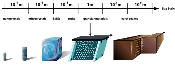
What do nanocrystals have in common with the earth’s crust?

The deformation properties of nanocrystals resemble those in the earth’s crust according to a Nov. 17, 2015 news item on Nanowerk,

Apparently, size doesn’t always matter. An extensive study by an interdisciplinary research group suggests that the deformation properties of nanocrystals are not much different from those of the Earth’s crust.

“When solid materials such as nanocrystals, bulk metallic glasses, rocks, or granular materials are slowly deformed by compression or shear, they slip intermittently with slip-avalanches similar to earthquakes,” explained Karin Dahmen, a professor of physics at the University of Illinois at Urbana-Champaign. “Typically these systems are studied separately. But we found that the scaling behavior of their slip statistics agree across a surprisingly wide range of different length scales and material structures.”

There’s an illustration accompanying the research,

Courtesy of the University of Illinois

Caption: When solid materials such as nanocrystals, bulk metallic glasses, rocks, or granular materials are slowly deformed by compression or shear, they slip intermittently with slip-avalanches similar to earthquakes. Credit: University of Illinois

A Nov. 17, 2015 University of Illinois news release (also on EurekAlert) by Rick Kubetz, which originated the news item, provides more detail,

“Identifying agreement in aspects of the slip statistics is important, because it enables us to transfer results from one scale to another, from one material to another, from one stress to another, or from one strain rate to another,” stated Shivesh Pathak, a physics undergraduate at Illinois, and a co-author of the paper, “Universal Quake Statistics: From Compressed Nanocrystals to Earthquakes,” appearing in Scientific Reports. “The study shows how to identify and explain commonalities in the deformation mechanisms of different materials on different scales.

“The results provide new tools and methods to use the slip statistics to predict future materials deformation,” added Michael LeBlanc, a physics graduate student and co-author of the paper. “They also clarify which system parameters significantly affect the deformation behavior on long length scales. We expect the results to be useful for applications in materials testing, failure prediction, and hazard prevention.”

Researchers representing a broad a range of disciplines–including physics, geosciences, mechanical engineering, chemical engineering, and materials science–from the United States, Germany, and the Netherlands contributed to the study, comparing five different experimental systems, on several different scales, with model predictions.

As a solid is sheared, each weak spot is stuck until the local shear stress exceeds a random failure threshold. It then slips by a random amount until it re-sticks. The released stress is redistributed to all other weak spots. Thus, a slipping weak spot can trigger other spots to fail in a slip avalanche.

Using tools from the theory of phase transitions, such as the renormalization group, one can show that the slip statistics of the model do not depend on the details of the system.

“Although these systems span 13 decades in length scale, they all show the same scaling behavior for their slip size distributions and other statistical properties,” stated Pathak. “Their size distributions follow the same simple (power law) function, multiplied with the same exponential cutoff.”

The cutoff, which is the largest slip or earthquake size, grows with applied force for materials spanning length scales from nanometers to kilometers. The dependence of the size of the largest slip or quake on stress reflects “tuned critical” behavior, rather than so-called self-organized criticality, which would imply stress-independence.

“The agreement of the scaling properties of the slip statistics across scales does not imply the predictability of individual slips or earthquakes,” LeBlanc said. “Rather, it implies that we can predict the scaling behavior of average properties of the slip statistics and the probability of slips of a certain size, including their dependence on stress and strain-rate.”

Here’s a link to and a citation for the paper,

Universal Quake Statistics: From Compressed Nanocrystals to Earthquakes by Jonathan T. Uhl, Shivesh Pathak, Danijel Schorlemmer, Xin Liu, Ryan Swindeman, Braden A. W. Brinkman, Michael LeBlanc, Georgios Tsekenis, Nir Friedman, Robert Behringer, Dmitry Denisov, Peter Schall, Xiaojun Gu, Wendelin J. Wright, Todd Hufnagel, Andrew Jennings, Julia R. Greer, P. K. Liaw, Thorsten Becker, Georg Dresen, & Karin A. Dahmen.  Scientific Reports 5, Article number: 16493 (2015)  doi:10.1038/srep16493 Published online: 17 November 2015

This is an open access paper.

One final comment, this story reminds me of a few other pieces of research featured here, which focus on repeating patterns in nature. The research was mentioned in an Aug. 27, 2015 posting about white dwarf stars and heartbeats and in an April 14, 2015 posting about gold nanoparticles and their resemblance to the Milky Way. You can also find more in the Wikipedia entry titled ‘Patterns in nature‘.

Fish skin for wound healing

A Feb. 11, 2015 news item on Nanowerk features Chinese research on tilapia fish skin and possible applications for wound healing (Note: A link has been removed),

With a low price tag and mild flavor, tilapia has become a staple dinnertime fish for many Americans. Now it could have another use: helping to heal our wounds. In the journal ACS Applied Materials & Interfaces (“Development of Biomimetic Tilapia Collagen Nanofibers for Skin Regeneration through Inducing Keratinocytes Differentiation and Collagen Synthesis of Dermal Fibroblasts”), scientists have shown that a protein found in this fish can promote skin repair in rats without an immune reaction, suggesting possible future use for human patients.

A Feb. 11, 2015 American Chemical Society (ACS) news release, which originated the news item, provides a few more details about the work,

Jiao Sun, Xiumei Mo and colleagues explain that applying collagen — a major structural protein in animals — to wounds can help encourage skin to heal faster.  But when the protein dressing comes from mammals such as cows and pigs, it has the potential to transmit conditions such as foot-and-mouth disease. Searching for an alternative source of collagen, scientists recently turned to the ocean. Sun’s team wanted to test fish collagen’s potential as a more benign wound treatment.

The researchers developed nanofibers from tilapia collagen and used them to cover skin wounds on rats. The rats with the nanofiber dressing healed faster than those without it. In addition, lab tests on cells suggested that the fish collagen was not likely to cause an immune reaction. The researchers conclude that it could be a good candidate to develop for clinical use.

Here’s a link to and a citation for the paper,

Development of Biomimetic Tilapia Collagen Nanofibers for Skin Regeneration through Inducing Keratinocytes Differentiation and Collagen Synthesis of Dermal Fibroblasts by Tian Zhou, Nanping Wang, Yang Xue, Tingting Ding, Xin Liu, Xiumei Mo, and Jiao Sun. ACS Appl. Mater. Interfaces, 2015, 7 (5), pp 3253–3262 DOI: 10.1021/am507990m Publication Date (Web): January 19, 2015

Copyright © 2015 American Chemical Society

This article is behind a paywall.

A ‘glass jaw’ might turn out to be a good thing

I don’t know if the phrase ‘glass jaw’ is used much any more but it was a term for someone who couldn’t ‘take’ a punch to the jaw (i.e., the person was instantly rendered unconscious or helplessly groggy). If scientists at Missouri University of Science and Technology (Missouri S&T)  have their way, the phrase ‘glass jaw’ will have a new meaning as per the July 26, 2012 news item on ScienceDaily,

Researchers at Missouri University of Science and Technology have developed a type of glass implant that could one day be used to repair injured bones in the arms, legs and other areas of the body that are most subject to the stresses of weight.

This marks the first time researchers have shown a glass implant strong enough to bear weight can also integrate with bone and promote bone growth, says lead researcher Dr. Mohamed N. Rahaman, professor of materials science and engineering at Missouri S&T.

The July 26, 2013 Missouri S&T news release by Andrew Careaga, which originated the news item, describes the work leading to this latest research,

In previous work, the Missouri S&T researchers developed a glass implant strong enough to handle the weight and pressure of repetitive movement, such as walking or lifting. In their most recent study, published in the journal Acta Biomaterialia, the research team reported that the glass implant, in the form of a porous scaffolding, also integrates with bone and promotes bone growth.

This combination of strength and bone growth opens new possibilities for bone repair, says Rahaman, who also directs Missouri S&T’s Center for Biomedical Science and Engineering, where the research was conducted.

The news release then goes on to describe one of the problems with using synthetic materials for bone repair and explains how this latest research addresses the issue,

Conventional approaches to structural bone repair involve either the use of a porous metal, which does not reliably heal bone, or a bone allograft from a cadaver. Both approaches are costly and carry risks, Rahaman says. He thinks the type of glass implant developed in his center could provide a more feasible approach for repairing injured bones. The glass is bioactive, which means that it reacts when implanted in living tissue and convert to a bone-like material.

In their latest research, Rahaman and his colleagues implanted bioactive glass scaffolds into sections of the calvarial bones (skullcaps) of laboratory rats, then examined how well the glass integrated with the surrounding bone and how quickly new bone grew into the scaffold. The scaffolds are manufactured in Rahaman’s lab through a process known as robocasting – a computer-controlled technique to manufacture materials from ceramic slurries, layer by layer – to ensure uniform structure for the porous material.

In previous studies by the Missouri S&T researchers, porous scaffolds of the silicate glass, known as 13-93, were found to have the same strength properties as cortical bone. Cortical bones are those outer bones of the body that bear the most weight and undergo the most repetitive stress. They include the long bones of the arms and legs.

But what Rahaman and his colleagues didn’t know was how well the silicate 13-93 bioactive glass scaffolds would integrate with bone or how quickly bone would grow into the scaffolding.

“You can have the strongest material in the world, but it also must encourage bone growth in a reasonable amount of time,” says Rahaman. He considers three to six months to be a reasonable time frame for completely regenerating an injured bone into one strong enough to bear weight.

In their studies, the S&T researchers found that the bioactive glass scaffolds bonded quickly to bone and promoted a significant amount of new bone growth within six weeks.

While the skullcap is not a load-bearing bone, it is primarily a cortical bone. The purpose of this research was to demonstrate how well this type of glass scaffolding – already shown to be strong – would interact with cortical bone.

Rahaman and his fellow researchers in the Center for Biomedical Science and Engineering are now experimenting with true load-bearing bones. They are now testing the silicate 13-93 implants in the femurs (leg bones) of laboratory rats.

In the future, Rahaman plans to experiment with modified glass scaffolds to see how well they enhance certain attributes within bone. For instance, doping the glass with copper should promote the growth of blood vessels or capillaries within the new bone, while doping the glass with silver will give it antibacterial properties.

It’s exciting work but they are years from human clinical trials. Still, for those who want to explore further, here’s a link to and a citation for the published paper,

Enhanced bone regeneration in rat calvarial defects implanted with surface-modified and BMP-loaded bioactive glass (13-93) scaffolds by Xin Liua, Mohamed N. Rahaman, Yongxing Liu, B. Sonny Bal, and Lynda F. Bonewald. Acta Biomaterialia, July 2013 issue (Volume 9, Issue 7)  http://dx.doi.org/10.1016/j.actbio.2013.03.039

This paper is behind a paywall.