Tag Archives: Yu Zhang

Smart dental implant resists bacterial growth and generates own electricity

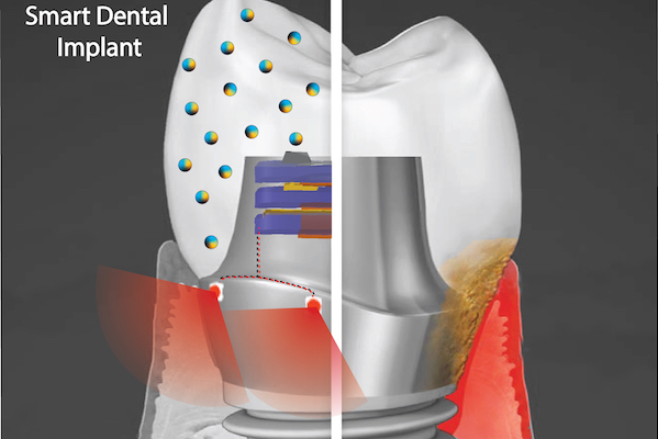
A “smart” dental implant could improve upon current devices by employing biofilm-resisting nanoparticles and a light powered by biomechanical forces to promote health of the surrounding gum tissue. (Image: Courtesy of Albert Kim)

A September 9, 2021 news item on ScienceDaily announces research into ‘smart’ dental implants,

More than 3 million people in America have dental implants, used to replace a tooth lost to decay, gum disease, or injury. Implants represent a leap of progress over dentures or bridges, fitting much more securely and designed to last 20 years or more.

But often implants fall short of that expectation, instead needing replacement in five to 10 years due to local inflammation or gum disease, necessitating a repeat of a costly and invasive procedure for patients.

“We wanted to address this issue, and so we came up with an innovative new implant,” says Geelsu Hwang, an assistant professor in the University of Pennsylvania School of Dental Medicine, who has a background in engineering that he brings to his research on oral health issues.

The novel implant would implement two key technologies, Hwang says. One is a nanoparticle-infused material that resists bacterial colonization. And the second is an embedded light source to conduct phototherapy, powered by the natural motions of the mouth, such as chewing or toothbrushing. In a paper in the journal ACS Applied Materials & Interfaces and a 2020 paper in the journal Advanced Healthcare Materials, Hwang and colleagues lay out their platform, which could one day be integrated not only into dental implants but other technologies, such as joint replacements, as well.

A September 9, 2021 University of Pennsylvania news release (also on EurekAlert), which originated the news item, provides more technical details about the proposed technology,

“Phototherapy can address a diverse set of health issues,” says Hwang. “But once a biomaterial is implanted, it’s not practical to replace or recharge a battery. We are using a piezoelectric material, which can generate electrical power from natural oral motions to supply a light that can conduct phototherapy, and we find that it can successfully protect gingival tissue from bacterial challenge.”

In the paper, the material the researchers explored was barium titanate (BTO), which has piezoelectric properties that are leveraged in applications such as capacitators and transistors, but has not yet been explored as a foundation for anti-infectious implantable biomaterials. To test its potential as the foundation for a dental implant, the team first used discs embedded with nanoparticles of BTO and exposed them to Streptococcus mutans, a primary component of the bacterial biofilm responsible for tooth decay commonly known as dental plaque. They found that the discs resisted biofilm formation in a dose-dependent manner. Discs with higher concentrations of BTO were better at preventing biofilms from binding.

While earlier studies had suggested that BTO might kill bacteria outright using reactive oxygen species generated by light-catalyzed or electric polarization reactions, Hwang and colleagues did not find this to be the case due to the short-lived efficacy and off-target effects of these approaches. Instead, the material generates enhanced negative surface charge that repels the negatively charged cell walls of bacteria. It’s likely that this repulsion effect would be long-lasting, the researchers say.

“We wanted an implant material that could resist bacterial growth for a long time because bacterial challenges are not a one-time threat,” Hwang says.

The power-generating property of the material was sustained and in tests over time the material did not leach. It also demonstrated a level of mechanical strength comparable to other materials used in dental applications.

Finally, the material did not harm normal gingival tissue in the researchers’ experiments, supporting the idea that this could be used without ill effect in the mouth.

The technology is a finalist in the Science Center’s research accelerator program, the QED Proof-of-Concept program. As one of 12 finalists, Hwang and colleagues will receive guidance from experts in commercialization. If the project advances to be one of three finalists, the group has the potential to receive up to $200,000 in funding.

In future work, the team hopes to continue to refine the “smart” dental implant system, testing new material types and perhaps even using assymetric properties on each side of the implant components, one that encourages tissue integration on the side facing the gums and one that resists bacterial formation on the side facing the rest of the mouth.

“We hope to further develop the implant system and eventually see it commercialized so it can be used in the dental field,” Hwang says.

Here’s a link to and a citation for the paper,

Bimodal Nanocomposite Platform with Antibiofilm and Self-Powering Functionalities for Biomedical Applications by Atul Dhall, Sayemul Islam, Moonchul Park, Yu Zhang, Albert Kim, and Geelsu Hwang. ACS Appl. Mater. Interfaces 2021, 13, 34, 40379–40391 DOI: https://doi.org/10.1021/acsami.1c11791 Publication Date:August 18, 2021 Copyright © 2021 American Chemical Society

This paper is behind a paywall.

The work from 2020, mentioned in the news release, laid groundwork for the latest paper.

Human Oral Motion-Powered Smart Dental Implant (SDI) for In Situ Ambulatory Photo-biomodulation Therapy by Moonchul Park, Sayemul Islam, Hye-Eun Kim, Jonathan Korosto, Markus B. Blatz, Geelsu Hwang, and Albert Kim. Adv. Healthcare Mater. 2020, 9, 2000658 DOI: 10.1002/adhm.202000658 First published: 01 July 2020 © 2020 WILEY-VCH Verlag GmbH & Co. KGaA, WeinheimHuman

This paper is behind a paywall.

Nanophotonics transforms Raman spectroscopy at Rice University (US)

This new technique for sensing molecules is intriguing. From a July 15, 2014 news item on Azonano,

Nanophotonics experts at Rice University [Texas, US] have created a unique sensor that amplifies the optical signature of molecules by about 100 billion times. Newly published tests found the device could accurately identify the composition and structure of individual molecules containing fewer than 20 atoms.

The new imaging method, which is described this week in the journal Nature Communications, uses a form of Raman spectroscopy in combination with an intricate but mass reproducible optical amplifier. Researchers at Rice’s Laboratory for Nanophotonics (LANP) said the single-molecule sensor is about 10 times more powerful that previously reported devices.

A July 15, 2014 Rice University news release (also on EurekAlert), which originated the news item, provides more detail about the research,

“Ours and other research groups have been designing single-molecule sensors for several years, but this new approach offers advantages over any previously reported method,” said LANP Director Naomi Halas, the lead scientist on the study. “The ideal single-molecule sensor would be able to identify an unknown molecule — even a very small one — without any prior information about that molecule’s structure or composition. That’s not possible with current technology, but this new technique has that potential.”

The optical sensor uses Raman spectroscopy, a technique pioneered in the 1930s that blossomed after the advent of lasers in the 1960s. When light strikes a molecule, most of its photons bounce off or pass directly through, but a tiny fraction — fewer than one in a trillion — are absorbed and re-emitted into another energy level that differs from their initial level. By measuring and analyzing these re-emitted photons through Raman spectroscopy, scientists can decipher the types of atoms in a molecule as well as their structural arrangement.

Scientists have created a number of techniques to boost Raman signals. In the new study, LANP graduate student Yu Zhang used one of these, a two-coherent-laser technique called “coherent anti-Stokes Raman spectroscopy,” or CARS. By using CARS in conjunction with a light amplifier made of four tiny gold nanodiscs, Halas and Zhang were able to measure single molecules in a powerful new way. LANP has dubbed the new technique “surface-enhanced CARS,” or SECARS.

“The two-coherent-laser setup in SECARS is important because the second laser provides further amplification,” Zhang said. “In a conventional single-laser setup, photons go through two steps of absorption and re-emission, and the optical signatures are usually amplified around 100 million to 10 billion times. By adding a second laser that is coherent with the first one, the SECARS technique employs a more complex multiphoton process.”

Zhang said the additional amplification gives SECARS the potential to address most unknown samples. That’s an added advantage over current techniques for single-molecule sensing, which generally require a prior knowledge about a molecule’s resonant frequency before it can be accurately measured.

Another key component of the SECARS process is the device’s optical amplifier, which contains four tiny gold discs in a precise diamond-shaped arrangement. The gap in the center of the four discs is about 15 nanometers wide. Owing to an optical effect called a “Fano resonance,” the optical signatures of molecules caught in that gap are dramatically amplified because of the efficient light harvesting and signal scattering properties of the four-disc structure.

Fano resonance requires a special geometric arrangement of the discs, and one of LANP’s specialties is the design, production and analysis of Fano-resonant plasmonic structures like the four-disc “quadrumer.” In previous LANP research, other geometric disc structures were used to create powerful optical processors.

Zhang said the quadrumer amplifiers are a key to SECARS, in part because they are created with standard e-beam lithographic techniques, which means they can be easily mass-produced.

“A 15-nanometer gap may sound small, but the gap in most competing devices is on the order of 1 nanometer,” Zhang said. “Our design is much more robust because even the smallest defect in a one-nanometer device can have significant effects. Moreover, the larger gap also results in a larger target area, the area where measurements take place. The target area in our device is hundreds of times larger than the target area in a one-nanometer device, and we can measure molecules anywhere in that target area, not just in the exact center.”

Halas, the Stanley C. Moore Professor in Electrical and Computer Engineering and a professor of biomedical engineering, chemistry, physics and astronomy at Rice, said the potential applications for SECARS include chemical and biological sensing as well as metamaterials research. She said scientific labs are likely be the first beneficiaries of the technology.

“Amplification is important for sensing small molecules because the smaller the molecule, the weaker the optical signature,” Halas said. “This amplification method is the most powerful yet demonstrated, and it could prove useful in experiments where existing techniques can’t provide reliable data.”

Here’s a link to and a citation for the paper,

Coherent anti-Stokes Raman scattering with single-molecule sensitivity using a plasmonic Fano resonance by Yu Zhang, Yu-Rong Zhen, Oara Neumann, Jared K. Day, Peter Nordlander & Naomi J. Halas. Nature Communications 5, Article number: 4424 doi:10.1038/ncomms5424 Published 14 July 2014

This paper is behind a paywall.