Tag Archives: Yuzi Liu

Alloy nanoparticles make better catalysts

A Jan. 4, 2021 news item on Nanowerk describes new insights into nanoscale catalysts derived from work at the US Argonne National Laboratory,

Catalysts are integral to countless aspects of modern society. By speeding up important chemical reactions, catalysts support industrial manufacturing and reduce harmful emissions. They also increase efficiency in chemical processes for applications ranging from batteries and transportation to beer and laundry detergent.

As significant as catalysts are, the way they work is often a mystery to scientists. Understanding catalytic processes can help scientists develop more efficient and cost-effective catalysts. In a recent study, scientists from University of Illinois Chicago (UIC) and the U.S. Department of Energy’s (DOE) Argonne National Laboratory discovered that, during a chemical reaction that often quickly degrades catalytic materials, a certain type of catalyst displays exceptionally high stability and durability.

The catalysts in this study are alloy nanoparticles, or nanosized particles made up of multiple metallic elements, such as cobalt, nickel, copper and platinum. These nanoparticles could have multiple practical applications, including water-splitting to generate hydrogen in fuel cells; reduction of carbon dioxide by capturing and converting it into useful materials like methanol; more efficient reactions in biosensors to detect substances in the body; and solar cells that produce heat, electricity and fuel more effectively.

A January 4, 2021 Argonne National Laboratory news release (also on EurekAlert) by Savannah Mitchem fills in some details,

In this study, the scientists investigated “high-entropy” (highly stable) alloy nanoparticles. The team of researchers, led by Reza Shahbazian-Yassar at UIC, used Argonne’s Center for Nanoscale Materials (CNM), a DOE Office of Science user facility, to characterize the particles’ compositions during oxidation, a process that degrades the material and reduces its usefulness in catalytic reactions.

“Using gas flow transmission electron microscopy (TEM) at CNM, we can capture the whole oxidation process in real time and at very high resolution,” said scientist Bob Song from UIC, a lead scientist on the study. “We found that the high-entropy alloy nanoparticles are able to resist oxidation much better than general metal particles.”

To perform the TEM, the scientists embedded the nanoparticles into a silicon nitride membrane and flowed different types of gas through a channel over the particles. A beam of electrons probed the reactions between the particles and the gas, revealing the low rate of oxidation and the migration of certain metals — iron, cobalt, nickel and copper — to the particles’ surfaces during the process.

“Our objective was to understand how fast high-entropy materials react with oxygen and how the chemistry of nanoparticles evolves during such a reaction,” said Shahbazian-Yassar, UIC professor of mechanical and industrial engineering at the College of Engineering.

According to Shahbazian-Yassar, the discoveries made in this research could benefit many energy storage and conversion technologies, such as fuel cells, lithium-air batteries, supercapacitors and catalyst materials. The nanoparticles could also be used to develop corrosion-resistant and high-temperature materials.

“This was a successful showcase of how CNM’s capabilities and services can meet the needs of our collaborators,” said Argonne’s Yuzi Liu, a scientist at CNM. “We have state-of-the-art facilities, and we want to deliver state-of-the-art science as well.”

Here’s a link to and a citation for the paper,

In Situ Oxidation Studies of High-Entropy Alloy Nanoparticles by Boao Song, Yong Yang, Muztoba Rabbani, Timothy T. Yang, Kun He, Xiaobing Hu, Yifei Yuan, Pankaj Ghildiyal, Vinayak P. Dravid, Michael R. Zachariah, Wissam A. Saidi, Yuzi Liu, and Reza Shahbazian-Yassar. ACS Nano 2020, 14, 11, 15131–15143 DOI: https://doi.org/10.1021/acsnano.0c05250 Publication Date:October 20, 2020 Copyright © 2020 American Chemical Society

This paper is behind a paywall.

Below is one of my favourite types of video work, a ‘blob video’ from the University of Illinois showing the alloy nanoparticles as they oxidate,
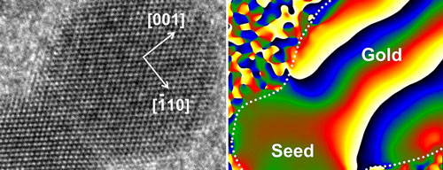
Video of transmission electron microscopy, performed at Argonne’s CNM, showing the oxidation of high-entropy nanoparticles in air at 400 °C, sped up by a factor of four. The oxidation process is depicted by the dissolution of the edges of the nanoparticles in the video. (Image by University of Illinois.)

Dumbbells at the nanoscale according to researchers at the (US) Argonne National Laboratory

Researchers at the US Dept. of Energy’s Argonne National Laboratory are providing new insight into how nanoparticles ‘grow’. From a Dec. 5, 2014 news item on Nanowerk,

Like snowflakes, nanoparticles come in a wide variety of shapes and sizes. The geometry of a nanoparticle is often as influential as its chemical makeup in determining how it behaves, from its catalytic properties to its potential as a semiconductor component.

Thanks to a new study from the U.S. Department of Energy’s (DOE) Argonne National Laboratory, researchers are closer to understanding the process by which nanoparticles made of more than one material – called heterostructured nanoparticles – form. This process, known as heterogeneous nucleation, is the same mechanism by which beads of condensation form on a windowpane.

The scientists have provided an image which illustrates their findings,

This picture combines a transmission electron microscope image of a nanodumbbell with a gold domain oriented in direction. The seed and gold domains in the dumbbell in the image on the right are identified by geometric phase analysis. Image credit: Soon Gu Kwon.

This picture combines a transmission electron microscope image of a nanodumbbell with a gold domain oriented in direction. The seed and gold domains in the dumbbell in the image on the right are identified by geometric phase analysis. Image credit: Soon Gu Kwon.

A Dec. 4, 2014 Argonne National Laboratory news release by Jared Sagoff, which originated the news item, describes the structures being examined and the reason for doing so,

Heterostructured nanoparticles can be used as catalysts and in advanced energy conversion and storage systems. Typically, these nanoparticles are created from tiny “seeds” of one material, on top of which another material is grown.  In this study, the Argonne researchers noticed that the differences in the atomic arrangements of the two materials have a big impact on the shape of the resulting nanoparticle.

“Before we started this experiment, it wasn’t entirely clear what’s happening at the interface when one material grows on another,” said nanoscientist Elena Shevchenko of Argonne Center for Nanoscale Materials, a DOE Office of Science user facility.

In this study, the researchers observed the formation of a nanoparticle consisting of platinum and gold.  The researchers started with a platinum seed and grew gold around it. Initially, the gold covered the platinum seed’s surface uniformly, creating a type of nanoparticle known as “core-shell.” However, as more gold was deposited, it started to grow unevenly, creating a dumbbell-like structure.

Thanks to state-of-the-art X-ray analysis provided by Argonne’s Advanced Photon Source (APS), a DOE Office of Science user facility, the researchers identified the cause of the dumbbell formation as “lattice mismatch,” in which the spacing between the atoms in the two materials doesn’t align.

“Essentially, you can think of lattice mismatch as having a row of smaller boxes on the bottom layer and larger boxes on the top layer.  When you try to fit the larger boxes into the space for a smaller box, it creates an immense strain,” said Argonne physicist Byeongdu Lee.

While the lattice mismatch is only fractions of a nanometer, the effect accumulates as layer after layer of gold forms on the platinum. The mismatch can be handled by the first two layers of gold atoms – creating the core-shell effect – but afterwards it proves too much to overcome. “The arrangement of atoms is the same in the two materials, but the distance between atoms is different,” said Argonne postdoctoral researcher Soon Gu Kwon. “Eventually, this becomes unstable, and the growth of the gold becomes unevenly distributed.”

As the gold continues to accumulate on one side of the seed nanoparticle, small quantities “slide” down the side of the nanoparticle like grains of sand rolling down the side of a sand hill, creating the dumbbell shape.

The advantage of the Argonne study comes from the researchers’ ability to perform in situ observations of the material in realistic conditions using the APS. “This is the first time anyone has been able to study the kinetics of this heterogeneous nucleation process of nanoparticles in real-time under realistic conditions,” said Argonne physicist Byeongdu Lee. “The combination of two X-ray techniques gave us the ability to observe the material at both the atomic level and the nanoscale, which gave us a good view of how the nanoparticles form and transform.” All conclusions made based on the X-ray studies were further confirmed using atomic-resolution microscopy in the group of Professor Robert Klie of the University of Illinois at Chicago.

This analysis of nanoparticle formation will help to lay the groundwork for the formation of new materials with different and controllable properties, according to Shevchenko. “In order to design materials, you have to understand how these processes happen at a very basic level,” she said.

Here’s a link to and a citation for the paper,

Heterogeneous nucleation and shape transformation of multicomponent metallic nanostructures by Soon Gu Kwon, Galyna Krylova, Patrick J. Phillips, Robert F. Klie, Soma Chattopadhyay, Tomohiro Shibata, Emilio E. Bunel, Yuzi Liu, Vitali B. Prakapenka, Byeongdu Lee, & Elena V. Shevchenko. Nature Materials (2014) doi:10.1038/nmat4115 Published online 02 November 2014

This paper is behind a paywall but there is a free preview via ReadCube Access.

Self-assembling chains of nanoparticles

The Argonne National Laboratory (US) has announced that their researchers have for the first time watched nanoparticles assemble into chains in real-time. From the Apr. 20, 2013 news item on Nanowerk (Note: Links have been removed),

In a new study performed at the Center for Nanoscale Materials at the U.S. Department of Energy’s (DOE) Argonne National Laboratory, researchers have for the first time seen the self-assembly of nanoparticle chains in situ, that is, in place as it occurs in real-time (“In Situ Visualization of Self-Assembly of Charged Gold Nanoparticles”).

The Apr. 19, 2013 Argonne National Laboratory press release by Jared Sagoff, which originated the news item, provides more detail,

The scientists exposed a tiny liquid “cell” or pouch that contained gold nanoparticles covered with a positively charged coating to an intense beam of electrons generated with a transmission electron microscope. Some of the electrons that penetrated the outside of the cell became trapped in the fluid medium in the cell. These “hydrated” electrons attracted the positively charged nanoparticles, which in time reduced the intensity of charge of the positive coating.

As the hydrated electrons reduced the coating’s positive charge, the nanoparticles no longer repelled each other as strongly.  Instead, their newfound relative attraction led the nanoparticles to “jump around” and eventually stick together in long chains. This self-assembly of nanoparticle chains had been detected before in different studies, but this technique allowed researchers, for the first time, to observe the phenomenon as it occurred.

“The moment-to-moment behavior of nanoparticles is something that’s not yet entirely understood by the scientific community,” said Argonne nanoscientist Yuzi Liu, the study’s lead author. “The potential of nanoparticles in all sorts of different applications and devices – from tiny machines to harvesters of new sources of energy – requires us to bring all of our resources to bear to look at how they function on the most basic physical levels.”

Self-assembly is particularly interesting to scientists because it could lead to new materials that could be used to develop new, energy-relevant technologies. “When we look at self-assembly, we’re looking to use nature as a springboard into man-made materials,” said Argonne nanoscientist Tijana Rajh, who directed the group that carried out the study.

Because the particles under study were so tiny – just a few dozen nanometers in diameter – an optical microscope would not have been able to resolve, or see, individual nanoparticles. By using the liquid cell in the transmission electron microscope at the Center for Nanoscale Materials, Liu and his colleagues could create short movies showing the quick movement of the nanoparticles as their coatings contacted the hydrated electrons.

Here’s a video of the self-assembling nanoparticles, provided by the Argonne National Laboratory,

Here’s a link to and citation for the paper,

In Situ Visualization of Self-Assembly of Charged Gold Nanoparticles by Yuzi Liu, Xiao-Min Lin, Yugang Sun, and Tijana Rajh. J. Am. Chem. Soc., [Journal of the American Chemical Socieyt] 2013, 135 (10), pp 3764–3767
DOI: 10.1021/ja312620e Publication Date (Web): February 22, 2013
Copyright © 2013 American Chemical Society


Gold nanoparticle self-assembly visualization at the Argonne National Laboratory (US)

There’s a Mar. 13, 2013 news item on phys.org which seems to have been written by someone who’s very technical,

The self-assembly of gold nanoparticles (Au NPs) coated with specific organic ions in water was observed by Center for Nanoscale Materials staff in the Nanobio Interfaces, Electronic & Magnetic Materials & Devices, and Nanophotonics groups at the Argonne National Laboratory using in situ transmission electron microscopy (TEM) equipped with a liquid cell. The Au NPs formed one-dimensional chains within a few minutes.

The originating March 2013 article is on an Argonne National Laboratory’s Center for Nanoscale Materials page,

The self-assembly of NPs attracts intense attention for its potential application in the fabrication of hybrid systems with collective properties from different types of materials. The observations provided here clearly elucidate the complex mechanism of charged NP self-assembly processes. They also paint a cautionary tale on using TEM in situ cells to imitate self-assembly processes in actual solution environments. [emphasis mine]

The hydrated electrons formed in radiolysis of water decrease the overall positive charge of cetyltrimethylammonium (CTA)-coated Au NPs. The NPs also were coated with negative citrate ions. (With citrate alone, however, the Au NPs remained steady in the liquid cell regardless of electron-beam intensity). The anisotropic attractive interactions, including dipolar and Van der Waals interactions, overcome the repulsion among the NPs and induce the assembly of NPs. The spatial segregation of different sizes of NPs as a result of electric field gradients within the cell was observed as well.

I’m not sure why the observations paint a cautionary tale. Perhaps a reader could enlighten me?

The researchers also provided an image,

Cetyltrimethylammonium-ion-coated gold nanoparticles before (top) and after (bottom) 500 seconds of electron-beam exposure inside a TEM liquid cell at 200 kV. Scale bar: 100 nm. [downloaded from http://nano.anl.gov/news/highlights/2013_gold_nanoparticles.html]

Cetyltrimethylammonium-ion-coated gold nanoparticles before (top) and after (bottom) 500 seconds of electron-beam exposure inside a TEM liquid cell at 200 kV. Scale bar: 100 nm. [downloaded from http://nano.anl.gov/news/highlights/2013_gold_nanoparticles.html]

For anyone who can understand the technical explanations, here’s a citation and a link to the research paper,

In Situ Visualization of Self-Assembly of Charged Gold Nanoparticles by Yuzi Liu, Xiao-Min Lin, Yugang Sun, and Tijana Rajh. J. Am. Chem. Soc., 2013, 135 (10), pp 3764–3767 DOI: 10.1021/ja312620e Publication Date (Web): February 22, 2013

Copyright © 2013 American Chemical Society

The paper is behind a paywall.