Tag Archives: Zhao Qin

A lobster’s stretch and strength in a hydrogel

An MIT team has fabricated a hydrogel-based material that mimics the structure of the lobster’s underbelly, the toughest known hydrogel found in nature. Credits: Courtesy of the researchers

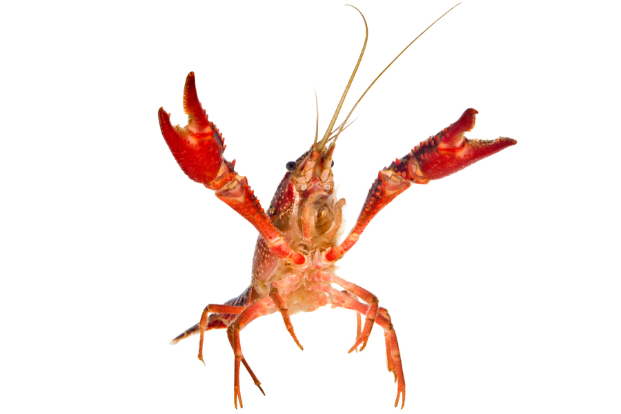
I love this lobster. In most photos, they’re food. This shows off the lobster as a living entity while showcasing its underbelly, which is what this story is all about. From an April 23, 2021 news item on phys.org (Note: A link has been removed),

A lobster’s underbelly is lined with a thin, translucent membrane that is both stretchy and surprisingly tough. This marine under-armor, as MIT [Massachusetts Institute of Technology] engineers reported in 2019, is made from the toughest known hydrogel in nature, which also happens to be highly flexible. This combination of strength and stretch helps shield a lobster as it scrabbles across the seafloor, while also allowing it to flex back and forth to swim.

Now a separate MIT team has fabricated a hydrogel-based material that mimics the structure of the lobster’s underbelly. The researchers ran the material through a battery of stretch and impact tests, and showed that, similar to the lobster underbelly, the synthetic material is remarkably “fatigue-resistant,” able to withstand repeated stretches and strains without tearing.

If the fabrication process could be significantly scaled up, materials made from nanofibrous hydrogels could be used to make stretchy and strong replacement tissues such as artificial tendons and ligaments.

The team’s results are published in the journal Matter. The paper’s MIT co-authors include postdocs Jiahua Ni and Shaoting Lin; graduate students Xinyue Liu and Yuchen Sun; professor of aeronautics and astronautics Raul Radovitzky; professor of chemistry Keith Nelson; mechanical engineering professor Xuanhe Zhao; and former research scientist David Veysset Ph.D. ’16, now at Stanford University; along with Zhao Qin, assistant professor at Syracuse University, and Alex Hsieh of the Army Research Laboratory.

An April 23, 2021 MIT news release (also on EurekAlert) by Jennifer Chu, which originated the news item, offers an overview of the groundwork for this latest research along with technical detail about the latest work,

Nature’s twist

In 2019, Lin and other members of Zhao’s group developed a new kind of fatigue-resistant material made from hydrogel — a gelatin-like class of materials made primarily of water and cross-linked polymers. They fabricated the material from ultrathin fibers of hydrogel, which aligned like many strands of gathered straw when the material was repeatedly stretched. This workout also happened to increase the hydrogel’s fatigue resistance.

“At that moment, we had a feeling nanofibers in hydrogels were important, and hoped to manipulate the fibril structures so that we could optimize fatigue resistance,” says Lin.

In their new study, the researchers combined a number of techniques to create stronger hydrogel nanofibers. The process starts with electrospinning, a fiber production technique that uses electric charges to draw ultrathin threads out of polymer solutions. The team used high-voltage charges to spin nanofibers from a polymer solution, to form a flat film of nanofibers, each measuring about 800 nanometers — a fraction of the diameter of a human hair.

They placed the film in a high-humidity chamber to weld the individual fibers into a sturdy, interconnected network, and then set the film in an incubator to crystallize the individual nanofibers at high temperatures, further strengthening the material.

They tested the film’s fatigue-resistance by placing it in a machine that stretched it repeatedly over tens of thousands of cycles. They also made notches in some films and observed how the cracks propagated as the films were stretched repeatedly. From these tests, they calculated that the nanofibrous films were 50 times more fatigue-resistant than the conventional nanofibrous hydrogels.

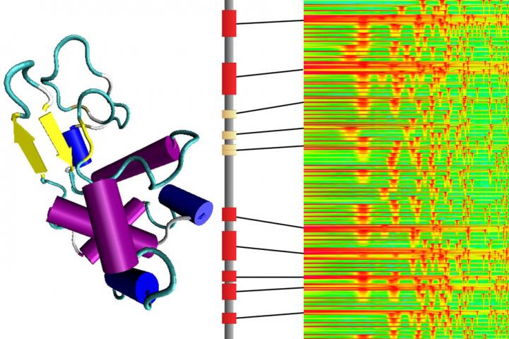
Around this time, they read with interest a study by Ming Guo, associate professor of mechanical engineering at MIT, who characterized the mechanical properties of a lobster’s underbelly. This protective membrane is made from thin sheets of chitin, a natural, fibrous material that is similar in makeup to the group’s hydrogel nanofibers.

Guo found that a cross-section of the lobster membrane revealed sheets of chitin stacked at 36-degree angles, similar to twisted plywood, or a spiral staircase. This rotating, layered configuration, known as a bouligand structure, enhanced the membrane’s properties of stretch and strength.

“We learned that this bouligand structure in the lobster underbelly has high mechanical performance, which motivated us to see if we could reproduce such structures in synthetic materials,” Lin says.

Angled architecture

Ni, Lin, and members of Zhao’s group teamed up with Nelson’s lab and Radovitzky’s group in MIT’s Institute for Soldier Nanotechnologies, and Qin’s lab at Syracuse University, to see if they could reproduce the lobster’s bouligand membrane structure using their synthetic, fatigue-resistant films.

“We prepared aligned nanofibers by electrospinning to mimic the chinic fibers existed in the lobster underbelly,” Ni says.

After electrospinning nanofibrous films, the researchers stacked each of five films in successive, 36-degree angles to form a single bouligand structure, which they then welded and crystallized to fortify the material. The final product measured 9 square centimeters and about 30 to 40 microns thick — about the size of a small piece of Scotch tape.

Stretch tests showed that the lobster-inspired material performed similarly to its natural counterpart, able to stretch repeatedly while resisting tears and cracks — a fatigue-resistance Lin attributes to the structure’s angled architecture.

“Intuitively, once a crack in the material propagates through one layer, it’s impeded by adjacent layers, where fibers are aligned at different angles,” Lin explains.

The team also subjected the material to microballistic impact tests with an experiment designed by Nelson’s group. They imaged the material as they shot it with microparticles at high velocity, and measured the particles’ speed before and after tearing through the material. The difference in velocity gave them a direct measurement of the material’s impact resistance, or the amount of energy it can absorb, which turned out to be a surprisingly tough 40 kilojoules per kilogram. This number is measured in the hydrated state.

“That means that a 5-millimeter steel ball launched at 200 meters per second would be arrested by 13 millimeters of the material,” Veysset says. “It is not as resistant as Kevlar, which would require 1 millimeter, but the material beats Kevlar in many other categories.”

It’s no surprise that the new material isn’t as tough as commercial antiballistic materials. It is, however, significantly sturdier than most other nanofibrous hydrogels such as gelatin and synthetic polymers like PVA. The material is also much stretchier than Kevlar. This combination of stretch and strength suggests that, if their fabrication can be sped up, and more films stacked in bouligand structures, nanofibrous hydrogels may serve as flexible and tough artificial tissues.

“For a hydrogel material to be a load-bearing artificial tissue, both strength and deformability are required,” Lin says. “Our material design could achieve these two properties.”

If you have the time and the interest, do check out the April 23, 2021 MIT news release, which features a couple of informative GIFs.

Here’s a link to and a citation for the paper,

Strong fatigue-resistant nanofibrous hydrogels inspired by lobster underbelly by Jiahua Ni, Shaoting Lin, Zhao Qin, David Veysset, Xinyue Liu, Yuchen Sun, Alex J. Hsieh, Raul Radovitzky, Keith A. Nelson, Xuanhe Zhao. Matter, 2021; DOI: 10.1016/j.matt.2021.03.023 Published April 23, 2021

This paper is behind a paywall.

Sonifying proteins to make music and brand new proteins

Markus Buehler at the Massachusetts Institute of Technology (MIT) has been working with music and science for a number of years. My December 9, 2011 posting, Music, math, and spiderwebs, was the first one here featuring his work. My November 28, 2012 posting, Producing stronger silk musically, was a followup to Buehler’s previous work.

A June 28, 2019 news item on Azonano provides a recent update,

Composers string notes of different pitch and duration together to create music. Similarly, cells join amino acids with different characteristics together to make proteins.

Now, researchers have bridged these two seemingly disparate processes by translating protein sequences into musical compositions and then using artificial intelligence to convert the sounds into brand-new proteins. …

Caption: Researchers at MIT have developed a system for converting the molecular structures of proteins, the basic building blocks of all living beings, into audible sound that resembles musical passages. Then, reversing the process, they can introduce some variations into the music and convert it back into new proteins never before seen in nature. Credit: Zhao Qin and Francisco Martin-Martinez

A June 26, 2019 American Chemical Society (ACS) news release, which originated the news item, provides more detail and a video,

To make proteins, cellular structures called ribosomes add one of 20 different amino acids to a growing chain in combinations specified by the genetic blueprint. The properties of the amino acids and the complex shapes into which the resulting proteins fold determine how the molecule will work in the body. To better understand a protein’s architecture, and possibly design new ones with desired features, Markus Buehler and colleagues wanted to find a way to translate a protein’s amino acid sequence into music.

The researchers transposed the unique natural vibrational frequencies of each amino acid into sound frequencies that humans can hear. In this way, they generated a scale consisting of 20 unique tones. Unlike musical notes, however, each amino acid tone consisted of the overlay of many different frequencies –– similar to a chord. Buehler and colleagues then translated several proteins into audio compositions, with the duration of each tone specified by the different 3D structures that make up the molecule. Finally, the researchers used artificial intelligence to recognize specific musical patterns that corresponded to certain protein architectures. The computer then generated scores and translated them into new-to-nature proteins. In addition to being a tool for protein design and for investigating disease mutations, the method could be helpful for explaining protein structure to broad audiences, the researchers say. They even developed an Android app [Amino Acid Synthesizer] to allow people to create their own bio-based musical compositions.

Here’s the ACS video,

A June 26, 2019 MIT news release (also on EurekAlert) provides some specifics and the MIT news release includes two embedded audio files,

Want to create a brand new type of protein that might have useful properties? No problem. Just hum a few bars.

In a surprising marriage of science and art, researchers at MIT have developed a system for converting the molecular structures of proteins, the basic building blocks of all living beings, into audible sound that resembles musical passages. Then, reversing the process, they can introduce some variations into the music and convert it back into new proteins never before seen in nature.

Although it’s not quite as simple as humming a new protein into existence, the new system comes close. It provides a systematic way of translating a protein’s sequence of amino acids into a musical sequence, using the physical properties of the molecules to determine the sounds. Although the sounds are transposed in order to bring them within the audible range for humans, the tones and their relationships are based on the actual vibrational frequencies of each amino acid molecule itself, computed using theories from quantum chemistry.

The system was developed by Markus Buehler, the McAfee Professor of Engineering and head of the Department of Civil and Environmental Engineering at MIT, along with postdoc Chi Hua Yu and two others. As described in the journal ACS Nano, the system translates the 20 types of amino acids, the building blocks that join together in chains to form all proteins, into a 20-tone scale. Any protein’s long sequence of amino acids then becomes a sequence of notes.

While such a scale sounds unfamiliar to people accustomed to Western musical traditions, listeners can readily recognize the relationships and differences after familiarizing themselves with the sounds. Buehler says that after listening to the resulting melodies, he is now able to distinguish certain amino acid sequences that correspond to proteins with specific structural functions. “That’s a beta sheet,” he might say, or “that’s an alpha helix.”

Learning the language of proteins

The whole concept, Buehler explains, is to get a better handle on understanding proteins and their vast array of variations. Proteins make up the structural material of skin, bone, and muscle, but are also enzymes, signaling chemicals, molecular switches, and a host of other functional materials that make up the machinery of all living things. But their structures, including the way they fold themselves into the shapes that often determine their functions, are exceedingly complicated. “They have their own language, and we don’t know how it works,” he says. “We don’t know what makes a silk protein a silk protein or what patterns reflect the functions found in an enzyme. We don’t know the code.”

By translating that language into a different form that humans are particularly well-attuned to, and that allows different aspects of the information to be encoded in different dimensions — pitch, volume, and duration — Buehler and his team hope to glean new insights into the relationships and differences between different families of proteins and their variations, and use this as a way of exploring the many possible tweaks and modifications of their structure and function. As with music, the structure of proteins is hierarchical, with different levels of structure at different scales of length or time.

The team then used an artificial intelligence system to study the catalog of melodies produced by a wide variety of different proteins. They had the AI system introduce slight changes in the musical sequence or create completely new sequences, and then translated the sounds back into proteins that correspond to the modified or newly designed versions. With this process they were able to create variations of existing proteins — for example of one found in spider silk, one of nature’s strongest materials — thus making new proteins unlike any produced by evolution.

Although the researchers themselves may not know the underlying rules, “the AI has learned the language of how proteins are designed,” and it can encode it to create variations of existing versions, or completely new protein designs, Buehler says. Given that there are “trillions and trillions” of potential combinations, he says, when it comes to creating new proteins “you wouldn’t be able to do it from scratch, but that’s what the AI can do.”

“Composing” new proteins

By using such a system, he says training the AI system with a set of data for a particular class of proteins might take a few days, but it can then produce a design for a new variant within microseconds. “No other method comes close,” he says. “The shortcoming is the model doesn’t tell us what’s really going on inside. We just know it works.

This way of encoding structure into music does reflect a deeper reality. “When you look at a molecule in a textbook, it’s static,” Buehler says. “But it’s not static at all. It’s moving and vibrating. Every bit of matter is a set of vibrations. And we can use this concept as a way of describing matter.”

The method does not yet allow for any kind of directed modifications — any changes in properties such as mechanical strength, elasticity, or chemical reactivity will be essentially random. “You still need to do the experiment,” he says. When a new protein variant is produced, “there’s no way to predict what it will do.”

The team also created musical compositions developed from the sounds of amino acids, which define this new 20-tone musical scale. The art pieces they constructed consist entirely of the sounds generated from amino acids. “There are no synthetic or natural instruments used, showing how this new source of sounds can be utilized as a creative platform,” Buehler says. Musical motifs derived from both naturally existing proteins and AI-generated proteins are used throughout the examples, and all the sounds, including some that resemble bass or snare drums, are also generated from the sounds of amino acids.

The researchers have created a free Android smartphone app, called Amino Acid Synthesizer, to play the sounds of amino acids and record protein sequences as musical compositions.

Here’s a link to and a citation for the paper,

A Self-Consistent Sonification Method to Translate Amino Acid Sequences into Musical Compositions and Application in Protein Design Using Artificial Intelligence by Chi-Hua Yu, Zhao Qin, Francisco J. Martin-Martinez, Markus J. Buehler. ACS Nano 2019 XXXXXXXXXX-XXX DOI: https://doi.org/10.1021/acsnano.9b02180 Publication Date:June 26, 2019 Copyright © 2019 American Chemical Society

This paper is behind a paywall.

ETA October 23, 2019 1000 hours: Ooops! I almost forgot the link to the Aminot Acid Synthesizer.

Worm-inspired gel material and soft robots

The Nereis virens worm inspired new research out of the MIT Laboratory for Atomistic and Molecular Mechanics. Its jaw is made of soft organic material, but is as strong as harder materials such as human dentin. Photo: Alexander Semenov/Wikimedia Commons

What an amazing worm! Here’s more about robots inspired by the Nereis virens worm in a March 20, 2017 news item on Nanowerk,

A new material that naturally adapts to changing environments was inspired by the strength, stability, and mechanical performance of the jaw of a marine worm. The protein material, which was designed and modeled by researchers from the Laboratory for Atomistic and Molecular Mechanics (LAMM) in the Department of Civil and Environmental Engineering (CEE) [at the Massachusetts Institute of Technology {MIT}], and synthesized in collaboration with the Air Force Research Lab (AFRL) at Wright-Patterson Air Force Base, Ohio, expands and contracts based on changing pH levels and ion concentrations. It was developed by studying how the jaw of Nereis virens, a sand worm, forms and adapts in different environments.

The resulting pH- and ion-sensitive material is able to respond and react to its environment. Understanding this naturally-occurring process can be particularly helpful for active control of the motion or deformation of actuators for soft robotics and sensors without using external power supply or complex electronic controlling devices. It could also be used to build autonomous structures.

A March 20, 2017 MIT news release, which originated the news item, provides more detail,

“The ability of dramatically altering the material properties, by changing its hierarchical structure starting at the chemical level, offers exciting new opportunities to tune the material, and to build upon the natural material design towards new engineering applications,” wrote Markus J. Buehler, the McAfee Professor of Engineering, head of CEE, and senior author of the paper.

The research, recently published in ACS Nano, shows that depending on the ions and pH levels in the environment, the protein material expands and contracts into different geometric patterns. When the conditions change again, the material reverts back to its original shape. This makes it particularly useful for smart composite materials with tunable mechanics and self-powered roboticists that use pH value and ion condition to change the material stiffness or generate functional deformations.

Finding inspiration in the strong, stable jaw of a marine worm

In order to create bio-inspired materials that can be used for soft robotics, sensors, and other uses — such as that inspired by the Nereis — engineers and scientists at LAMM and AFRL needed to first understand how these materials form in the Nereis worm, and how they ultimately behave in various environments. This understanding involved the development of a model that encompasses all different length scales from the atomic level, and is able to predict the material behavior. This model helps to fully understand the Nereis worm and its exceptional strength.

“Working with AFRL gave us the opportunity to pair our atomistic simulations with experiments,” said CEE research scientist Francisco Martin-Martinez. AFRL experimentally synthesized a hydrogel, a gel-like material made mostly of water, which is composed of recombinant Nvjp-1 protein responsible for the structural stability and impressive mechanical performance of the Nereis jaw. The hydrogel was used to test how the protein shrinks and changes behavior based on pH and ions in the environment.

The Nereis jaw is mostly made of organic matter, meaning it is a soft protein material with a consistency similar to gelatin. In spite of this, its strength, which has been reported to have a hardness ranging between 0.4 and 0.8 gigapascals (GPa), is similar to that of harder materials like human dentin. “It’s quite remarkable that this soft protein material, with a consistency akin to Jell-O, can be as strong as calcified minerals that are found in human dentin and harder materials such as bones,” Buehler said.

At MIT, the researchers looked at the makeup of the Nereis jaw on a molecular scale to see what makes the jaw so strong and adaptive. At this scale, the metal-coordinated crosslinks, the presence of metal in its molecular structure, provide a molecular network that makes the material stronger and at the same time make the molecular bond more dynamic, and ultimately able to respond to changing conditions. At the macroscopic scale, these dynamic metal-protein bonds result in an expansion/contraction behavior.

Combining the protein structural studies from AFRL with the molecular understanding from LAMM, Buehler, Martin-Martinez, CEE Research Scientist Zhao Qin, and former PhD student Chia-Ching Chou ’15, created a multiscale model that is able to predict the mechanical behavior of materials that contain this protein in various environments. “These atomistic simulations help us to visualize the atomic arrangements and molecular conformations that underlay the mechanical performance of these materials,” Martin-Martinez said.

Specifically, using this model the research team was able to design, test, and visualize how different molecular networks change and adapt to various pH levels, taking into account the biological and mechanical properties.

By looking at the molecular and biological makeup of a the Nereis virens and using the predictive model of the mechanical behavior of the resulting protein material, the LAMM researchers were able to more fully understand the protein material at different scales and provide a comprehensive understanding of how such protein materials form and behave in differing pH settings. This understanding guides new material designs for soft robots and sensors.

Identifying the link between environmental properties and movement in the material

The predictive model explained how the pH sensitive materials change shape and behavior, which the researchers used for designing new PH-changing geometric structures. Depending on the original geometric shape tested in the protein material and the properties surrounding it, the LAMM researchers found that the material either spirals or takes a Cypraea shell-like shape when the pH levels are changed. These are only some examples of the potential that this new material could have for developing soft robots, sensors, and autonomous structures.

Using the predictive model, the research team found that the material not only changes form, but it also reverts back to its original shape when the pH levels change. At the molecular level, histidine amino acids present in the protein bind strongly to the ions in the environment. This very local chemical reaction between amino acids and metal ions has an effect in the overall conformation of the protein at a larger scale. When environmental conditions change, the histidine-metal interactions change accordingly, which affect the protein conformation and in turn the material response.

“Changing the pH or changing the ions is like flipping a switch. You switch it on or off, depending on what environment you select, and the hydrogel expands or contracts” said Martin-Martinez.

LAMM found that at the molecular level, the structure of the protein material is strengthened when the environment contains zinc ions and certain pH levels. This creates more stable metal-coordinated crosslinks in the material’s molecular structure, which makes the molecules more dynamic and flexible.

This insight into the material’s design and its flexibility is extremely useful for environments with changing pH levels. Its response of changing its figure to changing acidity levels could be used for soft robotics. “Most soft robotics require power supply to drive the motion and to be controlled by complex electronic devices. Our work toward designing of multifunctional material may provide another pathway to directly control the material property and deformation without electronic devices,” said Qin.

By studying and modeling the molecular makeup and the behavior of the primary protein responsible for the mechanical properties ideal for Nereis jaw performance, the LAMM researchers are able to link environmental properties to movement in the material and have a more comprehensive understanding of the strength of the Nereis jaw.

Here’s link to and a citation for the paper,

Ion Effect and Metal-Coordinated Cross-Linking for Multiscale Design of Nereis Jaw Inspired Mechanomutable Materials by Chia-Ching Chou, Francisco J. Martin-Martinez, Zhao Qin, Patrick B. Dennis, Maneesh K. Gupta, Rajesh R. Naik, and Markus J. Buehler. ACS Nano, 2017, 11 (2), pp 1858–1868 DOI: 10.1021/acsnano.6b07878 Publication Date (Web): February 6, 2017

Copyright © 2017 American Chemical Society

This paper is behind a paywall.

Fusing graphene flakes for 3D graphene structures that are 10x as strong as steel

A Jan. 6, 2017 news item on Nanowerk describes how geometry may have as much or more to do with the strength of 3D graphene structures than the graphene used to create them,

A team of researchers at MIT [Massachusetts Institute of Technology] has designed one of the strongest lightweight materials known, by compressing and fusing flakes of graphene, a two-dimensional form of carbon. The new material, a sponge-like configuration with a density of just 5 percent, can have a strength 10 times that of steel.

In its two-dimensional form, graphene is thought to be the strongest of all known materials. But researchers until now have had a hard time translating that two-dimensional strength into useful three-dimensional materials.

The new findings show that the crucial aspect of the new 3-D forms has more to do with their unusual geometrical configuration than with the material itself, which suggests that similar strong, lightweight materials could be made from a variety of materials by creating similar geometric features.

The findings are being reported today [Jan. 6, 2017\ in the journal Science Advances, in a paper by Markus Buehler, the head of MIT’s Department of Civil and Environmental Engineering (CEE) and the McAfee Professor of Engineering; Zhao Qin, a CEE research scientist; Gang Seob Jung, a graduate student; and Min Jeong Kang MEng ’16, a recent graduate.

A Jan. 6, 2017 MIT news release (also on EurekAlert), which originated the news item, describes the research in more detail,

Other groups had suggested the possibility of such lightweight structures, but lab experiments so far had failed to match predictions, with some results exhibiting several orders of magnitude less strength than expected. The MIT team decided to solve the mystery by analyzing the material’s behavior down to the level of individual atoms within the structure. They were able to produce a mathematical framework that very closely matches experimental observations.

Two-dimensional materials — basically flat sheets that are just one atom in thickness but can be indefinitely large in the other dimensions — have exceptional strength as well as unique electrical properties. But because of their extraordinary thinness, “they are not very useful for making 3-D materials that could be used in vehicles, buildings, or devices,” Buehler says. “What we’ve done is to realize the wish of translating these 2-D materials into three-dimensional structures.”

The team was able to compress small flakes of graphene using a combination of heat and pressure. This process produced a strong, stable structure whose form resembles that of some corals and microscopic creatures called diatoms. These shapes, which have an enormous surface area in proportion to their volume, proved to be remarkably strong. “Once we created these 3-D structures, we wanted to see what’s the limit — what’s the strongest possible material we can produce,” says Qin. To do that, they created a variety of 3-D models and then subjected them to various tests. In computational simulations, which mimic the loading conditions in the tensile and compression tests performed in a tensile loading machine, “one of our samples has 5 percent the density of steel, but 10 times the strength,” Qin says.

Buehler says that what happens to their 3-D graphene material, which is composed of curved surfaces under deformation, resembles what would happen with sheets of paper. Paper has little strength along its length and width, and can be easily crumpled up. But when made into certain shapes, for example rolled into a tube, suddenly the strength along the length of the tube is much greater and can support substantial weight. Similarly, the geometric arrangement of the graphene flakes after treatment naturally forms a very strong configuration.

The new configurations have been made in the lab using a high-resolution, multimaterial 3-D printer. They were mechanically tested for their tensile and compressive properties, and their mechanical response under loading was simulated using the team’s theoretical models. The results from the experiments and simulations matched accurately.

The new, more accurate results, based on atomistic computational modeling by the MIT team, ruled out a possibility proposed previously by other teams: that it might be possible to make 3-D graphene structures so lightweight that they would actually be lighter than air, and could be used as a durable replacement for helium in balloons. The current work shows, however, that at such low densities, the material would not have sufficient strength and would collapse from the surrounding air pressure.

But many other possible applications of the material could eventually be feasible, the researchers say, for uses that require a combination of extreme strength and light weight. “You could either use the real graphene material or use the geometry we discovered with other materials, like polymers or metals,” Buehler says, to gain similar advantages of strength combined with advantages in cost, processing methods, or other material properties (such as transparency or electrical conductivity).

“You can replace the material itself with anything,” Buehler says. “The geometry is the dominant factor. It’s something that has the potential to transfer to many things.”

The unusual geometric shapes that graphene naturally forms under heat and pressure look something like a Nerf ball — round, but full of holes. These shapes, known as gyroids, are so complex that “actually making them using conventional manufacturing methods is probably impossible,” Buehler says. The team used 3-D-printed models of the structure, enlarged to thousands of times their natural size, for testing purposes.

For actual synthesis, the researchers say, one possibility is to use the polymer or metal particles as templates, coat them with graphene by chemical vapor deposit before heat and pressure treatments, and then chemically or physically remove the polymer or metal phases to leave 3-D graphene in the gyroid form. For this, the computational model given in the current study provides a guideline to evaluate the mechanical quality of the synthesis output.

The same geometry could even be applied to large-scale structural materials, they suggest. For example, concrete for a structure such a bridge might be made with this porous geometry, providing comparable strength with a fraction of the weight. This approach would have the additional benefit of providing good insulation because of the large amount of enclosed airspace within it.

Because the shape is riddled with very tiny pore spaces, the material might also find application in some filtration systems, for either water or chemical processing. The mathematical descriptions derived by this group could facilitate the development of a variety of applications, the researchers say.

“This is an inspiring study on the mechanics of 3-D graphene assembly,” says Huajian Gao, a professor of engineering at Brown University, who was not involved in this work. “The combination of computational modeling with 3-D-printing-based experiments used in this paper is a powerful new approach in engineering research. It is impressive to see the scaling laws initially derived from nanoscale simulations resurface in macroscale experiments under the help of 3-D printing,” he says.

This work, Gao says, “shows a promising direction of bringing the strength of 2-D materials and the power of material architecture design together.”

There’s a video describing the work,

Here’s a link to and a citation for the paper,

The mechanics and design of a lightweight three-dimensional graphene assembly by Zhao Qin, Gang Seob Jung, Min Jeong Kang, and Markus J. Buehler. Science Advances  06 Jan 2017: Vol. 3, no. 1, e1601536 DOI: 10.1126/sciadv.1601536  04 January 2017

This paper appears to be open access.

Powering up your graphene implants so you don’t get fried in the process

A Sept. 23, 2016 news item on phys.org describes a way of making graphene-based medical implants safer,

In the future, our health may be monitored and maintained by tiny sensors and drug dispensers, deployed within the body and made from graphene—one of the strongest, lightest materials in the world. Graphene is composed of a single sheet of carbon atoms, linked together like razor-thin chicken wire, and its properties may be tuned in countless ways, making it a versatile material for tiny, next-generation implants.

But graphene is incredibly stiff, whereas biological tissue is soft. Because of this, any power applied to operate a graphene implant could precipitously heat up and fry surrounding cells.

Now, engineers from MIT [Massachusetts Institute of Technology] and Tsinghua University in Beijing have precisely simulated how electrical power may generate heat between a single layer of graphene and a simple cell membrane. While direct contact between the two layers inevitably overheats and kills the cell, the researchers found they could prevent this effect with a very thin, in-between layer of water.

A Sept. 23, 2016 MIT news release by Emily Chu, which originated the news item, provides more technical details,

By tuning the thickness of this intermediate water layer, the researchers could carefully control the amount of heat transferred between graphene and biological tissue. They also identified the critical power to apply to the graphene layer, without frying the cell membrane. …

Co-author Zhao Qin, a research scientist in MIT’s Department of Civil and Environmental Engineering (CEE), says the team’s simulations may help guide the development of graphene implants and their optimal power requirements.

“We’ve provided a lot of insight, like what’s the critical power we can accept that will not fry the cell,” Qin says. “But sometimes we might want to intentionally increase the temperature, because for some biomedical applications, we want to kill cells like cancer cells. This work can also be used as guidance [for those efforts.]”

Sandwich model

Typically, heat travels between two materials via vibrations in each material’s atoms. These atoms are always vibrating, at frequencies that depend on the properties of their materials. As a surface heats up, its atoms vibrate even more, causing collisions with other atoms and transferring heat in the process.

The researchers sought to accurately characterize the way heat travels, at the level of individual atoms, between graphene and biological tissue. To do this, they considered the simplest interface, comprising a small, 500-nanometer-square sheet of graphene and a simple cell membrane, separated by a thin layer of water.

“In the body, water is everywhere, and the outer surface of membranes will always like to interact with water, so you cannot totally remove it,” Qin says. “So we came up with a sandwich model for graphene, water, and membrane, that is a crystal clear system for seeing the thermal conductance between these two materials.”

Qin’s colleagues at Tsinghua University had previously developed a model to precisely simulate the interactions between atoms in graphene and water, using density functional theory — a computational modeling technique that considers the structure of an atom’s electrons in determining how that atom will interact with other atoms.

However, to apply this modeling technique to the group’s sandwich model, which comprised about half a million atoms, would have required an incredible amount of computational power. Instead, Qin and his colleagues used classical molecular dynamics — a mathematical technique based on a “force field” potential function, or a simplified version of the interactions between atoms — that enabled them to efficiently calculate interactions within larger atomic systems.

The researchers then built an atom-level sandwich model of graphene, water, and a cell membrane, based on the group’s simplified force field. They carried out molecular dynamics simulations in which they changed the amount of power applied to the graphene, as well as the thickness of the intermediate water layer, and observed the amount of heat that carried over from the graphene to the cell membrane.

Watery crystals

Because the stiffness of graphene and biological tissue is so different, Qin and his colleagues expected that heat would conduct rather poorly between the two materials, building up steeply in the graphene before flooding and overheating the cell membrane. However, the intermediate water layer helped dissipate this heat, easing its conduction and preventing a temperature spike in the cell membrane.

Looking more closely at the interactions within this interface, the researchers made a surprising discovery: Within the sandwich model, the water, pressed against graphene’s chicken-wire pattern, morphed into a similar crystal-like structure.

“Graphene’s lattice acts like a template to guide the water to form network structures,” Qin explains. “The water acts more like a solid material and makes the stiffness transition from graphene and membrane less abrupt. We think this helps heat to conduct from graphene to the membrane side.”

The group varied the thickness of the intermediate water layer in simulations, and found that a 1-nanometer-wide layer of water helped to dissipate heat very effectively. In terms of the power applied to the system, they calculated that about a megawatt of power per meter squared, applied in tiny, microsecond bursts, was the most power that could be applied to the interface without overheating the cell membrane.

Qin says going forward, implant designers can use the group’s model and simulations to determine the critical power requirements for graphene devices of different dimensions. As for how they might practically control the thickness of the intermediate water layer, he says graphene’s surface may be modified to attract a particular number of water molecules.

“I think graphene provides a very promising candidate for implantable devices,” Qin says. “Our calculations can provide knowledge for designing these devices in the future, for specific applications, like sensors, monitors, and other biomedical applications.”

This research was supported in part by the MIT International Science and Technology Initiative (MISTI): MIT-China Seed Fund, the National Natural Science Foundation of China, DARPA [US Defense Advanced Research Projects Agency], the Department of Defense (DoD) Office of Naval Research, the DoD Multidisciplinary Research Initiatives program, the MIT Energy Initiative, and the National Science Foundation.

Here’s a link to and a citation for the paper,

Intercalated water layers promote thermal dissipation at bio–nano interfaces by Yanlei Wang, Zhao Qin, Markus J. Buehler, & Zhiping Xu. Nature Communications 7, Article number: 12854 doi:10.1038/ncomms12854 Published 23 September 2016

This paper is open access.