Monthly Archives: July 2015

Nanocellulose as a biosensor

While nanocellulose always makes my antennae quiver (for anyone unfamiliar with the phrase, it means something along the lines of ‘attracts my attention’), it’s the collaboration which intrigues me most about this research. From a July 23, 2015 news item on Azonano (Note: A link has been removed),

An international team led by the ICREA Prof Arben Merkoçi has just developed new sensing platforms based on bacterial cellulose nanopaper. These novel platforms are simple, low cost and easy to produce and present outstanding properties that make them ideal for optical (bio)sensing applications. …

ICN2 [Catalan Institute of Nanoscience and Nanotechnology; Spain] researchers are going a step further in the development of simple, low cost and easy to produce biosensors. In an article published in ACS Nano they recently reported various innovative nanopaper-based optical sensing platforms. To achieve this endeavour the corresponding author ICREA Prof Arben Merkoçi, Group Leader at ICN2 and the first author, Dr Eden Morales-Narváez (from ICN2) and Hamed Golmohammadi (visiting researcher at ICN2), established an international collaboration with the Shahid Chamran University (Iran), the Gorgan University of Agricultural Sciences and Natural Resources (Iran) and the Academy of Sciences of the Czech Republic. [emphases mine]

Spain, Iran, and the Czech Republic. That’s an interesting combination of countries.

A July 23, 2015 ICN2 press release, which originated the news item, provides more explanations and detail,

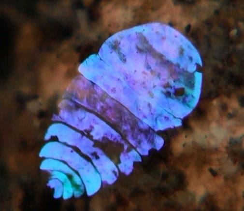
Cellulose is simple, naturally abundant and low cost. However, cellulose fibres ranging at the nanoscale exhibit extraordinary properties such as flexibility, high crystallinity, biodegradability and optical transparency, among others. The nanomaterial can be extracted from plant cellulose pulp or synthetized by non-pathogenic bacteria. Currently, nanocellulose is under active research to develop a myriad of applications including filtration, wound dressing, pollution removal approaches or flexible and transparent electronics, whereas it has been scarcely explored for optical (bio)sensing applications.

The research team led by ICREA Prof Arben Merkoçi seeks to design, fabricate, and test simple, disposable and versatile sensing platforms based on this material. They designed different bacterial cellulose nanopaper based optical sensing platforms. In the article, the authors describe how the material can be tuned to exhibit plasmonic or photoluminescent properties that can be exploited for sensing applications. Specifically, they have prepared two types of plasmonic nanopaper and two types of photoluminescent nanopaper using different optically active nanomaterials.

The researchers took advantage of the optical transparency, porosity, hydrophilicity, and amenability to chemical modification of the material. The bacterial cellulose employed throughout this research was obtained using a bottom-up approach and it is shown that it can be easily turned into useful devices for sensing applications using wax printing or simple punch tools. The scientific team also demonstrates how these novel sensing platforms can be modulated to detect biologically relevant analytes such as cyanide and pathogens among others.

According to the authors, this class of platforms will prove valuable for displaying analytical information in diverse fields such as diagnostics, environmental monitoring and food safety. Moreover, since bacterial cellulose is flexible, lightweight, biocompatible and biodegradable, the proposed composites could be used as wearable optical sensors and could even be integrated into novel theranostic devices. In general, paper-based sensors are known to be simple, portable, disposable, low power-consuming and inexpensive devices that might be exploited in medicine, detection of explosives or hazardous compounds and environmental studies.

Here’s a link to and a citation for the paper,

Nanopaper as an Optical Sensing Platform by Eden Morales-Narváez, Hamed Golmohammadi, Tina Naghdi, Hossein Yousefi, Uliana Kostiv, Daniel Horák, Nahid Pourreza, and Arben Merkoçi.ACS Nano, Article ASAP DOI: 10.1021/acsnano.5b03097 Publication Date (Web): July 2, 2015
Copyright © 2015 American Chemical Society

This paper is behind a paywall.

Sea sapphires: now you see them, now you don’t and more about structural colour/color

The structural colour of the sea sapphire

 Scientists are studying the disappearing act of this ocean-dwelling copepod. Credit: Kaj Maney, www.liquidguru.com Courtesy: American Chemical Society


Scientists are studying the disappearing act of this ocean-dwelling copepod.
Credit: Kaj Maney, www.liquidguru.com Courtesy: American Chemical Society

Now, you’ve seen a sea sapphire. Here’s more about them and the interest they hold for experts in photonics, from a July 15, 2015 news item on ScienceDaily,

Sapphirina, or sea sapphire, has been called “the most beautiful animal you’ve never seen,” and it could be one of the most magical. Some of the tiny, little-known copepods appear to flash in and out of brilliantly colored blue, violet or red existence. Now scientists are figuring out the trick to their hues and their invisibility. The findings appear in the Journal of the American Chemical Society and could inspire the next generation of optical technologies.

A July 15, 2015 American Chemical Society (ACS) news release, which originated the news item, provides more detail,

Copepods are tiny aquatic crustaceans that live in both fresh and salt water. Some males of the ocean-dwelling Sapphirina genus display striking, iridescent colors that scientists think play a role in communication and mate recognition. The shimmering animals’ colors result when light bounces off of the thin, hexagonal crystal plates that cover their backs. These plates also help them vanish, if only fleetingly. Scientists didn’t know specifically what factors contributed to creating different shades. Scientists at the Weizmann Institute [Israel] and the Interuniversity Institute for Marine Sciences in Eilat [Israel] wanted to investigate the matter.

The researchers measured the light reflectance — which determines color — of live Sapphirina males and the spacing between crystal layers. They found that changes of reflectance depended on the thickness of the spacing. And for at least one particular species, when light hits an animal at a 45-degree angle, reflectance shifts out of the visible light range and into the ultraviolet, and it practically disappears. Their results could help inform the design of artificial photonic crystal structures, which have many potential uses in reflective coatings, optical mirrors and optical displays.

To sum this up, the colour and the invisibility properties are due to thin, hexagonal crystal plates and the spacing of these plates, in other words, structural colour, which is usually achieved at the nanoscale.

Here’s a link to and a citation for the paper,

Structural Basis for the Brilliant Colors of the Sapphirinid Copepods by Dvir Gur, Ben Leshem, Maria Pierantoni, Viviana Farstey, Dan Oron, Steve Weiner, and Lia Addadi. J. Am. Chem. Soc., 2015, 137 (26), pp 8408–8411 DOI: 10.1021/jacs.5b05289 Publication Date (Web): June 22, 2015

Copyright © 2015 American Chemical Society

This paper is behind a paywall.

For anyone who’s interested, Lynn Kimlicka has a nice explanation of structural colour in a July 22, 2015 posting on the Something About Science blog where she discusses some recent research iridescence in bird feathers and synthetic melanin. She also shares a picture of her budgie and its iridescent feathers. The ‘melanin’ research was mentioned here in a May 19, 2015 posting where I also provide a link to a great 2013 piece on structural throughout the animal and plant kingdoms by Cristina Luiggi for The Scientist.

Understanding how nanostructures can affect optical properties could be leading to new ways of managing light. A July 23, 2015 news item on ScienceDaily describes a project at the University of Delaware dedicated to “changing the color of light,”

Researchers at the University of Delaware have received a $1 million grant from the W.M. Keck Foundation to explore a new idea that could improve solar cells, medical imaging and even cancer treatments. Simply put, they want to change the color of light.

A July 23, 2015 University of Delaware (UD) news release, which originated the news item, provides more information about the proposed research,

“A ray of light contains millions and millions of individual units of light called photons,” says project leader Matthew Doty. “The energy of each photon is directly related to the color of the light — a photon of red light has less energy than a photon of blue light. You can’t simply turn a red photon into a blue one, but you can combine the energy from two or more red photons to make one blue photon.”

This process, called “photon upconversion,” isn’t new, Doty says. However, the UD team’s approach to it is.

They want to design a new kind of semiconductor nanostructure that will act like a ratchet. It will absorb two red photons, one after the other, to push an electron into an excited state when it can emit a single high-energy (blue) photon.

These nanostructures will be so teeny they can only be viewed when magnified a million times under a high-powered electron microscope.

“Think of the electrons in this structure as if they were at a water park,” Doty says. “The first red photon has only enough energy to push an electron half-way up the ladder of the water slide. The second red photon pushes it the rest of the way up. Then the electron goes down the slide, releasing all of that energy in a single process, with the emission of the blue photon. The trick is to make sure the electron doesn’t slip down the ladder before the second photon arrives. The semiconductor ratchet structure is how we trap the electron in the middle of the ladder until the second photon arrives to push it the rest of the way up.”

The UD team will develop new semiconductor structures containing multiple layers of different materials, such as aluminum arsenide and gallium bismuth arsenide, each only a few nanometers thick. This “tailored landscape” will control the flow of electrons into states with varying potential energy, turning once-wasted photons into useful energy.

The UD team has shown theoretically that their semiconductors could reach an upconversion efficiency of 86 percent, which would be a vast improvement over the 36 percent efficiency demonstrated by today’s best materials. What’s more, Doty says, the amount of light absorbed and energy emitted by the structures could be customized for a variety of applications, from lightbulbs to laser-guided surgery.

How do you even begin to make structures so tiny they can only be seen with an electron microscope? In one technique the UD team will use, called molecular beam epitaxy, nanostructures will be built by depositing layers of atoms one at a time. Each structure will be tested to see how well it absorbs and emits light, and the results will be used to tailor the structure to improve performance.

The researchers also will develop a milk-like solution filled with millions of identical individual nanoparticles, each one containing multiple layers of different materials. The multiple layers of this structure, like multiple candy shells in an M&M, will implement the photon ratchet idea. Through such work, the team envisions a future upconversion “paint” that could be easily applied to solar cells, windows and other commercial products.

Improving medical tests and treatments

While the initial focus of the three-year project will be on improving solar energy harvesting, the team also will explore biomedical applications.

A number of diagnostic tests and medical treatments, ranging from CT [computed tomography] and PET [positron emission tomography] scans to chemotherapy, rely on the release of fluorescent dyes and pharmaceutical drugs. Ideally, such payloads are delivered both at specific disease sites and at specific times, but this is hard to control in practice.

The UD team aims to develop an upconversion nanoparticle that can be triggered by light to release its payload. The goal is to achieve the controlled release of drug therapies even deep within diseased human tissue while reducing the peripheral damage to normal tissue by minimizing the laser power required.

“This is high-risk, high-reward research,” Doty says. “High-risk because we don’t yet have proof-of-concept data. High-reward because it has such a huge potential impact in renewable energy to medicine. It’s amazing to think that this same technology could be used to harvest more solar energy and to treat cancer. We’re excited to get started!”

That’s it for structural colour/color today.

Quantum and classical physics may be closer than we thought

It seems that a key theory about the boundary between the quantum world and our own macro world has been disproved and I think the July 21, 2015 news item on Nanotechnology Now says it better,

Quantum theory is one of the great achievements of 20th century science, yet physicists have struggled to find a clear boundary between our everyday world and what Albert Einstein called the “spooky” features of the quantum world, including cats that could be both alive and dead, and photons that can communicate with each other across space instantaneously.

For the past 60 years, the best guide to that boundary has been a theorem called Bell’s Inequality, but now a new paper shows that Bell’s Inequality is not the guidepost it was believed to be, which means that as the world of quantum computing brings quantum strangeness closer to our daily lives, we understand the frontiers of that world less well than scientists have thought.

In the new paper, published in the July 20 [2015] edition of Optica, University of Rochester [New York state, US] researchers show that a classical beam of light that would be expected to obey Bell’s Inequality can fail this test in the lab, if the beam is properly prepared to have a particular feature: entanglement.

A July 21, 2015 University of Rochester news release, which originated the news item, reveals more about the boundary and the research,

Not only does Bell’s test not serve to define the boundary, the new findings don’t push the boundary deeper into the quantum realm but do just the opposite. They show that some features of the real world must share a key ingredient of the quantum domain. This key ingredient is called entanglement, exactly the feature of quantum physics that Einstein labeled as spooky. According to Joseph Eberly, professor of physics and one of the paper’s authors, it now appears that Bell’s test only distinguishes those systems that are entangled from those that are not. It does not distinguish whether they are “classical” or quantum. In the forthcoming paper the Rochester researchers explain how entanglement can be found in something as ordinary as a beam of light.

Eberly explained that “it takes two to tangle.” For example, think about two hands clapping regularly. What you can be sure of is that when the right hand is moving to the right, the left hand is moving to the left, and vice versa. But if you were asked to guess without listening or looking whether at some moment the right hand was moving to the right, or maybe to the left, you wouldn’t know. But you would still know that whatever the right hand was doing at that time, the left hand would be doing the opposite. The ability to know for sure about a common property without knowing anything for sure about an individual property is the essence of perfect entanglement.

Eberly added that many think of entanglement as a quantum feature because “Schrodinger coined the term ‘entanglement’ to refer to his famous cat scenario.” But their experiment shows that some features of the “real” world must share a key ingredient of Schrodinger’s Cat domain: entanglement.

The existence of classical entanglement was pointed out in 1980, but Eberly explained that it didn’t seem a very interesting concept, so it wasn’t fully explored. As opposed to quantum entanglement, classical entanglement happens within one system. The effect is all local: there is no action at a distance, none of the “spookiness.”

With this result, Eberly and his colleagues have shown experimentally “that the border is not where it’s usually thought to be, and moreover that Bell’s Inequalities should no longer be used to define the boundary.”

Here’s a link to and a citation for the paper,

Shifting the quantum-classical boundary: theory and experiment for statistically classical optical fields by Xiao-Feng Qian, Bethany Little, John C. Howell, and J. H. Eberly. Optica Vol. 2, Issue 7, pp. 611-615 (2015) •doi: 10.1364/OPTICA.2.000611

This paper is open access.

Nanomaterials and UV (ultraviolet) light for environmental cleanups

I think this is the first time I’ve seen anything about a technology that removes toxic materials from both water and soil; it’s usually one or the other. A July 22, 2015 news item on Nanowerk makes the announcement (Note: A link has been removed),

Many human-made pollutants in the environment resist degradation through natural processes, and disrupt hormonal and other systems in mammals and other animals. Removing these toxic materials — which include pesticides and endocrine disruptors such as bisphenol A (BPA) — with existing methods is often expensive and time-consuming.

In a new paper published this week in Nature Communications (“Nanoparticles with photoinduced precipitation for the extraction of pollutants from water and soil”), researchers from MIT [Massachusetts Institute of Technology] and the Federal University of Goiás in Brazil demonstrate a novel method for using nanoparticles and ultraviolet (UV) light to quickly isolate and extract a variety of contaminants from soil and water.

A July 21, 2015 MIT news release by Jonathan Mingle, which originated the news item, describes the inspiration and the research in more detail,

Ferdinand Brandl and Nicolas Bertrand, the two lead authors, are former postdocs in the laboratory of Robert Langer, the David H. Koch Institute Professor at MIT’s Koch Institute for Integrative Cancer Research. (Eliana Martins Lima, of the Federal University of Goiás, is the other co-author.) Both Brandl and Bertrand are trained as pharmacists, and describe their discovery as a happy accident: They initially sought to develop nanoparticles that could be used to deliver drugs to cancer cells.

Brandl had previously synthesized polymers that could be cleaved apart by exposure to UV light. But he and Bertrand came to question their suitability for drug delivery, since UV light can be damaging to tissue and cells, and doesn’t penetrate through the skin. When they learned that UV light was used to disinfect water in certain treatment plants, they began to ask a different question.

“We thought if they are already using UV light, maybe they could use our particles as well,” Brandl says. “Then we came up with the idea to use our particles to remove toxic chemicals, pollutants, or hormones from water, because we saw that the particles aggregate once you irradiate them with UV light.”

A trap for ‘water-fearing’ pollution

The researchers synthesized polymers from polyethylene glycol, a widely used compound found in laxatives, toothpaste, and eye drops and approved by the Food and Drug Administration as a food additive, and polylactic acid, a biodegradable plastic used in compostable cups and glassware.

Nanoparticles made from these polymers have a hydrophobic core and a hydrophilic shell. Due to molecular-scale forces, in a solution hydrophobic pollutant molecules move toward the hydrophobic nanoparticles, and adsorb onto their surface, where they effectively become “trapped.” This same phenomenon is at work when spaghetti sauce stains the surface of plastic containers, turning them red: In that case, both the plastic and the oil-based sauce are hydrophobic and interact together.

If left alone, these nanomaterials would remain suspended and dispersed evenly in water. But when exposed to UV light, the stabilizing outer shell of the particles is shed, and — now “enriched” by the pollutants — they form larger aggregates that can then be removed through filtration, sedimentation, or other methods.

The researchers used the method to extract phthalates, hormone-disrupting chemicals used to soften plastics, from wastewater; BPA, another endocrine-disrupting synthetic compound widely used in plastic bottles and other resinous consumer goods, from thermal printing paper samples; and polycyclic aromatic hydrocarbons, carcinogenic compounds formed from incomplete combustion of fuels, from contaminated soil.

The process is irreversible and the polymers are biodegradable, minimizing the risks of leaving toxic secondary products to persist in, say, a body of water. “Once they switch to this macro situation where they’re big clumps,” Bertrand says, “you won’t be able to bring them back to the nano state again.”

The fundamental breakthrough, according to the researchers, was confirming that small molecules do indeed adsorb passively onto the surface of nanoparticles.

“To the best of our knowledge, it is the first time that the interactions of small molecules with pre-formed nanoparticles can be directly measured,” they write in Nature Communications.

Nano cleansing

Even more exciting, they say, is the wide range of potential uses, from environmental remediation to medical analysis.

The polymers are synthesized at room temperature, and don’t need to be specially prepared to target specific compounds; they are broadly applicable to all kinds of hydrophobic chemicals and molecules.

“The interactions we exploit to remove the pollutants are non-specific,” Brandl says. “We can remove hormones, BPA, and pesticides that are all present in the same sample, and we can do this in one step.”

And the nanoparticles’ high surface-area-to-volume ratio means that only a small amount is needed to remove a relatively large quantity of pollutants. The technique could thus offer potential for the cost-effective cleanup of contaminated water and soil on a wider scale.

“From the applied perspective, we showed in a system that the adsorption of small molecules on the surface of the nanoparticles can be used for extraction of any kind,” Bertrand says. “It opens the door for many other applications down the line.”

This approach could possibly be further developed, he speculates, to replace the widespread use of organic solvents for everything from decaffeinating coffee to making paint thinners. Bertrand cites DDT, banned for use as a pesticide in the U.S. since 1972 but still widely used in other parts of the world, as another example of a persistent pollutant that could potentially be remediated using these nanomaterials. “And for analytical applications where you don’t need as much volume to purify or concentrate, this might be interesting,” Bertrand says, offering the example of a cheap testing kit for urine analysis of medical patients.

The study also suggests the broader potential for adapting nanoscale drug-delivery techniques developed for use in environmental remediation.

“That we can apply some of the highly sophisticated, high-precision tools developed for the pharmaceutical industry, and now look at the use of these technologies in broader terms, is phenomenal,” says Frank Gu, an assistant professor of chemical engineering at the University of Waterloo in Canada, and an expert in nanoengineering for health care and medical applications.

“When you think about field deployment, that’s far down the road, but this paper offers a really exciting opportunity to crack a problem that is persistently present,” says Gu, who was not involved in the research. “If you take the normal conventional civil engineering or chemical engineering approach to treating it, it just won’t touch it. That’s where the most exciting part is.”

The researchers have made this illustration of their work available,

Nanoparticles that lose their stability upon irradiation with light have been designed to extract endocrine disruptors, pesticides, and other contaminants from water and soils. The system exploits the large surface-to-volume ratio of nanoparticles, while the photoinduced precipitation ensures nanomaterials are not released in the environment. Image: Nicolas Bertrand Courtesy: MIT

Nanoparticles that lose their stability upon irradiation with light have been designed to extract endocrine disruptors, pesticides, and other contaminants from water and soils. The system exploits the large surface-to-volume ratio of nanoparticles, while the photoinduced precipitation ensures nanomaterials are not released in the environment.
Image: Nicolas Bertrand Courtesy: MIT

Here’s a link to and a citation for the paper,

Nanoparticles with photoinduced precipitation for the extraction of pollutants from water and soil by Ferdinand Brandl, Nicolas Bertrand, Eliana Martins Lima & Robert Langer. Nature Communications 6, Article number: 7765 doi:10.1038/ncomms8765 Published 21 July 2015

This paper is open access.

US Navy invests in graphene

More usually, I feature research from DARPA (Defense Advanced Research Progects Agency) which I think belongs to the US Army and the US Air Force Research Office. The US Navy has featured here only once before (a Nov. 1, 2011 posting) and even then it was tangentially. I think it’s long past time that the US Navy gets some attention.

A July 22, 2015 news item on Nanowerk explains the Navy’s interest in electricity and graphene,

The U.S. Navy distributes electricity aboard most of its ships like a power company. It relies on conductors, transformers and other bulky infrastructure.

The setup works, but with powerful next generation weapons on the horizon and the omnipresent goal of energy efficiency, the Navy is seeking alternatives to conventional power control systems.

One option involves using graphene, which, since its discovery in 2004, has become the material of choice for researchers working to improve everything from solar cells to smartphone batteries.

Accordingly, the Office of Naval Research has awarded University at Buffalo engineers an $800,000 grant to develop narrow strips of graphene called nanoribbons that may someday revolutionize how power is controlled in ships, smartphones and other electronic devices.

A July 20, 2015 University of Buffalo news release by Cory Nealon, which originated the news item, expands on the theme,

“We need to develop new nanomaterials capable of handling greater amounts of energy densities in much smaller devices. Graphene nanoribbons show remarkable promise in this endeavor,” says Cemal Basaran, PhD, a professor in UB’s Department of Civil, Structural and Environmental Engineering, School of Engineering and Applied Sciences, and the grant’s principal investigator.

Graphene is a single layer of carbon atoms packed together like a honeycomb. It is extremely thin, light and strong. It’s also the best known conductor of heat and electricity.

“The beauty of graphene is that it can be grown like biological organisms as opposed to manufacturing materials with traditional techniques,” says Basaran, director of UB’s Electronic Packaging Laboratory and a researcher in UB’s New York State Center of Excellence in Materials Informatics. “These bio-inspired materials allow us to control their atomic organizations like controlling genetic DNA makeup of a lab-grown cell.”

While promising, researchers are just beginning to understand graphene and its potential uses. One area of interest is power control systems.

Like overhead power lines, most ships rely on copper or other metals to move electricity. Unfortunately, this process is relatively inefficient; electrons bash into each other and create heat in a process called Joule heating.

“You lose a great deal of energy that way,” Basaran says. “With graphene, you avoid those collisions because it conducts electricity in a different process, known as semi-ballistic conduction. It’s like a high-speed bullet train versus bumper cars.”

Another limitation of metal-based power distribution is the bulky infrastructure – transistors, copper wires, transformers, etc. – needed to move electricity. Whether in a ship or tablet computer, the components take up space and add weight.

Graphene nanoribbons offer a potential solution because they can act as both a conductor (instead of copper) and semiconductor (instead of silicon). Moreover, their ability to withstand failure under extreme energy loads is roughly 1,000 times greater than copper.

That bodes well for the Navy, which, like segments of the automotive industry, is pivoting toward electric vehicles.

It recently launched an all-electric destroyer; the ship’s propellers and drive shafts are turned by electric motors, as opposed to being connected to combustion engines. The integrated power-generation and distribution system may also be used to fire next generation weapons, such as railguns and powerful lasers. And the automation has allowed the Navy to reduce the ship’s crew, which places fewer sailors in potentially dangerous situations.

Graphene nanoribbons could improve these systems by making them more robust and energy-efficient, Basaran said. He and a team of researchers will:

·         Design complex simulations that examine how graphene nanoribbons can be used as a power switch.

·         Explore how adding hydrogen and other elements, a process known as “doping,” to graphene nanoribbons could improve their performance.

·         Investigate graphene nanoribbons’ failure limit under high power loads and try to find ways to improve it.

The research will be performed over the next four years.

I was particularly intrigued by the caption for this image included with the news release,

The technology may lead to more powerful weapons, energy savings and reduced crew numbers [Downloaded from http://www.buffalo.edu/news/releases/2015/07/021.html]

The technology may lead to more powerful weapons, energy savings and reduced crew numbers [Downloaded from http://www.buffalo.edu/news/releases/2015/07/021.html]

Presumably “reduced crew numbers’ means fewer jobs. I wonder if they’ll figure out that people without jobs are without money to pay taxes to fund these projects.

Science and music festivals such as Latitude 2015 and some Guerilla Science

Science has been gaining prominence at music festivals in Britain if nowhere else. I wrote about the Glastonbury Festival’s foray into science in a July 12, 2011 posting which featured the Guerilla Science group tent and mentioned other of the festival’s science and technology efforts over the years. More recently, I noticed that Stephen Hawking was scheduled for the 2015 Glastonbury Festival (he had to cancel due to personal reasons).

The 2015 Latitude Festival seems to have more luck with its science-themed events. according to a July 22, 2015 posting by Suzi Gage for the Guardian’s science blogs,

Why do people go to music festivals? When I was 18 years old and heading to Reading festival the answer was very much ‘to listen to Pulp and Beck in a field while drinking overpriced beer and definitely not trying to sneak a hip flask on to the site’. But I’ve grown up since then, and so, it seems, have festivals.

At Latitude this weekend, I probably only watched a handful of bands. Not to say that the musical lineup wasn’t great, but there was so much more on offer that caught my attention. The Wellcome Trust funded a large number of talks, interactive sessions and demos that appeared both in their ‘hub’, a tiny tent on the outskirts of the festival, but also in the Literary Tent at the heart of the festival and at other locations across the site.

The programming of the science content was imaginative, often pairing a scientist with an author who had written on a similar topic. This was effective in that it allowed a discussion, but kept it from becoming too technical or full of jargon.

Dr Robin Carhart-Harris, an expert in psychedelics, was paired with Zoe Cormier, author of ‘Sex Drugs and Rock and Roll’ in the Literary Tent, to discuss the use of psychedelics as ‘medicine for the soul’. [emphasis mine] Robin was very measured in his description of the trials he has been involved with at Imperial College London, being clear that while preliminary findings about psilocybin in treatment-resistant depression might be exciting, there’s a long way to go in such research. Talking about drugs at a festival is always going to be a crowd pleaser, but both Robin and Zoe never sensationalized.

A highlight for me was a session organised by The Psychologist magazine, featuring Professor Sarah-Jayne Blakemore and Fiona Neil, author of The Good Girl. Entitled ‘Being Young Never Gets Old’, it claimed to ‘debunk’ teenagers. …

Gage’s piece is a good read and I find it interesting she makes no comment about a literary tent at a music festival. I don’t know of a music festival in Canada that would feature literature or literature and science together.

Guerilla Science

I highlighted Zoe Cormier’s name as a participant (born in Canada and living in London, England) as she is a founder of Guerilla Science, the group I mentioned earlier with regard to the Glastonbury Festival. A science communicator with some fairly outrageous events under her belt, her and her co-founder’s ‘guerilla’ approach to science is exciting. I mentioned their annual Secret Garden event in a Aug. 1, 2012 posting where they sang and danced the Higgs Boson and otherwise celebrated elementary particles. The 2015 Secret Garden Party featured rest, noise, and neuroscience. (Perhaps it’s not too early to plan attendance at the 2016 Secret Garden Party?) Here’s an excerpt from this year’s lineup found in Louis’ July 15, 2015 posting on the Guerilla Science website,

Friday [July 24, 2015]

….

12:00 – Rest & Noise Shorts

Crash, bang, shush, zzz… four short talks about rest and noise from artist Zach Walker, psychologist Will Lawn and neuroscientists Ed Bracey and Melissa Ellamil.

13.00 Speed, Synapse… Go!

Two teams go head-to-head in a competition to see whose neurotransmitters can move the fastest. What happens when cocaine, marijuana and ketamine are introduced? Join us for some fast and furious neuroscientific gameplay.

15.00 Craft a Connectome

Help us transform the Guerilla Science tent into a giant model brain with a tangle of woolen connections. Neuroscientists Julia Huntenburg and Melissa Ellamil will be on hand to conduct our connectome and coax it into a resting state.

16.00 Sound, Fire and Water

We test out our new toy: a fire organ that visualises sound in flames! Join engineers from Buro Happold and artist Zach Walker as we make fire, water and cornstarch dance and jump to the beat.

Saturday [July 25, 2015]

11.00 Hearing the Voice

Philosopher Sam Wilkinson explores the idea of the brain as a hypothesis testing machine. He asks whether thinking about the mind in this way can help explain mental illness, hallucinations and the voices in our heads.

15.00 – The Unquiet Mind

Hallucinations are our contact with the unreal but are also a window into human nature. Neuroscientist and clinical psychologist Vaughan Bell reveals what they tell us about brain function and the limits of human experience.

Sunday [July 26, 2015]

12.00 Phantom Terrains

Frank Swain and Daniel Jones present their project to listen in to wireless networks. By streaming wi-fi signals to a pair of hearing aids, Frank can hear the changing landscapes of data that silently surround us.

13.00 Rest and Nose

Join chemists Rose Gray and Alex Bour and neuroscientist Ed Bracey to explore the links between relaxation, rest and sense of smell. Create a perfume to lull yourself to sleep, help you unwind and evoke a peaceful place or time.

..

For anyone interested in Guerilla Science, this is their website. They do organize events year round.

Michelangelo, clinical anatomy, mathematics, the Golden Ratio, and a myth

I would have thought an article about Michelangelo, mathematics, and the Golden Ratio would be in a journal dedicated to the arts or mathematics or possibly both. Not even my tenth guess would  have been Clinical Anatomy. As for the myth, not everyone subscribes to the Golden Ratio theory of beauty.

A July 20, 2015 Wiley Periodicals press release (also on EurekAlert) announces the publication of the research,

New research provides mathematical evidence that Michelangelo used the Golden Ratio of 1.6 when painting The Creation of Adam on the ceiling of the Sistine Chapel. The Golden Ratio is found when you divide a line into two parts so that the longer part divided by the smaller part is equal to the whole length divided by the longer part.

The Golden Ratio has been linked with greater structural efficiency and has puzzled scientists for centuries due to its frequent occurrence in nature–for example in snail shells and flower petals. The Golden Ratio can also be found in a variety of works by architects and designers, in famous musical compositions, and in the creations of many artists.

The findings suggest that the beauty and harmony found in the works of Michelangelo may not be based solely on his anatomical knowledge. He likely knew that anatomical structures incorporating the Golden Ratio offer greater structural efficiency and, therefore, he used it to enhance the aesthetic quality of his works.

“We believe that this discovery will bring a new dimension to the great work of Michelangelo,” said Dr. Deivis de Campos, author of the Clinical Anatomy study.

Here’s a link to and a citation for the paper,

More than a neuroanatomical representation in The Creation of Adam by Michelangelo Buonarroti, a representation of the Golden Ratio by Deivis De Campos, Tais Malysz,  João Antonio Bonatto-Costa, Geraldo Pereira Jotz, Lino Pinto De Oliveira Junior, and Andrea Oxley da Rocha. Clinical Anatomy DOI: 10.1002/ca.22580 Article first published online: 17 JUL 2015

© 2015 Wiley Periodicals, Inc.

This paper is open access.

Golden Ratio myth

One final comment, it seems not everyone is convinced that the Golden Ratio plays an important role in design, art, and architecture according to an April 13, 2015 article by John Brownlee for Fast Company titled: The Golden Ratio: Design’s Biggest Myth,

In the world of art, architecture, and design, the golden ratio has earned a tremendous reputation. Greats like Le Corbusier and Salvador Dalí have used the number in their work. The Parthenon, the Pyramids at Giza, the paintings of Michelangelo, the Mona Lisa, even the Apple logo are all said to incorporate it.

It’s bullshit. The golden ratio’s aesthetic bona fides are an urban legend, a myth, a design unicorn. Many designers don’t use it, and if they do, they vastly discount its importance. There’s also no science to really back it up. Those who believe the golden ratio is the hidden math behind beauty are falling for a 150-year-old scam.

Fascinating, non?

Putting the speed on spin, spintronics that is

This is for physics fans, if you plan on looking at the published paper. Otherwise, the July 20, 2015 news item on ScienceDaily is more accessible to the rest of us,

In a tremendous boost for spintronic technologies, EPFL scientists have shown that electrons can jump through spins much faster than previously thought.

Electrons spin around atoms, but also spin around themselves, and can cross over from one spin state to another. A property which can be exploited for next-generation hard drives. However, “spin cross-over” has been considered too slow to be efficient. Using ultrafast measurements, EPFL scientists have now shown for the first time that electrons can cross spins at least 100,000 times faster than previously thought. Aside for its enormous implications for fundamental physics, the finding can also propel the field of spintronics forward. …

A July 20, 2015 EPFL press release on EurekAlert, which originated the news item, provides context for the research,

The rules of spin

Although difficult to describe in everyday terms, electron spin can be loosely compared to the rotation of a planet or a spinning top around its axis. Electrons can spin in different manners referred to as “spin states” and designated by the numbers 0, 1/2, 1, 3/2, 2 etc. During chemical reactions, electrons can cross from one spin state to another, e.g. from 0 to 1 or 1/2 to 3/2.

Spin cross-over is already used in many technologies, e.g. optical light-emitting devices (OLED), energy conversion systems, and cancer phototherapy. Most prominently, spin cross-over is the basis of the fledgling field of spintronics. The problem is that spin cross-over has been thought to be too slow to be efficient enough in circuits.

Spin cross-over is extremely fast

The lab of Majed Chergui at EPFL has now demonstrated that spin cross-over is considerably faster than previously thought. Using the highest time-resolution technology in the world, the lab was able to “see” electrons crossing through four spin states within 50 quadrillionths of a second — or 50 femtoseconds.

“Time resolution has always been a limitation,” says Chergui. “Over the years, labs have used techniques that could only measure spin changes to a billionth to a millionth of a second. So they thought that spin cross-over happened in this timeframe.”

Chergui’s lab focused on materials that show much promise in spintronics applications. In these materials, electrons jump through four spin-states: from 0 to 1 to 2. In 2009, Chergui’s lab pushed the boundaries of time resolution to show that this 0-2 “jump” can happen within 150 femtoseconds — suggesting that it was a direct event. Despite this, the community still maintained that such spin cross-overs go through intermediate steps.

But Chergui had his doubts. Working with his postdoc Gerald Auböck, they used the lab’s world-recognized expertise in ultrafast spectroscopy to “crank up” the time resolution. Briefly, a laser shines on the material sample under investigation, causing its electrons to move. Another laser measures their spin changes over time in the ultraviolet light range.

The finding essentially demolishes the notion of intermediate steps between spin jumps, as it does not allow enough time for them: only 50 quadrillionths of a second to go from the “0” to the “2” spin state. This is the first study to ever push time resolution to this limit in the ultraviolet domain. “This probably means that it’s even faster,” says Chergui. “But, more importantly, that it is a direct process.”

From observation to explanation

With profound implications for both technology and fundamental physics and chemistry, the study is an observation without an explanation. Chergui believes that the key is electrons shuttling back-and-forth between the iron atom at the center of the material’s molecules and its surrounding elements. “When the laser light shines on the atom, it changes the electron’s spin angle, affecting the entire spin dynamics in the molecule.”

It is now up to theoreticians to develop a new model for ultrafast spin changes. On the experimental side of things, Chergui’s lab is now focusing on actually observing electrons shuttling inside the molecules. This will require even more sophisticated approaches, such as core-level spectroscopy. Nonetheless, the study challenges ideas about spin cross-over, and might offer long-awaited solutions to the limitations of spintronics.

Here’s a link to and citation for the paper,

Sub-50-fs photoinduced spin crossover in [Fe(bpy)3]2+ by Gerald Auböck & Majed Chergui. Nature Chemistry (2015) doi:10.1038/nchem.2305 Published online 20 July 2015

This paper is behind a paywall.

Sticky-flares nanotechnology to track and observe RNA (ribonucleic acid) regulation

I like the name ‘sticky-flares’ and had hoped there was an amusing story about its origins. Ah well, perhaps I’ll have better luck next time.

This work comes out of Chad Mirkin’s lab at Northwestern University (Chicago, US) according to a July 21, 2015 news item on Azonano,

RNA [ribonucleic acid] is a fundamental ingredient in all known forms of life — so when RNA goes awry, a lot can go wrong. RNA misregulation plays a critical role in the development of many disorders, such as mental disability, autism and cancer.

A new technology — called “Sticky-flares” — developed by nanomedicine experts at Northwestern University offers the first real-time method to track and observe the dynamics of RNA distribution as it is transported inside living cells.

A July 20, 2015 Northwestern University news release by Erin Spain, which originated the news item, describes the research in a little more detail also including information about predecessor technology,

Sticky-flares have the potential to help scientists understand the complexities of RNA better than any analytical technique to date and observe and study the biological and medical significance of RNA misregulation.

Previous technologies made it possible to attain static snapshots of RNA location, but that isn’t enough to understand the complexities of RNA transport and localization within a cell. Instead of analyzing snapshots of RNA to try to understand functioning, Sticky-flares help create an experience that is more like watching live-streaming video.

“This is very exciting because much of the RNA in cells has very specific quantities and localization, and both are critical to the cell’s function, but until this development it has been very difficult, and often impossible, to probe both attributes of RNA in a live cell,” said Chad A. Mirkin, a nanomedicine expert and corresponding author of the study. “We hope that many more researchers will be able to use this platform to increase our understanding of RNA function inside cells.”

Sticky-flares are tiny spherical nucleic acid gold nanoparticle conjugates that can enter living cells and target and transfer a fluorescent reporter or “tracking device” to RNA transcripts. This fluorescent labeling can be tracked via fluorescence microscopy as it is transported throughout the cell, including the nucleus.

In the … paper, the scientists explain how they used Sticky-flares to quantify β–actin mRNA in HeLa cells (the oldest and most commonly used human cell line) as well as to follow the real-time transport of β–actin mRNA in mouse embryonic fibroblasts.

Sticky-flares are built upon another technology from Mirkin’s group called NanoFlares, which was the first genetic-based approach that is able to detect live circulating tumor cells out of the complex matrix that is human blood.

NanoFlares have been very useful for researchers that operate in the arena of quantifying gene expression. AuraSense, Inc., a biotechnology company that licensed the NanoFlare technology from Northwestern University, and EMD-Millipore, another biotech company, have commercialized NanoFlares. There are now more than 1,700 commercial forms of NanoFlares sold under the SmartFlareä name in more than 230 countries.

The Sticky-flare is designed to address limitations of SmartFlares, most notably their inability to track RNA location and enter the nucleus. The Northwestern team believes Sticky-flares are poised to become a valuable tool for researchers who desire to understand the function of RNA in live cells.

Based on the paragraph about the precursor technology’s commercial success , I gather they are excited about similar possibilities for sticky-flares.

Here’s a link to and a citation for the paper,

Quantification and real-time tracking of RNA in live cells using Sticky-flares by William E. Briley, Madison H. Bondy, Pratik S. Randeria, Torin J. Dupper, and Chad A. Mirkin. Published online before print July 20, 2015, doi: 10.1073/pnas.1510581112 PNAS July 20, 2015

This paper is behind a paywall.

X-raying fungus on paper to conserve memory

Civilization is based on memory. Our libraries and archives serve as memories of how things are made, why we use certain materials rather than others, how the human body is put together, what the weather patterns have been, etc. For centuries we have preserved our memories on paper. While this has many advantages, there are some drawbacks including fungus infestations.

A July 21, 2015 news item on ScienceDaily describes how a technique used to x-ray rocks has provided insights into paper and its fungal infestations,

Believe it or not: X-ray works a lot better on rocks than on paper. This has been a problem for conservators trying to save historical books and letters from the ravages of time and fungi. They frankly did not know what they were up against once the telltale signs of vandals such as Dothidales or Pleosporales started to spot the surface of their priceless documents

Now Diwaker Jha, an imaging specialist from Department of Chemistry, University of Copenhagen, has managed to adapt methods developed to investigate interiors of rocks to work on paper too, thus getting a first look at how fungus goes about infesting paper. …

A July 21, 2015 University of Copenhagen press release (also on EurekAlert), which originated the news item, expands on the theme,

This is good news for paper conservators and others who wish to study soft materials with X-ray tomography. “Rocks are easy because they are hard. The X-ray images show a very good contrast between the solid and the pores or channels, which are filled with low density materials such as air or fluids. In this case, however, paper and fungi, both are soft and carbon based, which makes them difficult to distinguish,” says Diwaker.

Diwaker Jha is a PhD student in the NanoGeoScience group, which is a part of the Nano-Science Center at Department of Chemistry. He investigates methods to improve imaging techniques used by chemists and physicists to investigate how fluids move in natural porous materials. At a recent conference, he was presenting an analysis method he developed for X-ray tomography data, for which he was awarded the Presidential Scholar Award by the Microscopy Society of America. And this sparked interest with a conservator in the audience.

Hanna Szczepanowska works as a research conservator with the Smithsonian Institution in the USA. She had been wondering how fungi interact with the paper. Does it sit on the surface, or does it burrow deeper? If they are surface dwellers, it should be easy to just brush them off, but no such luck, says Jha.

“As it turns out, microscopic fungi that infest paper grow very much the same way as mushrooms on a forest floor. However, unlike mushrooms, where the fruiting body emerges out of the soil to the surface, here the fruiting bodies can be embedded within the paper fibres, making it difficult to isolate them. This is not great news for conservators because the prevalent surface cleaning approaches are not adequate,” explains Diwaker Jha.

In working out a way to see into the paper, Jha investigated a 17th century letter on a handmade sheet and a 1920 engraving on machine-made paper. Compared with mushrooms, these fungi are thousands of times smaller, which required an advanced X-ray imaging technique available at the European Synchrotron Radiation Facility (ESRF), Grenoble, France. The technique is very similar to medical tomography (CT scanning) done at hospitals but in Grenoble the X-ray is produced by electrons accelerated to about the speed of light in an 844 meter long circular tube. A handy comparison: “If I were to use medical X-ray tomography to look at an Olympic village, I would be able to make out only the stadium. With the synchrotron based X-ray tomography, I would be able to distinguish individual blades of grass on the field..”

Diwaker hopes that conservators will be able to use the new insight to develop conservation strategies not just for paper artefacts but for combating biodegradation on a host of other types of cultural heritage materials. And that the developed methods can be extended to other studies related to soft matter.

Here’s a citation and a link for the paper,

Morphology and characterization of Dematiaceous fungi on a cellulose paper substrate using synchrotron X-ray microtomography, scanning electron microscopy and confocal laser scanning microscopy in the context of cultural heritage by H. M. Szczepanowska, D. Jha, and Th. G. Mathia. Anal. At. Spectrom. (Journal of Analystical Atomic Spetrometry), 2015,30, 651-657 DOI: 10.1039/C4JA00337C First published online 27 Nov 2014

This paper is behind a paywall. By the way, it is part of something the journal calls a themed collection:  Synchrotron radiation and neutrons in art and archaeology. Clicking on the ‘themed collection’ link will give you a view of the collection, i.e., titles, authors and brief abstracts.