I don’t know the dates for the last ‘golden’ age of electronics but I can certainly understand why these Japanese researchers are excited about their work. In any event, I think the ‘golden age’ is more of a play on words. From a June 25, 2019 news item on Nanowerk (Note: A link has been removed),
One way that heat damages electronic equipment is it makes components expand at different rates, resulting in forces that cause micro-cracking and distortion. Plastic components and circuit boards are particularly prone to damage due to changes in volume during heating and cooling cycles. But if a material could be incorporated into the components that compensates for the expansion, the stresses would be reduced and their lifetime increased.
Everybody knows one material that behaves like this: liquid water expands when it freezes and ice contracts when it melts. But liquid water and electronics don’t mix well – instead, what’s needed is a solid with “negative thermal expansion” (NTE).
Although such materials have been known since the 1960s, a number of challenges had to be overcome before the concept would be broadly useful and commercially viable. In terms of both materials and function, these efforts have only had limited success.
The experimental materials had been produced under specialized laboratory conditions using expensive equipment; and even then, the temperature and pressure ranges in which they would exhibit NTE were well outside normal everyday conditions.
Moreover, the amount they expanded and contracted depended on the direction, which induced internal stresses that changed their structure, meaning that the NTE property would not last longer than a few heating and cooling cycles.
A research team led by Koshi Takenaka of Nagoya University has succeeded in overcoming these materials-engineering challenges (APL Materials, “Valence fluctuations and giant isotropic negative thermal expansion in Sm1–xRxS (R = Y, La, Ce, Pr, Nd)”).
Inspired by the series of work by Noriaki Sato, also of Nagoya University – whose discovery last year of superconductivity in quasicrystals was considered one of the top ten physics discoveries of the year by Physics World magazine – Professor Takenaka took the rare earth element samarium and its sulfide, samarium monosulfide (SmS), which is known to change phase from the “black phase” to the smaller-volume “golden phase”. The problem was to tune the range of temperatures at which the phase transition occurs. The team’s solution was to replace a small proportion of samarium atoms with another rare earth element, giving Sm1-xRxS, where “R” is any one of the rare earth elements cerium (Ce), neodymium (Nd), praseodymium (Pr) or yttrium (Y). The fraction x the team used was typically 0.2, except for yttrium. These materials showed “giant negative thermal expansion” of up to 8% at ordinary room pressure and a useful range of temperatures (around 150 degrees) including at room temperature and above … . Cerium is the star candidate here because it is relatively cheap.
The nature of the phase transition is such that the materials can be powdered into very small crystal sizes around a micron on a side without losing their negative expansion property. This broadens the industrial applications, particularly within electronics.
While the Nagoya University group’s engineering achievement is impressive, how the negative expansion works is fascinating from a fundamental physics viewpoint. During the black-golden transition, the crystal structure stays the same but the atoms get closer together: the unit cell size becomes smaller because (as is very likely but perhaps not yet 100% certain) the electron structure of the samarium atoms changes and makes them smaller – a process of intra-atomic charge transfer called a “valence transition” or “valence fluctuation” within the samarium atoms … . “My impression,” says Professor Takenaka, “is that the correlation between the lattice volume and the electron structure of samarium is experimentally verified for this class of sulfides.”
More specifically, in the black (lower temperature) phase, the electron configuration of the samarium atoms is (4f)6, meaning that in their outermost shell they have 6 electrons in the f orbitals (with s, p and d orbitals filled); while in the golden phase the electronic configuration is (4f)5(5d)1 -an electron has moved out of a 4f orbital into a 5d orbital. Although a “higher” shell is starting to be occupied, it turns out – through a quirk of the Pauli Exclusion Principle – that the second case gives a smaller atom size, leading to a smaller crystal size and negative expansion.
But this is only part of the fundamental picture. In the black phase, samarium sulfide and its doped offshoots are insulators – they do not conduct electricity; while in the golden phase they turn into conductors (i.e. metals). This is suggesting that during the black-golden phase transition the band structure of the whole crystal is influencing the valance transition within the samarium atoms. Although nobody has done the theoretical calculations for the doped samarium sulfides made by Professor Takenaka’s group, a previous theoretical study has indicated that when electrons leave the samarium atoms’ f orbital, they leave behind a positively charged “hole” which itself interacts repulsively with holes in the crystal’s conduction band, affecting their exchange interaction. This becomes a cooperative effect that then drives the valence transition in the samarium atoms. The exact mechanism, though, is not well understood.
Nevertheless, the Nagoya University-led group’s achievement is one of engineering, not pure physics. “What is important for many engineers is the ability to use the material to reduce device failure due to thermal expansion,” explains Professor Takenaka. “In short, in a certain temperature range – the temperature range in which the intended device operates, typically an interval of dozens of degrees or more – the volume needs to gradually decrease with a rise in temperature and increase as the temperature falls. Of course, I also know that volume expansion on cooling during a phase transition [like water freezing] is a common case for many materials. However, if the volume changes in a very narrow temperature range, there is no engineering value. The present achievement is the result of material engineering, not pure physics.”
Perhaps it even heralds a new “golden” age for electronics.
I worked in a company for a data communications company that produced hardware and network management software. From a hardware perspective, heat was an enemy which distorted your circuit boards and cost you significant money not only for replacements but also when you included fans to keep the equipment cool (or as cool as possible).
Enough with the reminiscences, here’s a link to and a citation for the paper,
Caption: The CRISPR Journal delivers groundbreaking multidisciplinary research, advances, and commentary on CRISPR, the extraordinary technology that gives scientists the power to cure disease and sculpt evolution. Credit: Mary Ann Liebert, Inc., publishers
The CRISPR Journal’s publisher, Mary Ann Liebert, Inc., released two notices about their special issue on ethics. I found this October 10, 2019 media alert on EurekAlert a little more informative than the other one,
Highlights from this Issue:
1. Human Germline Genome Editing: An Assessment In the opening Perspective of the special issue on The Ethics of Human Genome Editing, Stanford Law professor Henry Greely argues that germline editing is not inherently bad or unethical, but the technology is unlikely to be particularly useful, at least in the near future. Greely takes issue with the notion that the human genome is “the heritage of humanity” – the equivalent of The Ark of the Covenant that “cannot be allowed to fall into the wrong hands.” He contrasts germline editing with the practical applications of preimplantation genetic testing and somatic gene therapy. Exceptions for germline editing might be found in the cases of rare couples where both partners have the same recessive disorder or one is homozygous for a dominant disease.
…
2. Pick Six: Democratic Governance of Germline Editing Two international commissions, organized by the World Health Organization, the U.S. National Academies, and the Royal Society, have been launched to provide recommendations for the governance of human germline editing, prompted by the actions of He Jiankui and the 2018 CRISPR babies reports. In this Perspective, Jasanoff, Hurlbut, and Saha [Sheila Jasanoff, Harvard University {Cambridge, MA}, J. Benjamin Hurlbut, Arizona State University {Tempe, AZ}, and Krishanu Saha, University of Wisconsin-Madison] argue that such an approach is “premature and problematic.” Global democratic governance “demands a new mechanism for active, sustained reflection by scientists” in partnership with scholars from other disciplines and the public. The authors present six recommendations to promote democratic governance.
…
3. Just Say No to a Moratorium In March 2019, Eric Lander, Francoise Baylis [emphasis mine], and colleagues issued a call for a temporary global moratorium on heritable genome editing. In this Perspective, Kerry Macintosh, author of Enhanced Beings, offers three reasons she opposes the imposition of a moratorium: the danger of a temporary ban becoming permanent; a disincentive to support appropriate research to make the technology safer and more effective; and the potential stigmatization of children born with edited genomes. Nations should regulate germline editing for safety and efficacy only, Macintosh says, without distinguishing between therapeutic applications and enhancement.
…
4. Who Speaks for Future Children? Law professor Bartha Knoppers and Erika Kleiderman write that the recent calls for a moratorium on germline editing “may create an illusion of control over rogue science and stifle the necessary international debate surrounding an ethically responsible translational path forward.” Focusing efforts on enforcing current laws and fostering public dialogue is a better route, the authors suggest.
…
5. The Daunting Economics of Therapeutic Genome Editing Ten years after the first gene editing clinical trial got underway, gene therapy is experiencing a renaissance. Recent approvals for some gene therapy drugs have been accompanied by exorbitant price tags, in one case exceeding $2 million. Looking ahead, Wilson [Ross C. Wilson, PhD, Innovative Genomics Institute, University of California, Berkeley] and Carroll [Dana Carroll, PhD, Department of Biochemistry, University of Utah School of Medicine] ask whether CRISPR can make good on its promise as “a great leveler” and “democratizing force in biomedicine”. They write: “Therapeutic genome editing must avoid several pitfalls that could substantially limit access to its transformative potential, especially in the developing world.” The costs of drug manufacture, testing, and delivery will have to come down to make the benefits of genome editing available to those most in need.
…
6. The Demand for Germline Editing: View from a Fertility Clinic A common argument against human germline editing is that there is already a safe, proven technology to help couples have a healthy biological child — preimplantation genetic testing (PGT). In this Perspective, Manuel Viotti and colleagues from a leading IVF clinic in California strive to calculate the likely occurrence of cases where germline editing might offer couples opportunities to have a healthy biological child where PGT would not be applicable. The numbers are very small indeed.
…
7. Brave New World in the CRISPR Debate In any discussion or warnings of designer babies and future dystopian societies based on genetic or reproductive technologies, exhibit A is invariably Aldous Huxley’s iconic 1932 novel, Brave New World. Indeed, David Baltimore referred to the novel at both of the international genome editing summits. In this Perspective, Derek So dissects the misuse of Brave New World, particularly regarding genome editing technology, enhancement, and eugenics. So [even offers a few less celebrated, but potentially more appropriate, examples from the sci-fi literature.
…
I highlighted Françoise Baylis’ name as she has been mentioned on this blog a few times and, if you’re curious, there’s an opportunity to hear her speak in Toronto (Ontario) tonight, Thursday, October 17, 2019. You can find out where and exactly when in my October 14, 2019 posting, under the first subheading, ‘… on the future of life forms …’.
The October 15, 2019 news release on EurekAlert offers much the same information but also includes this link to the journal issue where you can read it for free,
The Ethics of Human Genome Editing is the subject of intensive discussion and debate in a special issue of The CRISPR Journal, a new peer-reviewed journal from Mary Ann Liebert, Inc., publishers. Click here) to read the full-text issue free on The CRISPR Journal.
The issue contains 11 articles: nine Perspectives and two research articles on issues including human rights for the unborn, the economics of gene editing therapies, the pros and cons of a moratorium on genome editing, the real-world cases where germline editing could provide medical utility, and (on a lighter note) the use and misuse of “Brave New World.”
…
It looks like a very interesting and comprehensive lineup of topics related to ethics and editing the human germline. FYI, I covered the story about the CRISPR twins, Lulu and Nana, here in a November 28, 2018 posting, about the time the news first broke.
Researchers at the University of British Columbia (UBC; Canada) have discovered a way to turn cotton waste into a potentially higher value product. An October 15, 2019 UBC news release makes the announcement (Note: Links have been removed),
In the materials engineering labs at UBC, surrounded by Bunsen burners, microscopes and spinning machines, professor Frank Ko and research scientist Addie Bahi have developed a simple process for converting waste cotton into much higher-value nanofibres.
These fibres are the building blocks of advanced products like surgical implants, antibacterial wound dressings and fuel cell batteries.
“More than 28 million tonnes of cotton are produced worldwide each year, but very little of that is actually recycled after its useful life,” explains Bahi, a materials engineer who previously worked on recycling waste in the United Kingdom. “We wanted to find a viable way to break down waste cotton and convert it into a value-added product. This is one of the first successful attempts to make nanofibres from fabric scraps – previous research has focused on using a ready cellulose base to make nanofibres.”
Compared to conventional fibres, nanofibres are extremely thin (a nanofibre can be 500 times smaller than the width of the human hair) and so have a high surface-to-volume ratio. This makes them ideal for use in applications ranging from sensors and filtration (think gas sensors and water filters) to protective clothing, tissue engineering and energy storage. Ko and Bahi developed their process in collaboration with ecologyst, a B.C.-based company that manufactures sustainable outdoor apparel, and with the participation of materials engineering student Kosuke Ayama.
They chopped down waste cotton fabric supplied by ecologyst into tiny strips and soaked it in a chemical bath to remove all additives and artificial dyes from the fabric. The resulting gossamer-thin material was then fed to an electrospinning machine to produce very fine, smooth nanofibres. These can be further processed into various finished products.
“The process itself is relatively simple, but what we’re thrilled about is that we’ve proved you can extract a high-value product from something that would normally go to landfill, where it will eventually be incinerated. It’s estimated that only a fraction of cotton clothing is recycled. The more product we can re-process, the better it will be for the environment,” said lead researcher Frank Ko, a Canada Research Chair in advanced fibrous materials in UBC’s faculty of applied science.
The process Bahi and Ko developed is lab-scale, supported by a grant from the Natural Sciences and Engineering Research Council of Canada. In the future, the pair hope to refine and scale up their process and eventually share their methods with industry partners.
“We started with cotton because it’s one of the most popular fabrics for clothing,” said Bahi. “Once we’re able to develop the process further, we can look at converting other textiles into value-added materials. Achieving zero waste [emphasis mine] for the fashion and textile industries is extremely challenging – this is simply one of the many first steps towards that goal.”
The researchers have a 30 sec. video illustrating the need to recycle cotton materials,
You can find the researchers’ industrial partner, ecologyst here.
At the mention of ‘zero waste’, I was reminded of an upcoming conference, Oct. 30 -31, 2019 in Vancouver (Canada) where UBC is located. It’s called the 2019 Zero Waste Conference and, oddly,there’s no mention of Ko or Bahi or Ayama or ecologyst on the speakers’ list. Maybe I was looking at the wrong list or the organizers didn’t have enough lead time to add more speakers.
One final comment, I wish there was a little more science (i.e., more technical details) in the news release.
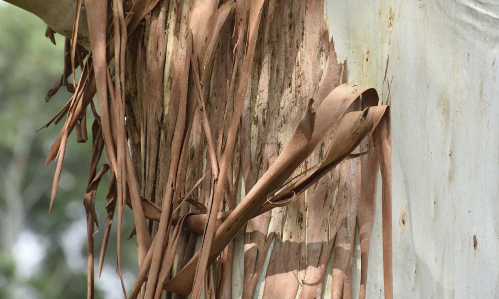
Caption: Eucalyptus bark extract has never been used to synthesise graphene sheets before. Courtesy: RMIT University
It’s been quite educational reading a June 24, 2019 news item on Nanowerk about deriving graphene from Eucalyptus bark (Note: Links have been removed),
Graphene is the thinnest and strongest material known to humans. It’s also flexible, transparent and conducts heat and electricity 10 times better than copper, making it ideal for anything from flexible nanoelectronics to better fuel cells.
The new approach by researchers from RMIT University (Australia) and the National Institute of Technology, Warangal (India), uses Eucalyptus bark extract and is cheaper and more sustainable than current synthesis methods (ACS Sustainable Chemistry & Engineering, “Novel and Highly Efficient Strategy for the Green Synthesis of Soluble Graphene by Aqueous Polyphenol Extracts of Eucalyptus Bark and Its Applications in High-Performance Supercapacitors”).
RMIT lead researcher, Distinguished Professor Suresh Bhargava, said the new method could reduce the cost of production from $USD100 per gram to a staggering $USD0.5 per gram.
“Eucalyptus bark extract has never been used to synthesise graphene sheets before and we are thrilled to find that it not only works, it’s in fact a superior method, both in terms of safety and overall cost,” said Bhargava.
“Our approach could bring down the cost of making graphene from around $USD100 per gram to just 50 cents, increasing it availability to industries globally and enabling the development of an array of vital new technologies.”
Graphene’s distinctive features make it a transformative material that could be used in the development of flexible electronics, more powerful computer chips and better solar panels, water filters and bio-sensors.
Professor Vishnu Shanker from the National Institute of Technology, Warangal, said the ‘green’ chemistry avoided the use of toxic reagents, potentially opening the door to the application of graphene not only for electronic devices but also biocompatible materials.
“Working collaboratively with RMIT’s Centre for Advanced Materials and Industrial Chemistry we’re harnessing the power of collective intelligence to make these discoveries,” he said.
A novel approach to graphene synthesis:
Chemical reduction is the most common method for synthesising graphene oxide as it allows for the production of graphene at a low cost in bulk quantities.
This method however relies on reducing agents that are dangerous to both people and the environment.
When tested in the application of a supercapacitor, the ‘green’ graphene produced using this method matched the quality and performance characteristics of traditionally-produced graphene without the toxic reagents.
Bhargava said the abundance of eucalyptus trees in Australia made it a cheap and accessible resource for producing graphene locally.
“Graphene is a remarkable material with great potential in many applications due to its chemical and physical properties and there’s a growing demand for economical and environmentally friendly large-scale production,” he said.
I’ve already written about October 2019 science and art/science events in Canada (see my Sept. 26, 2019 posting), but more event notices for Octoberhave come my way. These events are all art/science (or sciart as it’s sometimes called).
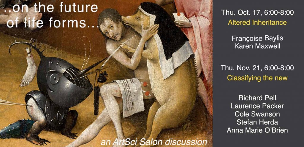
… on the future of life forms … a two-night (Oct./Nov.) discussion in Toronto, Canada
Here’s more from the ArtSci Salon’s October 3, 2019 announcement (received via email)
“…now they were perfecting a pigoon that could grow five or six kidneys at a time. Such a host animal could be reaped of its extra kidneys; then, rather than being destroyed, it could keep on living and grow more organs, much as a lobster could grow another claw to replace a missing one. That would be less wasteful, as it took a lot of food and care to grow a pigoon. A great deal of investment money had gone into OrganInc Farms…” (Margaret Atwood – Oryx & Crake 2003)
In Oryx and Crake Margaret Atwood describes a not-too-distant future where humans have perfected the art of fabricating and modifying a variety of creatures to improve and prolongue their own lives and wellbeing.
As Atwood has stated in various occasions, this is not science fiction.
It is in fact already happening. New forms of life appear not only as the product of lab fabrication or gene editing, but also as the result of toxic pollutants and climate change induced adaptation.
what to make of them?
how to cope with a world where extinction, adaptation and mutation risk to make traditional categories and taxonomies obsolete?
or not?
Join us to this two-parts series to discuss the ethics and implications of these transformations with artists, scientists and bioethicists.
this is a “double date”! Please, note the two dates please, RSVP here https://bit.ly/2AH1Pe8
Part 1 Thursday, October 17, 6:00-8:00 pm The Fields Institute for Research in Mathematical Sciences
Altered Inheritance: extinction, recreation or transformation? a dialogue and discussion on the implications of genome editing on humans and other organisms
with Francoise Baylis – Research Professor, Bioethicist, Dalhousie University
Karen Maxwell – Dept. of Biochemistry, Maxwell Lab, University of Toronto
emergent artists from OCADU [Ontario College of Art and Design University] and YorkU [York University, Toronto]
——————-
Part 2 Thursday, November 21, 6:00-8:00 pm The Fields Institute for Research in Mathematical Sciences
Classifying the new? why do we classify? what is it good for? what is the limit of taxonomy and classification in a transforming world?
with Richard Pell – Centre for PostNatural History, Pittsburgh, PA
Laurence Packer – Mellitologist, Professor of biology and environmental studies, York University
Stefan Herda – earth science artist
Cole Swanson – artist and educator (Art Foundation and Visual and Digital Arts, Humber college)
Anna Marie O’Brien – Frederickson, Rochman, and Sinton labs, University of Toronto
—————————
BIOS
Françoise Baylis is University Research Professor at Dalhousie University. She is a member of the Order of Canada and the Order of Nova Scotia, as well as a fellow of the Royal Society of Canada and of the Canadian Academy of Health Sciences. Baylis was one of the organizers of, and a key participant in, the 2015 International Summit on Human Gene Editing. She is a member of the WHO expert advisory committee on Developing Global Standards for Governance and Oversight of Human Genome Editing. Her new book “Altered Inheritance. CRISPR and the Ethics of Human Genome Editing” is published by Harvard University Press
Karen Maxwell is a research professor in the dept of biochemistry at the university of toronto, where she runs the Maxwell Lab. Among other topics, the lab’s three branches “Anti-CRISPR”, “Phage morons” and “Anti-Phage defences” study the interplay of phages with their bacterial hosts, with a focus on phage mediated bacterial virulence mechanisms and inhibitors of anti-phage bacterial defenses.
Richard Pell works at the intersections of science, engineering, and culture. He has worked in a variety of electronic media from documentary video to robotics to bioart to museum exhibition. He is the founder and director of the Center for PostNatural History (CPNH), an organization dedicated to the collection and exposition of life-forms that have been intentionally and heritably altered through domestication, selective breeding, tissue culture or genetic engineering. The CPNH operates a permanent museum in Pittsburgh, Pennsylvania, and produces traveling exhibitions that have appeared in science and art museums throughout Europe and the United States, including being the subject of a major exhibition at the Wellcome Collection in London.
Laurence Packer is a mellitologist, ie a scholar whose main subject of study is wild bees. his research primarily involves the systematics of the bee subfamily Xeromelissinae – an obscure, but fascinating group of bees, restricted to the New World south of central Mexico. he has also expended considerable energy leading the global campaign to barcode the bees of the world. his work is concerned with promulgating the importance of bees: for genetic reasons, it seems that bees are more extinction prone than are almost all other organisms
Stefan Herda‘s practice explores our troubling relationship to the natural world through drawing, sculpture and video. Inspired by the earth sciences, Herda’s work navigates the space between truth and fiction. His material and process-based investigations fuse elements of authenticity, façade, the natural and the manufactured together. He received his BAH from the University of Guelph in 2010. His work in both sculpture and video has been included in exhibitions nationally and has been featured by CBC Arts and Daily VICE. Recently, Stefan has held solo shows at Patel Projects (Toronto) and Wil Kucey Gallery (Toronto), participated in group shows such as Cultivars: Possible Worlds at InterAccess (Toronto) and was featured as one of 12 artists in the Cabinet Project at the University of Toronto
Cole Swanson is an artist and educator based in Toronto, Canada. He has exhibited in solo and group exhibitions across Canada and throughout international venues in North America, South America, Europe, and Asia. At the heart of recent work is a cross-disciplinary exploration of materials and their sociocultural and biological histories. Embedded within art media and commonplace resources are complex relations between nature and culture, humans and other agents, consumers and the consumed. Swanson has engaged in a broad material practice using sound, installation, painting, and sculpture to explore interspecies relationships.
Anna Marie O’Brien is a post doc in the Frederickson, Rochman, and Sinton labs at University of Toronto, working on duckweeds, microbes, urban contaminants, and phenotypes.her PhD work was at Davis, with thesis advisors Dr. Jeffrey Ross-Ibarra and Dr. Sharon Strauss. she also collaborated closely with Dr. Ruairidh Sawers at LANGEBIO-CINVESTAV in Guanajuato, Mexico.
The first highlighted speaker, Françoise Baylis, has been mentioned here twice before, in a May 17, 2019 posting (scroll down to the ‘Global plea for moratorium on heritable genome editing’ subheading) and in an April 26, 2019 posting (scroll down to the ‘Finally’ subheading, the second paragraph). Both postings touch on the topic of CRISPR (clustered regularly interspaced short palindromic repeats) and germline editing (genetic editing that will affect all of your descendents).
Cartooney in New Westminster (near Vancouver, Canada) starting October 18, 2019
I like physics but I love cartoons Stephen Hawking
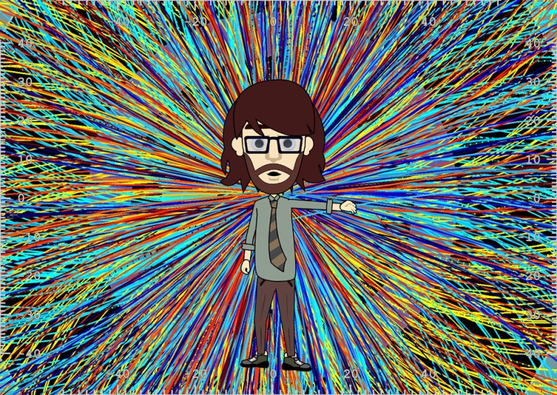
There you have it from one of the 20th/early 21st century’s most famous physicists. The quote is the opening line for the New Westminster (near Vancouver, Canada) New Media Gallery’s latest event webpage, Cartooney,
The impact of animated cartoons has been profound. In the early 20th century, we began exploiting the possibilities of the animated frame. The seven artists in this exhibition don’t create cartoons, they deconstruct those that already exist; from Looney Tunes, to The Simpsons to Charlie Brown. They exploit this potent material to reveal the inner and outer workings of our human world. The original cartoon is ever-present, haunting us with suggestive content.
The artists in this exhibition reframe our world. Here we are asked to consider the laws, systems and iconographies of the cartoon world while drawing parallels with our human world; physical laws, the laws of gravitation, matter + light, the physics of motion, and societal psychologies & behaviours. We are presented with fascinating catalogues and overlaying systems of symbolic language. The purposeful demolition of expectation in these works, mirrors the instabilities and dreams of modern life. They remind us that the pervasive medium of the cartoon can reflect and influence how we navigate the world. If there is a paradox here, it might be that dismantling a cartoon can throw open the doors of perception.
Artist Andy Holden becomes a cartoon avatar in Laws of Motion in a Cartoon Landscape.
Photograph By contributed [downloaded from https://www.newwestrecord.ca/entertainment/what-can-cartoons-tell-us-about-the-state-of-the-universe-find-out-in-new-west-1.23969740]
An Oct. 7, 2019 article in the New Westminster Record provides a few more details about the show,
The New Westminster New Media Gallery’s next exhibition is exploring the impact of animated cartoons.
Cartooney opens at the gallery on Friday, Oct. 18 and runs until Dec. 8 [2019], then again from Jan. 7 to Feb. 2 [2020].
Artist Kevin McCoy, one-half of the duo of Jennifer and Kevin McCoy, will be on hand for an artist talk on opening night, Friday, Oct. 18. The talk will run from 6:30 to 7:30 p.m., with a reception and open exhibition from 7:30 to 9 p.m.
…
Laws of Motion in a Cartoon Landscape, by Andy Holden (U.K.):
In his two-channel audiovisual installation, 57 minutes long, Holden becomes a cartoon avatar, giving both a lecture on cartoons and a cartoon lecture, describing how our world is best now understood as a cartoon. The project incorporates Greek philosophy, Stephen Hawking, critical theory, physics, art, the financial crisis and Donald Trump, while adapting 10 laws of cartoon physics to create a theory of the world and a prophetic glimpse of the world we live in.
…
CB-MMXVIII (I’ve been thinking of giving sleeping lessons), by Patten (U.K.):
In this multi-screen audiovisual installation, the artist duo Patten subjects Charlie Brown to all the digital stresses, distortions and manipulations available in 2018, testing his plasticity.
“Sampled texts from philosophy, science and critical theory criss-cross the screens and are linked with scrolling images related to the natural world, DNA, systems, multiples; all serving to influence our reading of the cartoon character and the texts,” says the release. The ambient soundtrack is a dramatically slowed down Linus and Lucy theme.
You can find the New Westminster New Media Gallery on the third floor at the Anvil Centre, 777 Columbia St. See www.newmediagallery.ca for more details.
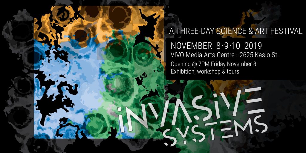
Collisions Festival: Invasive Systems in Vancouver, November 2019
Curiosity Collider, a Vancouver-based not-for-profit organization, will be hosting its inaugural art-science Collisions Festival: Invasive Systems at the VIVO Media Arts Centre from November 8 to 10, 2019. The festival features an art-science exhibition showcasing independent works and collaborative works by artist/scientist pairs, a hands-on DNA sonification workshop, an opening reception with performances, and guided discussions and tours.
Curated by Curiosity Collider’s Creative Director Char Hoyt, the theme of the festival focuses on the “invasive systems” that surround us – from technology and infections, to pollution and invasive species. “We want to create a space to explore the influence of the invasive aspects of our world on our inner and outer lives” said Char. “We will examine our observations from both scientific and artistic perspectives- are these influences beneficial, inevitable, or preventable?” Attendees can anticipate a deep dive into the delicate and complicated nature of how both living and inanimate things redefine our lives and environments – through visual art, multimedia installations, and interactive experiences.
“I am not a scientist and do not come from a family of scientists, but I have always appreciated knowing how things work, how things are connected and how things evolve – collaboration between art and science feel natural to me,” said Vancouver artist Dzee Lousie. “Both artists and scientists are curious, perform experiments and are driven by questions.” Dzee’s work Crossing, an interactive puzzle painting that examines how microbial colonies can impact our behaviours and processes in our body, is the result of a collaboration with UBC PhD candidate Linda Horianopoulos. “As scientists, we often want people to take notice of our work and engage with it. I think that art attracts people to do exactly that,” said Linda.
The sculptural work Invasion by Prince George artist Twyla Exner explores the remnants of technology. “My artworks propose hybrids of technological structures and living organisms. They take form as abandoned technologies that have sprouted with new life, clever artificialities that imitate nature, or biotechnological fixtures of the not-so-distant future,” Twyla shared. Like Dzee, she feels that artists and scientists share the sense of curiosity, experimentation, and creative problem solving. “Both art and science have the ability to tell stories and shape how people see and interpret the world around them.”
The festival is hosted in collaboration with the VIVO Media Arts Centre (2625 Kaslo Street, Vancouver, BC V5M 3G9). It will open on the evening of November 8th, with a reception and a live performance by local sound artist Edzi’u, during which her sculptural installation Moose are Life will be brought to life. On Saturday, artist Laara Cerman will co-host a DNA sonification workshop with scientist Scott Pownall. Their work Flora’s Song No. 1 in C Major – a hand-cranked music box that plays a tune created from the DNA of local invasive plants – will be on exhibit during the festival. The festival will also include tours by the curator at 3:30pm and guided discussions at 4pm on both Saturday and Sunday. Visit https://collisionsfestival2019.eventbrite.ca for festival tickets and http://bit.ly/collisionsfestival2019 for festival information.
Other participating artists and collaborating scientists include: Christian Dahlberg (Photography/ Painting / New Media Artist), Chris Dunnett (Multidisciplinary Artist), Garth Covernton (PhD Candidate, University of Victoria), Joanne Hastie (Artist / Mechanical Engineer), Kathryn Wadel (Interdisciplinary Mixed Media Artist), and Katrina Vera Wong (Artist / Writer / Editor).
Curiosity Collider and VIVO Media Arts Centre gratefully acknowledge the support of BC Arts Council, Canada Council for the Arts, City of Vancouver, Metro Vancouver Regional Cultural Project Grants Program, UBC Faculty of Science, and our printing sponsor Jukebox, for making Collisions Festival: Invasive Systems possible.
About Curiosity Collider Art-Science Foundation
Curiosity Collider Art-Science Foundation is a Vancouver based non-profit organization that is committed to providing opportunities for artists whose work expresses scientific concepts and scientists who collaborate with artists. We challenge the perception and experience of science in our culture, break down the walls between art and science, and engage our growing community to bringing life to the concepts that describe our world.
Are you curious about data sonification? Wondering how music theory and DNA sequences could converge to create a work of art and science? Join us to explore more!
In this DNA sonification workshop, participants will learn the process of DNA barcoding of invasive plant species, and how to sonify DNA sequences with basic music theory and MIDI freeware. Participants will also get hands-on experience in amplying specific genetic regions in plants through polymerase chain reaction (PCR), a step necessary in preparing samples for DNA barcoding.
This workshop will be led by artist Laara Cerman and scientist Scott Pownall, whose art-science collaborative work “Flora’s Song No. 1 in C Major” will be on exhibit during Collisions Festival: Invasive Systems. Laara and Scott will also share their process of working together, and how decisions were made to arrive at their collaborative work of art and science.
We acknowledge that Collisions Festival and its events take place on the traditional, ancestral, unceded territories of the xwməθkwəy̓əm (Musqueam), Skwxwú7mesh (Squamish), Stó:lō and Səl̓ílwətaʔ/Selilwitulh (Tsleil- Waututh) Nations. We are grateful for the opportunity to live and work on this land.
I asked the Curiosity Collider folks (@CCollider on Twitter) if you needed to bring any equipment or have any knowledge of music. The answer was: no, you don’t need to bring anything (unless you want to) and you don’t need to know about music.
Uncorked at Science World at TELUS World of Science in Vancouver on November 14, 2019
This is not a cheap night out. An October 10, 2019 article by Lindsay William-Ross for the Daily Hive website gives you reasons to go anyway (Note: Links have been removed),
A new wine-themed event will have Vancouverites swirling with nerdy glee. Uncorked: A Celebration of the Science of Wine is an evening of sipping and learning that will bring together world-renown winemakers, chefs, and science experts for an unforgettable event.
…
Participating wineries are:
Mission Hill Family Estate CedarCreek Estate Winery CheckMate Artisanal Winery Martin’s Lane Winery Road 13 Vineyards
The wines will be paired with bites from Chef Patrick Gayler from Mission Hill’s Terrace Restaurant and Chef Neil Taylor from CedarCreek’s new Home Block Restaurant.
Programming for the evening includes seminars on the science of blending wine, the science of aging wine, the role of technology at modern vineyards, and the science of soil and terroir.
…
Proceeds from Uncorked will support Science World’s On the Road program, which last year brought live science performances to 41,500 students throughout B.C. who otherwise might not have had a chance to visit TELUS World of Science.
Tickets are $89 and can be purchased here. You may also want to reserve some money for the silent auction. Don’t forget, it’s November 14, 2019 from 7 pm to 10 pm at Science World in Vancouver. You can find directions and a map here.
Years ago I was asked about carbon sequestration and nanotechnology and could not come up with any examples. At last I have something for the next time the question is asked. From a June 11, 2019 news item on ScienceDaily,
University of Colorado Boulder researchers have developed nanobio-hybrid organisms capable of using airborne carbon dioxide and nitrogen to produce a variety of plastics and fuels, a promising first step toward low-cost carbon sequestration and eco-friendly manufacturing for chemicals.
By using light-activated quantum dots to fire particular enzymes within microbial cells, the researchers were able to create “living factories” that eat harmful CO2 and convert it into useful products such as biodegradable plastic, gasoline, ammonia and biodiesel.
“The innovation is a testament to the power of biochemical processes,” said Prashant Nagpal, lead author of the research and an assistant professor in CU Boulder’s Department of Chemical and Biological Engineering. “We’re looking at a technique that could improve CO2 capture to combat climate change and one day even potentially replace carbon-intensive manufacturing for plastics and fuels.”
The project began in 2013, when Nagpal and his colleagues began exploring the broad potential of nanoscopic quantum dots, which are tiny semiconductors similar to those used in television sets. Quantum dots can be injected into cells passively and are designed to attach and self-assemble to desired enzymes and then activate these enzymes on command using specific wavelengths of light.
Nagpal wanted to see if quantum dots could act as a spark plug to fire particular enzymes within microbial cells that have the means to convert airborne CO2 and nitrogen, but do not do so naturally due to a lack of photosynthesis.
By diffusing the specially-tailored dots into the cells of common microbial species found in soil, Nagpal and his colleagues bridged the gap. Now, exposure to even small amounts of indirect sunlight would activate the microbes’ CO2 appetite, without a need for any source of energy or food to carry out the energy-intensive biochemical conversions.
“Each cell is making millions of these chemicals and we showed they could exceed their natural yield by close to 200 percent,” Nagpal said.
The microbes, which lie dormant in water, release their resulting product to the surface, where it can be skimmed off and harvested for manufacturing. Different combinations of dots and light produce different products: Green wavelengths cause the bacteria to consume nitrogen and produce ammonia while redder wavelengths make the microbes feast on CO2 to produce plastic instead.
The process also shows promising signs of being able to operate at scale. The study found that even when the microbial factories were activated consistently for hours at a time, they showed few signs of exhaustion or depletion, indicating that the cells can regenerate and thus limit the need for rotation.
“We were very surprised that it worked as elegantly as it did,” Nagpal said. “We’re just getting started with the synthetic applications.”
The ideal futuristic scenario, Nagpal said, would be to have single-family homes and businesses pipe their CO2 emissions directly to a nearby holding pond, where microbes would convert them to a bioplastic. The owners would be able to sell the resulting product for a small profit while essentially offsetting their own carbon footprint.
“Even if the margins are low and it can’t compete with petrochemicals on a pure cost basis, there is still societal benefit to doing this,” Nagpal said. “If we could convert even a small fraction of local ditch ponds, it would have a sizeable impact on the carbon output of towns. It wouldn’t be asking much for people to implement. Many already make beer at home, for example, and this is no more complicated.”
The focus now, he said, will shift to optimizing the conversion process and bringing on new undergraduate students. Nagpal is looking to convert the project into an undergraduate lab experiment in the fall semester, funded by a CU Boulder Engineering Excellence Fund grant. Nagpal credits his current students with sticking with the project over the course of many years.
“It has been a long journey and their work has been invaluable,” he said. “I think these results show that it was worth it.”
I do like to keep up with nanocellulose doings, especially when there’s some Canadian involvement, and an October 8, 2019 news item on Nanowerk alerted me to a newish application for the product,
Physiological parameters in our blood can be determined without painful punctures. Empa researchers are currently working with a Canadian team to develop flexible, biocompatible nanocellulose sensors that can be attached to the skin. The 3D-printed analytic chips made of renewable raw materials will even be biodegradable in future.
The idea of measuring parameters that are relevant for our health via the skin has already taken hold in medical diagnostics. Diabetics, for example, can painlessly determine their blood sugar level with a sensor instead of having to prick their fingers.
Nanocellulose is an inexpensive, renewable raw material, which can be obtained in form of crystals and fibers, for example from wood. However, the original appearance of a tree no longer has anything to do with the gelatinous substance, which can consist of cellulose nanocrystals and cellulose nanofibers. Other sources of the material are bacteria, algae or residues from agricultural production. Thus, nanocellulose is not only relatively easy and sustainable to obtain. Its mechanical properties also make the “super pudding” an interesting product. For instance, new composite materials based on nanocellulose can be developed that could be used as surface coatings, transparent packaging films or even to produce everyday objects like beverage bottles.
Researchers at Empa’s Cellulose & Wood Materials lab and Woo Soo Kim from the Simon Fraser University [SFU] in Burnaby, Canada, are also focusing on another feature of nanocellulose: biocompatibility. Since the material is obtained from natural resources, it is particularly suitable for biomedical research.
With the aim of producing biocompatible sensors that can measure important metabolic values, the researchers used nanocellulose as an “ink” in 3D printing processes. To make the sensors electrically conductive, the ink was mixed with silver nanowires. The researchers determined the exact ratio of nanocellulose and silver threads so that a three-dimensional network could form.
Just like spaghetti – only a wee bit smaller
It turned out that cellulose nanofibers are better suited than cellulose nanocrystals to produce a cross-linked matrix with the tiny silver wires. “Cellulose nanofibers are flexible similar to cooked spaghetti, but with a diameter of only about 20 nanometers and a length of just a few micrometers,” explains Empa researcher Gilberto Siqueira.
The team finally succeeded in developing sensors that measure medically relevant metabolic parameters such as the concentration of calcium, potassium and ammonium ions. The electrochemical skin sensor sends its results wirelessly to a computer for further data processing. The tiny biochemistry lab on the skin is only half a millimeter thin.
While the tiny biochemistry lab on the skin – which is only half a millimeter thin – is capable of determining ion concentrations specifically and reliably, the researchers are already working on an updated version. “In the future, we want to replace the silver [nano] particles with another conductive material, for example on the basis of carbon compounds,” Siqueira explains. This would make the medical nanocellulose sensor not only biocompatible, but also completely biodegradable.
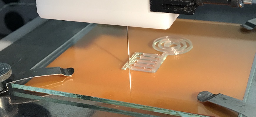
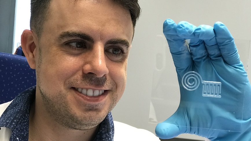
I like the images from Empa better than the ones from SFU,
Using a 3D printer, the nanocellulose “ink” is applied to a carrier plate. Silver particles provide the electrical conductivity of the material. Image: EmpaEmpa researcher Gilberto Siqueira demonstrates the newly printed nanocellulose circuit. After a subsequent drying, the material can be further processed. Image: Empa
SFU produced a news release about this work back in February 2019. Again, I prefer what the Swiss have done because they’re explaining/communicating the science, as well as , communicating benefits. From a February 13, 2019 SFU news release (Note: Links have been removed),
Simon Fraser University and Swiss researchers are developing an eco-friendly, 3D printable solution for producing wireless Internet-of-Things (IoT) sensors that can be used and disposed of without contaminating the environment. Their research has been published as the cover story in the February issue of the journal Advanced Electronic Materials.
SFU professor Woo Soo Kim is leading the research team’s discovery, which uses a wood-derived cellulose material to replace the plastics and polymeric materials currently used in electronics.
Additionally, 3D printing can give flexibility to add or embed functions onto 3D shapes or textiles, creating greater functionality.
“Our eco-friendly, 3D-printed cellulose sensors can wirelessly transmit data during their life, and then can be disposed without concern of environmental contamination,” says Kim, a professor in the School of Mechatronic Systems Engineering. The SFU research is being carried out at PowerTech Labs in Surrey, which houses several state-of-the-art 3D printers used to advance the research.
“This development will help to advance green electronics. For example, the waste from printed circuit boards is a hazardous source of contamination to the environment. If we are able to change the plastics in PCB to cellulose composite materials, recycling of metal components on the board could be collected in a much easier way.”
Kim’s research program spans two international collaborative projects, including the latest focusing on the eco-friendly cellulose material-based chemical sensors with collaborators from the Swiss Federal Laboratories for Materials Science.
He is also collaborating with a team of South Korean researchers from the Daegu Gyeongbuk Institute of Science and Technology’s (DGIST)’s department of Robotics Engineering, and PROTEM Co Inc, a technology-based company, for the development of printable conductive ink materials.
In this second project, researchers have developed a new breakthrough in the embossing process technology, one that can freely imprint fine circuit patterns on flexible polymer substrate, a necessary component of electronic products.
Embossing technology is applied for the mass imprinting of precise patterns at a low unit cost. However, Kim says it can only imprint circuit patterns that are imprinted beforehand on the pattern stamp, and the entire, costly stamp must be changed to put in different patterns.
The team succeeded in developing a precise location control system that can imprint patterns directly resulting in a new process technology. The result will have widespread implications for use in semiconductor processes, wearable devices and the display industry.
This paper was made available online back in December 2018 and then published in print in February 2019. As to why there’d be such large gaps between the paper’s publication dates and the two institution’s news/press releases, it’s a mystery to me. In any event, here’s a link to and a citation for the paper,
3D Printed Disposable Wireless Ion Sensors with Biocompatible Cellulose Composites by Taeil Kim, Chao Bao, Michael Hausmann, Gilberto Siqueira, Tanja Zimmermann, Woo Soo Kim. Advanced Electronic Materials DOI: https://doi.org/10.1002/aelm.201970007 First published online December 19, 2018. First published in print: 08 February 2019 (Adv. Electron. Mater. 2/2109) Volume 5, Issue 2 February 2019 1970007
CRISPR (clustered regularly interspaced short palindromic repeats) gene editing is usually paired with a virus (9, 12a, etc.) but this time scientists are using a gold nanoparticle. From a May 27, 2019 news item on Nanowerk (Note: Links have been removed),
Scientists at Fred Hutchinson Cancer Research Center took a step toward making gene therapy more practical by simplifying the way gene-editing instructions are delivered to cells. Using a gold nanoparticle instead of an inactivated virus, they safely delivered gene-editing tools in lab models of HIV and inherited blood disorders, as reported in Nature Materials (“Targeted homology-directed repair in blood stem and progenitor cells with CRISPR nanoformulations”).
It’s the first time that a gold nanoparticle loaded with CRISPR has been used to edit genes in a rare but powerful subset of blood stem cells, the source of all blood cells. The CRISPR-carrying gold nanoparticle led to successful gene editing in blood stem cells with no toxic effects.
“As gene therapies make their way through clinical trials and become available to patients, we need a more practical approach,” said senior author Dr. Jennifer Adair, an assistant member of the Clinical Research Division at Fred Hutch, adding that current methods of performing gene therapy are inaccessible to millions of people around the world. “I wanted to find something simpler, something that would passively deliver gene editing to blood stem cells.”
While CRISPR has made it faster and easier to precisely deliver genetic modifications to the genome, it still has challenges. Getting cells to accept CRISPR gene-editing tools involves a small electric shock that can damage and even kill the cells. And if precise gene edits are required, then additional molecules must be engineered to deliver them –adding cost and time.
Gold nanoparticles are a promising alternative because the surface of these tiny spheres (around 1 billionth the size of a grain of table salt) allows other molecules to easily stick to them and stay adhered.
“We engineered the gold nanoparticles to quickly cross the cell membrane, dodge cell organelles that seek to destroy them and go right to the cell nucleus to edit genes,” said Dr. Reza Shahbazi, a Fred Hutch postdoctoral researcher who has worked with gold nanoparticles for drug and gene delivery for seven years.
Shahbazi made the gold particles from laboratory-grade gold that is purified and comes as a liquid in a small lab bottle. He mixed the purified gold into a solution that causes the individual gold ions to form tiny particles, which the researchers then measured for size.
They found that a particular size – 19 nanometers wide – was the best for being big and sticky enough to add gene-editing materials to the surface of the particles, while still being small enough for cells to absorb them.
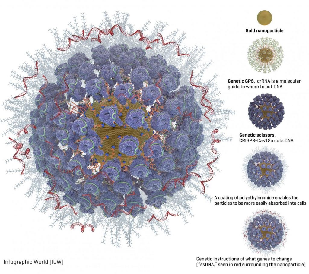
Packed onto the gold particles, the Fred Hutch team added these gene-editing components (diagram available [see below]):
A type of molecular guide called crRNA acts as a genetic GPS to show the CRISPR complex where in the genome to make the cut.
CRISPR nuclease protein, often called “genetic scissors,” makes the cut in the DNA. The CRISPR nuclease protein most often used is Cas9. But the Fred Hutch researchers also studied Cas12a (formerly called Cpf1) because Cas12a makes a staggered cut in DNA. The researchers hoped this would allow the cells to more efficiently repair the cut and while so doing embed the new genetic instructions into the cell. Another advantage of Cas12a over Cas9 is that it only requires one molecular guide, which is important because of space constraints on the nanoparticles. Cas9 requires two molecular guides.
Instructions for what genetic changes to make (“ssDNA”). The Fred Hutch team chose two inherited genetic changes that bestow protection from disease: CCR5, which protects against HIV, and gamma hemoglobin, which protects against blood disorders such as sickle cell disease and thalassemia.
A coating of a polyethylenimine swarms the surface of the particles to give them a more positive charge, which enables them to more readily be absorbed into cells. This is an improvement over another method of getting cells to take up gene editing tools, called electroporation, which involves lightly shocking the cells to get them to open and allow the genetic instructions to enter.
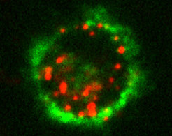
Then the researchers isolated blood stem cells with a protein marker on their surface called CD34. These CD34-positive cells contain the blood-making progenitor cells that give rise to the entire blood and immune system.
“These cells replenish blood in the body every day, making them a good candidate for one-time gene therapy because it will last a lifetime as the cells replace themselves,” Adair said.
Observing human blood stem cells in a lab dish, the researchers found that their fully loaded gold nanoparticles were taken up naturally by cells within six hours of being added and within 24 to 48 hours they could see gene editing happening. They observed that the Cas12a CRISPR protein partner was better at delivering very precise genetic edits to the cells than the more commonly used cas9 protein partner.
The gene-editing effect reached a peak eight weeks after the researchers injected the cells into mouse models; 22 weeks after injection the edited cells were still there. The Fred Hutch researchers also found edited cells in the bone marrow, spleen and thymus of the mouse models, a sign that the dividing blood cells in those organs could carry on the treatment without the mice having to be treated again.
“We believe we have a good candidate for two diseases — HIV and hemoglobinopathies — though we are also evaluating other disease targets where small genetic changes can have a big impact, as well as ways to make bigger genetic changes,” Adair said. “The next step is to increase how much gene editing happens in each cell, which is definitely doable. That will make it closer to being an effective therapy.”
In the study, the researchers report 10 to 20 percent of cells took on the gene edits, which is a promising start, but the researchers would like to aim for 50% or more of the cells being edited, which they believe will have a good chance of combatting these diseases.
###
Adair and Shahbazi are looking for commercial partners to develop the technology into therapies for people. They hope to begin clinical trials within a few years.
Here’s the diagram of a gold nanoparticle loaded with CRISPR,
Caption: Graphic of a fully loaded gold nanoparticle with CRISPR and other gene editing tools. Credit: Image courtesy of the Adair lab at Fred Hutch.
A June 3, 2019 news item on Nanowerk describes an inexpensive way to safely handle carbon nanotubes (CNTs), Note: A link has been removed,
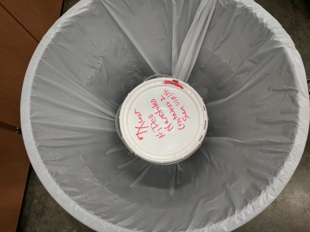
With a little practice, it doesn’t take much more than 10 minutes, a couple of bags and a big bucket to keep nanomaterials in their place.
The Rice University lab of chemist Andrew Barron works with bulk carbon nanotubes on a variety of projects. Years ago, members of the lab became concerned that nanotubes could escape into the air, and developed a cheap and clean method to keep them contained as they were transferred from large containers into jars for experimental use.
More recently Barron himself became concerned that too few labs around the world were employing best practices to handle nanomaterials. He decided to share what his Rice team had learned.
“There was a series of studies that said if you’re going to handle nanotubes, you really need to use safety protocols,” Barron said. “Then I saw a study that said many labs didn’t use any form of hood or containment system. In the U.S., it was really bad, and in Asia it was even worse. But there are a significant number of labs scaling up to use these materials at the kilogram scale without taking the proper precautions.”
The lab’s inexpensive method is detailed in an open-access paper in the Springer Nature journal SN Applied Sciences (“The safe handling of bulk low-density nanomaterials”).
…
Here’s a bag and a bucket,
Caption: A plastic bucket and a plastic bag contain a 5-gallon supply of carbon nanotubes in a lab at Rice University, the beginning of the process to safely transfer the nanotubes for experimental use. The Rice lab published its technique in SN Applied Sciences. Credit: Barron Research Group/Rice University
In bulk form, carbon nanotubes are fluffy and disperse easily if disturbed. The Rice lab typically stores the tubes in 5-gallon plastic buckets, and simply opening the lid is enough to send them flying because of their low density.
Varun Shenoy Gangoli, a research scientist in Barron’s lab, and Pavan Raja, a scientist with Rice’s Nanotechnology-Enabled Water Treatment center, developed for their own use a method that involves protecting the worker and sequestering loose tubes when removing smaller amounts of the material for use in experiments.
Full details are available in the paper, but the precautions include making sure workers are properly attired with long pants, long sleeves, lab coats, full goggles and face masks, along with two pairs of gloves duct-taped to the lab coat sleeves. The improvised glove bag involves a 25-gallon trash bin with a plastic bag taped to the rim. The unopened storage container is placed inside, and then the bin is covered with another transparent trash bag, with small holes cut in the top for access.
After transferring the nanotubes, acetone wipes are used to clean the gloves and more acetone is sprayed inside the barrel so settling nanotubes would stick to the surfaces. These can be recovered and returned to the storage container.
Barron said it took lab members time to learn to use the protocol efficiently, “but now they can get their samples in 5 to 10 minutes.” He’s sure other labs can and will enhance the technique for their own circumstances. He noted a poster presented at the Ninth Guadalupe Workshop on the proper handling of carbon nanotubes earned recognition and discussion among the world’s premier researchers in the field, noting the importance of the work for agencies in general.
“When we decided to write about this, we were originally just going to put it on the web and hope somebody would read it occasionally,” Barron said. “We couldn’t imagine who would publish it, but we heard that an editor at Springer Nature was really keen to have published articles like this.
“I think this is something people will use,” he said. “There’s nothing outrageous but it helps everybody, from high schools and colleges that are starting to use nanoparticles for experiments to small companies. That was the goal: Let’s provide a process that doesn’t cost thousands of dollars to install and allows you to transfer nanomaterials safely and on a large scale. Finally, publish said work in an open-access journal to maximize the reach across the globe.”
Here’s a link to and a citation for the paper,
The safe handling of bulk low-density nanomaterials by Varun Shenoy Gangoli, Pavan M. V. Raja, Gibran Liezer Esquenazi, Andrew R. Barron. SN Applied Sciences June 2019, 1:644 DOI: https://doi.org/10.1007/s42452-019-0647-5 First Online 25 May 2019
A May 28, 2019 news item on Nanowerk announced research targeting Langerham cells and the immune system (Note: A link has been removed),
Researchers at the Max Planck Institute of Colloids and Interfaces in Potsdam developed targeted nanoparticles that are taken up by certain immune cells of the human skin (ACS Central Science, “A specific, glycomimetic Langerin ligand for human Langerhans cell targeting”). These so-called Langerhans cells coordinate the immune response and alert the body when pathogens or tumors occur.
This new nanoparticle technology platform enables targeted drug delivery of vaccines or pharmaceuticals to Langerhans cells, triggering a controlled immune response to naturally eradicate the pathogen or tumor.
The skin is a particularly attractive place for the application of many drugs that affect the immune system, as the appropriate target cells lie directly beneath the skin. These Langerhans cells are able to elicit an immune reaction in the entire body of the patient after local application of an active substance.
Langerhans Cells – Experts of pathogen defense
To develop a targeted drug delivery system, which guides drugs directly to Langerhans cells, one can make use of their natural function: as professional, antigen-presenting cells they detect pathogens, internalize them and present components of these pathogens to effector cells of the immune system (T cells). For detection and uptake, Langerhans cells use receptors on their surface that search the environment for microbes. They especially recognize pathogens by the unique coating of sugar structures on their surface. Langerin, a protein of the C-type lectins family, is such a receptor on Langerhans cells that can detect viruses and bacteria. The specific expression of Langerin on Langerhans cells allows a targeted drug delivery encapsulated in nanoparticleswhile minimizing the side effects.
The research team of Dr. Christoph Rademacher at the Max Planck Institute of Colloids and Interfaces has now been able to exploit the knowledge of the underlying detection mechanisms with atomic resolution: “Based on our insight how immune cells recognize sugars, we developed a synthetic, sugar-like substance that enables nanoparticles to specifically bind to Langerhans cells”, says Dr. Christoph Rademacher. In collaboration with a scientific team from the Laboratory for Langerhans Cell Research of the Medical University of Innsbruck, nanoparticles have been developed that can be incorporated into Langerhans cells of the human skin through this interaction. The researchers thus lay the foundation for further developments, for example to deliver vaccines directly through the skin to the immune cells. “Imagine avoiding needles for vaccination in the future or directly activating the body’s immune system against infections and maybe even cancer”, adds Dr. Christoph Rademacher. Langerhans cells are responsible for activating the immune system systemically. Based on these findings, it may be possible in the future to develop novel vaccines against infections or immunotherapies for the treatment of cancer or autoimmune diseases.
The starting points for this work were the pioneering contributions from Ralph M. Steinman (Nobel Prize 2011) and other scientists who showed the potential of dendritic cells. Langerhans cells are one subset of these cells and are able to trigger an immune response. These findings were subsequently refined for use in cancer therapy. It has been shown that an immune response can be achieved via artificially introduced antigens. Later work confirmed these findings and also demonstrated that human Langerhans cells are also able to activate the immune system, which is particularly interesting for skin vaccination. Targeted delivery of immunomodulators to Langerhans cells would thus be desirable. However, this is often hindered or even prevented by the complex environment of the skin, especially by competing phagocytes in this tissue, such as macrophages. Consequently, pharmaceuticals not taken up by the Langerhans cells, but internalized into bystander cells may lead to unwanted side effects.
Recognition through synthetic sugars
Based on insights on the interaction between Langerin and its natural sugar ligands Christoph Rademacher and his team developed a synthetic ligand, which binds specifically to the receptor on Langerhans cells. For this purpose, synthetic sugars were produced in the laboratory and their interactions with the receptor were examined by nuclear magnetic resonance spectroscopy. With this method the researchers were able to determine which atoms of the ligand interact with which parts of the receptor. By using this structure-based approach they found out that a compound can be anchored and tested on these nanoparticles. These particles are liposomes, which have been used for many years in the clinic in the absence of such targeting ligands as a carrier for various drugs. The difference with existing systems is that the sugar-like ligand now allows specific binding to Langerhans cells. The investigations on these immune cells were carried out in collaboration with the research group of Assoz. Prof. Patrizia Stoitzner at the Langerhans Cell Research Laboratory of the Medical University of Innsbruck. Together they could show that the specific uptake of liposomes is possible even in the complex environment of human skin. The scientists used different methods such as flow cytometry and confocal microscopy for their findings.
These liposomal particles may now provide a common platform for researchers at the MPI of Colloids and Interfaces to work on the development of novel vaccines in the future.